Comparative Evaluation of Fracture Resistance of Root Obturated with Resilon and Gutta-Percha Using Two Different Techniques: An in Vitro Study
Vijayakumar L. Shiraguppi1, Hema Bindera Shekar2, Chandu Giriyapur Shivalingappa3, Niranjan Desai4, Antriksh Azad5
1 Reader, Department of Conservative Dentistry & Endodontics, M A Rangoonwala College of Dental Sciences and Research Centre, Pune, Maharashtra, India.
2 Reader, Department of Conservative Dentistry & Endodontics, Rishiraj College of Dental Sciences & Research Centre, Bhopal, Madhyapradesh, India.
3 Professor, Department of Prosthodontics, Rishiraj College of Dental Sciences & Research Centre, Bhopal, Madhyapradesh, India.
4 Reader, Department of Conservative Dentistry & Endodontics. Sinhgad Dental College and Hospital, STES Campus, Vadgaon BK Pune 41, Maharashtra, India.
5 Senior Lecturer, Department of Conservative Dentistry & Endodontics, Rishiraj College of Dental Sciences& Research centre, Bhopal, Madhyapradesh, India.
NAME, ADDRESS, E-MAIL ID OF THE CORRESPONDING AUTHOR: Dr. Hema Bindera Shekar, Reader, Department of Conservative Dentistry & Endodontics, Rishiraj College of Dental Sciences & Research Centre, Bhopal, Madhyapradesh, India.
E-mail: drhemabs@gmail.com.
Introduction:
Present study evaluated the fracture resistance of endodontically treated teeth filled with Gutta percha and a new resin based obturating material (Resilon).
Materials and Methods:
A total of 150 freshly extracted Mandibular premolar with fully formed apices were selected and decoronated at cemento-enamel junction (CEJ). Teeth were divided into Group A and Group B of 75 teeth each. In Group A canals were prepared up to # no 40 K file and Group B up to #no 80 K file. Both the groups were sub divided into five group of 15 teeth each as control group (unfilled canals), lateral condensation with Gutta-percha using AH 26 sealer, vertical condensation with Gutta-percha using AH 26 sealer, lateral condensation with Resilon using resilon sealer, vertical condensation with Resilon using resilon sealer. Each specimen was subjected to compressive load using Universal testing machine. The force required to fracture was recorded and data were analysed by ANOVA, Duncan’s test and student T test.
Result:
The result showed that there is statistically significant difference among experimental groups (p < 0.05). The groups with the Resilon material displayed higher mean fracture loads than the Gutta percha groups. No statistically significant differences were observed between different preparation techniques.
Conclusion:
Obturating the canals with the new resin-based obturation material increases the in vitro fracture resistance of endodontically treated teeth when compared with standard Gutta percha techniques.
Endodontic treatment, Lateral condensation, Obturation
Introduction
The fracture susceptibility of endodontically treated teeth are more common than the vital teeth. The reasons most often reported are the water loss [1], loss of collagen cross-linking [2], excessive pressure during obturation [3] and the removal of tooth structure during endodontic treatment [4]. The amount of remaining sound tooth structure and methods of canal preparation directly contributes to the strength of endodontically treated tooth. From a fracture mechanics point of view, the presence of structural defects, cracks, or canal irregularities are likely to play a major role in determining fracture strength [5], because an applied stress may be exponentially amplified at the tip of those defects [6]. Potential influencing factors for fracture susceptibility involves the dentin thickness, radius of canal curvature and external root morphology [7].
Obturation strains [8–10] and post placements [11] have been investigated as major cause of vertical root fracture. The excessive force during lateral compaction of the Gutta-percha caused 84% of vertical root fracture [12]. On the contrary use of NiTi spreader may minimize the potentials for vertical root fracture in curved canals during lateral compaction [13].
The use of Gutta-percha and root canal sealers for obturating root canal has remained the standard of care in endodontics, despite their inability to routinely achieve an impervious seal along the dentinal wall of the root canal [14,15]. Both total-etch and self-etch adhesives techniques are found experimentally to reduce apical and coronal leakage as it seals intraradicular dentin just before the obturation of root canals with gutta-percha [16–20]. However, these techniques have limitation due to the lack of copolymerization between the methacrylate-based dentin adhesives, the epoxy resin or zinc oxide eugenol-based root canal sealer, and gutta-percha [21].
Resin-based dental materials have been proposed as a means to reinforce an endodontically treated tooth with the use of adhesive sealers in the root canal system [22]. However, bonding agents and resins studied to date as root filling materials had problems in working properties, radiopacity and lack of re-treatability when used for endodontic purposes [23,24]. In recent years, an endodontic obturation material Resilon based on polyester chemistry which contains bioactive and radiopaque fillers has been developed and tested. Its performance and handling are similar to Gutta-percha. In addition, when used in conjunction with a resin-based sealant or bonding agent it forms a monoblock within the canals that bonds to the dentinal walls and strengthen the walls against fracture [25].
The purpose of the study was–
To compare the fracture resistance of endodontically treated tooth filled with Gutta-percha and Resilon obturating material.
To compare the lateral and vertical obturating technique in root fracture filled with Gutta-percha and Resilon obturating material.
To compare the effective reinforcing ability of Gutta percha and Resilon obturates material on different canal preparation.
Materials and Methods
One hundred fifty freshly extracted human mandibular premolars with fully formed apices, free of apical root resorption and caries were collected and were stored in 10% of formalin. The collected samples were cut at the CEJ with diamond disk. The working length was established with 10 no file, 1 mm short to the apex. Then 75 teeth were enlarged to the size 40 no and remaining 75 teeth were enlarged to the size 80 no. crown down preparation technique was carried out in all the teeth. Preparations were irrigated between uses of each succeeding file with 5.25% sodium hypochlorite (Novo Dental Products Ptv. Ltd. India). After preparation the entire specimens were flushed with the 17% EDTA (Prime Dental Product, India), to remove smear layer and canal were dried with paper points.
Teeth were divided in to two groups of 75 each.
Group A (canal preparation up to 40 no size)
Group B (canal preparation up to 80 no size)
Group A is sub divided as follows,
Group A:
A-0 ------- Control group. This group received no obturation; the canal opening was sealed with a temporary filling material (Cavit 3M ESPE, St Paul, MN, USA).
A-1 -------Lateral condensation with size 40 Gutta percha using AH 26 sealer. (Dentsply Pvt. Ltd, Delhi, India).
A-2 ------ vertical condensation with size 40 Gutta percha using AH 26 sealer.
A-3 ------ Lateral condensation with size 40 Resilon using Resilon sealer. (Pentron clinical Technologies, LLC Wallingford CT. USA)
A-4 ------ Vertical condensation with size 40 Resilon using Resilon sealer.
Group B is sub divided as follows,
B-0 ----- Control group. Canal without any obturation. Canals were sealed with cavit.
B-1 ----- Lateral condensation with size 80 Gutta percha using AH 26 sealer.
B-2 ----- vertical condensation with size 80 Gutta percha using AH 26 sealer.
B-3 ----- Lateral condensation with size 80 Resilon using Resilon sealer.
B-4 ----- Vertical condensation with size 80 Resilon using Resilon sealer.
Preparation for Mechanical Testing
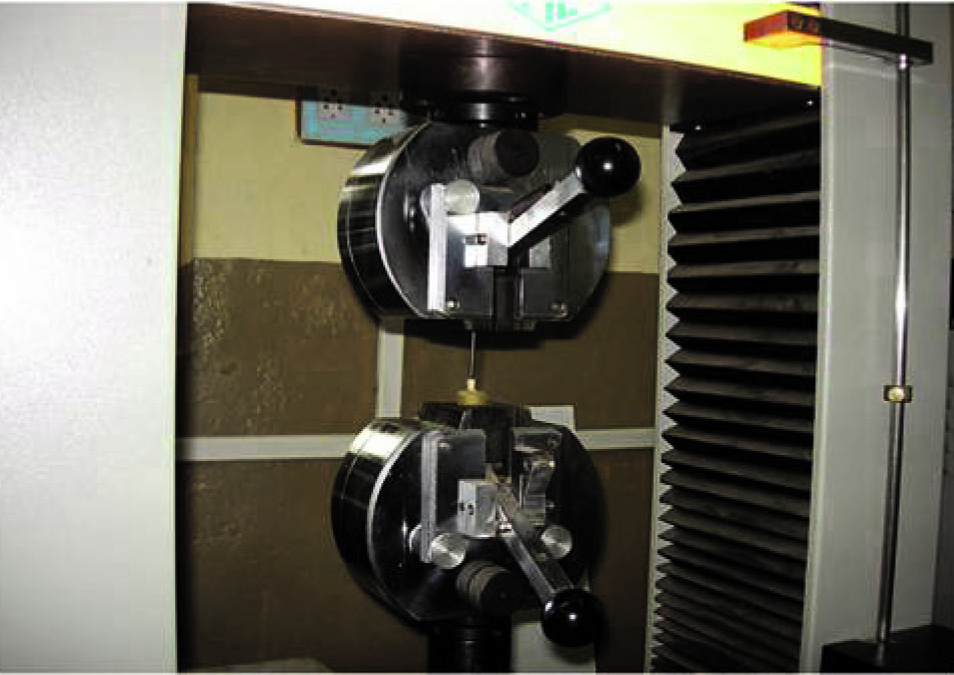
After two weeks, the root specimens were prepared for the mechanical testing. The apical root ends were embedded individually in phenolic rings with acrylic resin, leaving 9mm of each root exposed. A carbide bur was used to remove temporary material and to shape the root canal access to accept the loading fixture. The acrylic blocks were mounted with the vertically aligned roots in the Universal testing machine one at a time. The specimen were mounted and aligned to the loading fixture with a spherical tip of radius (r=2mm) on the centre of the canal opening [Table/Fig-1,2]. Each specimen was subjected to compressive load at a crosshead speed of 1mm/min until the fracture of root occurred [Table/Fig-3]. The force when fracture occurred was recorded in Newtons and data from all experimental groups were collected and statistically analysed using ANOVA & Duncan’s multiple comparison test. Intergroup comparison was done by student’s T-test.
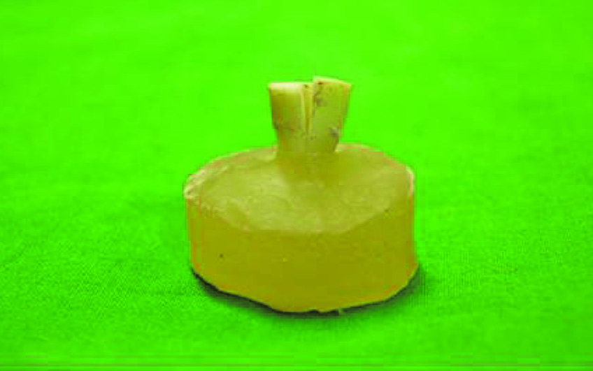
Close up view of the specimen
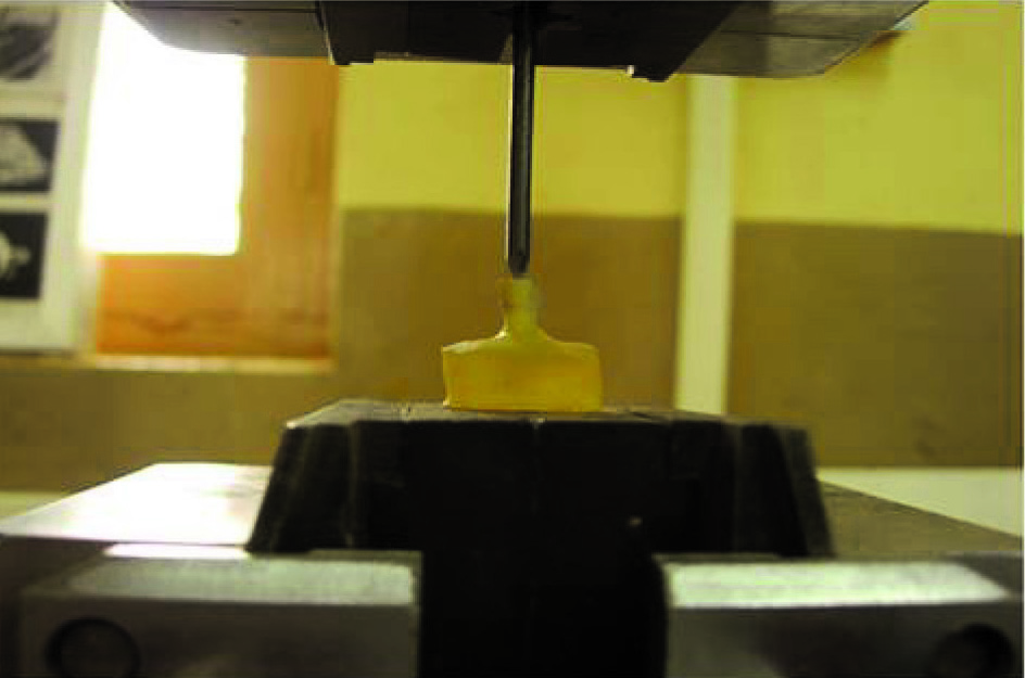
Tooth fracture under load
Result
Fracture resistance of tooth in groups: Data on the applied force were subjected to statistical evaluation. The comparison of group A with respect to applied force by ANOVA & Duncan’s multiple comparison test presented in [Table/Fig-4,5]. Pair wise comparison of five groups by student’s unpaired t-test is given in [Table/Fig-6]. Similar tests were done for Group B is shown in [Table/Fig-7,8,9]. Comparison of two groups (A & B) by student’s t-test is shown in [Table/Fig-10].
Comparison of five groups (A0, A1, A2, A3, and A4) instrumented with 40 k file with respect to applied force by one-way analysis of variance (ANOVA)
| SV | DF | SS | MSS | f-value | p-value | Signi. |
|---|
| Between Groups | 4 | 242.90 | 60.7251 | 143.6443 | 0.0001 | S |
| Within groups | 60 | 25.36 | 0.4227 | |
| Total | 64 | 268.27 | |
Pair wise comparison of five groups instrumented with 40 k file by Duncan’s multiple comparison test
| Groups | A0 | A1 | A2 | A3 | A4 |
|---|
| Means | 36.7380 | 31.8220 | 31.5850 | 32.1920 | 32.1540 |
| A0 | - | | | | |
| A1 | 0.0001* | - | | | |
| A2 | 0.0000* | 0.3567 | - | | |
| A3 | 0.0001* | 0.1756 | 0.0315* | - | |
| A4 | 0.0001* | 0.1976 | 0.0377* | 0.8807 | - |
*indicates significant at 5 level of significance (p<0.05)
Pair wise comparison of five groups instrumented with 40 k file by student’s unpaired t-test
| Group | Mean | SD | t-value | p-value | Signi. |
|---|
| A0 | 36.7385 | 1.0096 | 15.0430 | 0.0001 | S |
| A1 | 31.8215 | 0.6080 |
| A0 | 36.7385 | 1.0096 | 17.2239 | 0.0001 | S |
| A2 | 31.5846 | 0.3805 |
| A0 | 36.7385 | 1.0096 | 13.7715 | 0.0001 | S |
| A3 | 32.1923 | 0.6304 |
| A0 | 36.7385 | 1.0096 | 15.0777 | 0.0001 | S |
| A4 | 32.1538 | 0.4274 |
| A1 | 31.8215 | 0.6080 | 1.1911 | 0.2453 | NS |
| A2 | 31.5846 | 0.3805 |
| A1 | 31.8215 | 0.6080 | -1.5264 | 0.1400 | NS |
| A3 | 32.1923 | 0.6304 |
| A1 | 31.8215 | 0.6080 | -1.6122 | 0.1200 | NS |
| A4 | 32.1538 | 0.4274 |
| A2 | 31.5846 | 0.3805 | -2.9757 | 0.0066 | S |
| A3 | 32.1923 | 0.6304 |
| A2 | 31.5846 | 0.3805 | -3.5867 | 0.0015 | S |
| A4 | 32.1538 | 0.4274 |
| A3 | 32.1923 | 0.6304 | 0.1821 | 0.8571 | NS |
| A4 | 32.1538 | 0.4274 |
Comparison of five groups (B0, B1, B2, B3, and B4) instrumented with 80 k file with respect to applied force by one-way analysis of variance (ANOVA)
| SV | DF | SS | MSS | f-value | p-value | Signi. |
|---|
| Between Groups | 4 | 86.81 | 21.7019 | 82.1723 | 0.0001 | S |
| Within groups | 60 | 15.85 | 0.2641 | |
| Total | 64 | 102.65 |
Pair wise comparison of five groups instrumented with 80 k-file by Duncan’s multiple comparison tests
| Groups | B0 | B1 | B2 | B3 | B4 |
|---|
| Means | 30.0770 | 27.1150 | 26.9230 | 28.5000 | 27.5770 |
| B0 | - | | | | |
| B1 | 0.0001* | - | | | |
| B2 | 0.0000* | 0.3440 | - | | |
| B3 | 0.0001* | 0.0001* | 0.0001* | - | |
| B4 | 0.0001* | 0.0257* | 0.0028* | 0.0001* | - |
*indicates significant at 5 level of significance (p<0.05)
Pair wise comparison of five groups instrumented with 80 k file by student’s unpaired t-test
| Group | Mean | SD | t-value | p-value | Signi. |
|---|
| B0 | 30.0769 | 0.5718 | 13.5067 | 0.0001 | S |
| B1 | 27.1154 | 0.5460 |
| B0 | 30.0769 | 0.5718 | 14.0629 | 0.0001 | S |
| B2 | 26.9231 | 0.5718 |
| B0 | 30.0769 | 0.5718 | 8.4577 | 0.0001 | S |
| B3 | 28.5000 | 0.3536 |
| B0 | 30.0769 | 0.5718 | 11.9338 | 0.0001 | S |
| B4 | 27.5769 | 0.4935 |
| B1 | 27.1154 | 0.5460 | 0.8771 | 0.3892 | NS |
| B2 | 26.9231 | 0.5718 |
| B1 | 27.1154 | 0.5460 | -7.6752 | 0.0001 | S |
| B3 | 28.5000 | 0.3536 |
| B1 | 27.1154 | 0.5460 | -2.2611 | 0.0331 | S |
| B4 | 27.5769 | 0.4935 |
| B2 | 26.9231 | 0.5718 | -8.4577 | 0.0001 | S |
| B3 | 28.5000 | 0.3536 |
| B2 | 26.9231 | 0.5718 | -3.1211 | 0.0046 | S |
| B4 | 27.5769 | 0.4935 |
| B3 | 28.5000 | 0.3536 | 5.4820 | 0.0001 | S |
| B4 | 27.5769 | 0.4935 |
Comparison of two groups by student’s unpaired t-test with respect to applied force
| Group | Mean | SD | t-value | p-value | Signi. |
|---|
| A0 | 36.7385 | 1.0096 | 20.7014 | 0.0001 | S |
| B0 | 30.0769 | 0.5718 |
| A1 | 31.8215 | 0.6080 | 20.7656 | 0.0001 | S |
| B1 | 27.1154 | 0.5460 |
| A2 | 31.5846 | 0.3805 | 24.4728 | 0.0001 | S |
| B2 | 26.9231 | 0.5718 |
| A3 | 32.1923 | 0.6304 | 18.4184 | 0.0001 | S |
| B3 | 28.5000 | 0.3536 |
| A4 | 32.1538 | 0.4274 | 25.2753 | 0.0001 | S |
| B4 | 27.5769 | 0.4935 |
Comparison Between Materials
As compared to A1, A2 to A3, A4 & B1, B2 to B3, B4 by all the three tests there is statistical significant difference in fracture resistance of the root (p<0.05).
Overall results showed that the resilon increases the fracture resistance of the root compared to Gutta percha obturation.
Comparison Between Techniques
As compared to A1 to A2, A3 to A4 & B1 to B2 there is no significant difference in obturation techniques. But B3 to B4 showed the significant difference in obturation techniques.
Overall result showed that an obturation technique does not affect the fracture resistance of the tooth.
Discussion
The concept of dentin bonding with methylmethacrylate (MMA) tributyl borane (TBB), based resin sealer has shown promising results not only in restorative dentistry but also in endodontic treatment [26].
Many studies have suggested that as removal of tooth structure increases, fracture resistance of the tooth decreases. In endodontic therapy, instrumentation of root canal system is an inevitable step. Fracture susceptibility of the root is increased during lateral condensation procedure due to the wedging forces of the spreader and also due to excessive removal of dentin for the insertion of plugger in vertical condensation [27].
Lateral and vertical guttapercha techniques have ardent advocates in the endodontic community. As the techniques are so different and the potential for weakening the roots by different mechanisms so real, the study evaluated both techniques as well as the new obturating material Resilon’s potential for strengthening the roots.
The roots used were narrower in a mesiodistal direction, and the majority fractured in a buccolingual direction, which is in accordance with previous studies [28,29]. To enhance the bonding of the materials tested to the dentinal surfaces of the root, the specimens were rinsed with EDTA followed by NaOCl [30].
Result showed no significant differences between the lateral condensation and vertical condensation groups using the same material. This is because the canal dimensions were same in each group, the theoretical increased weakening effect of wedging effect of spreader load for lateral condensation was not borne out. In this study the fracture resistances of both the groups were found significantly different from the unfilled control group. The instrumented canals were not left unfilled in the clinical situation. Therefore, more invivo research is needed between the Resilon and Gutta percha groups.
The result of Resilon groups were significantly more resistant to fracture than were the Gutta percha groups because the adhesion of the Resilon between dental structures and resin based sealers is the result of a physicochemical interaction across the interface, allowing the union between filling material, sealer and root canal wall. The resin core, sealant and the dentinal wall all are “attached”, it appears logical that they have the potential to strengthen the walls against fracture [31]. This indicates that the monoblock concept is important not only to resist bacterial penetration through the material but also to hold the root together, thereby increasing the resistance to fracture [32].
The results regarding Resilon in our study were in accordance with some previous studies. Listed in [Table/Fig-11] see above.
Comparative evaluation with previous studies
| Author | year | Materials | Findings |
|---|
| Texiera FB et al., [27] | 2004 | Resilon ,Guttapercha | Resilon obturated tooth increased resistance to fracture. |
| Hammad M et al., [33] | 2007 | Resilon, EndoRez, guttapercha | Resilon ,endoRez increased fracture resistance. |
| Hengamesh A et al., [34] | 2013 | Resilon, guttapercha | Resilon increased fracture resistance. |
| Kiran halkai et al., [35] | 2014 | Resilon, guttapercha | Resilon increased fracture resistance than guttapercha |
| Present study | 2014 | Resilon, guttapercha | Resilon obturated tooth increased resistance to fracture than compared to guttapercha |
On the contrary, the traditional obturating material Gutta percha does not provide chemical bonding to the root canal wall, so recent research in obturation materials is focused on the introduction of resins into the cones and the sealer. This system creates a chemical bond with root canal structure that is maintained over time, therefore, representing a better option than gutta-percha [36].
Limitation of the Study
The static compressive force that gradually increased until breakage of specimen occurred was used in study which fundamentally differed in nature from the masticatory force. In addition periodontal ligament were not stimulated. Therefore clinical studies are necessary to evaluate these findings.
Conclusion
Under the conditions of this study, Resilon with vertical condensation technique increases the fracture resistance than Gutta percha and AH 26 sealer and to the lateral condensation technique.
*indicates significant at 5 level of significance (p<0.05)
*indicates significant at 5 level of significance (p<0.05)
[1]. Helfer AR, Melnick S, Schilder H, Determination of moisture content of vital and pulpless teethOral surg Oral Med Oral Pathol 1972 34:661-70. [Google Scholar]
[2]. Rivera EM, Yamauchi M, Site. Comparisons of dentin collagen cross-links from extracted human teethArch oral Biol 1993 38:541-46. [Google Scholar]
[3]. Holcomb JQ, Pilts D, Nicholls JI, Further investigation of spreader loads required to cause vertical root fracture during lateral condensationJ Endod 1987 13:277-84. [Google Scholar]
[4]. Sornkul E, Stannard JG, Strength of roots before and after endodontic treatment and restorationJ Endod 1992 18:440-43. [Google Scholar]
[5]. Trabert KC, Caput AA, Abou-Rass M, Tooth fracture: a comparison of endodontic and restorative treatmentsJ Endod 1978 4:341-45. [Google Scholar]
[6]. Bemder B, Freedland J B, Adult root fractureJADA 1983 107:413-19. [Google Scholar]
[7]. Reeh ES, Reduction in tooth stiffness as a result of endodontic restorative proceduresJ Endodo 1989 15:512-16. [Google Scholar]
[8]. Panitvisai P, Messer HH, Cuspal deflection in molars in relation to endodontic and restorative proceduresJ Endodon 1995 21:57-61. [Google Scholar]
[9]. Gutmann JL, The dentin-root complex; anatomic and biologic consideration in restoring endodontically treated teethJ prosthet Dent 1992 67:458-67. [Google Scholar]
[10]. Randow K, Glantz P, on contiliver loading of vital and non-vital teethActa odontol Scand 1986 44:271-77. [Google Scholar]
[11]. Wilox IR, Roskelley C, Sutton T, The relationship of root canal enlargement to finger-spreader induced vertical root fractureJ Endod 1997 23:533-34. [Google Scholar]
[12]. Tan BT, Messer HH, The quality of apical canal preparation using hand and rotary instruments with specific criteria for enlargement based on initial apical file sizeJ Endod 2002 28:658-64. [Google Scholar]
[13]. Portenies I, Lutz F, Barbakow F, Prepatation of the apical part of the root canal by the lightspeed and step-back techniquesIntl Endod J 1998 31:103-11. [Google Scholar]
[14]. Gdoutos EE, Fracture mechanics: an introduction 1993 Dordrect, BostonKluwer Academic Publisher:307 [Google Scholar]
[15]. Callister WD, Failur In: WD Callister, ed. Materials Science and engineering: an introduction 2003 6th ednNew York ChichesterWiley:192-245. [Google Scholar]
[16]. Lertchirakaran V, Palamara JE, Messer HH, Patterns of vertical root fracture: factors affecting stress distribution in the root canalJ Endod 2003 29:523-28. [Google Scholar]
[17]. Lertchirakarn V, Palamara J, Messer H, Load and strain during lateral condensation and vertical root fractureJ Endod 1999 25:99-104. [Google Scholar]
[18]. Pitts D, Matheng H, Nicholls J, An in vitro study of spreader loads required to cause vertical root fracture during lateral condensationJ Endod 1983 9:544-50. [Google Scholar]
[19]. Saw I, Messer H, Root strains associated with different obturation techniquesJ Endod 1995 21:314-20. [Google Scholar]
[20]. Ross R, A comparison of strains generated during placement of 5 endodontic postsJ Endod 1991 17:450-56. [Google Scholar]
[21]. Meister FJ, Lommel TJ, Gerstein H, Diagnosis and possible causes of vertical root fractureOral Surg Oral Med Oral Pathol 1980 49:243-53. [Google Scholar]
[22]. Schmidt KJ, Walker TL, Johnon JD, Nicoll BK, Comparison of nickel-titanium and stainless-steel spreaders penetration and accessory cone fit in carved canalsJ Endod 2000 26:42-44. [Google Scholar]
[23]. Lam Patsandra PS, Palamara Joseph EA, Messer Harold H, Fracture strength of tooth roots following canal preparation by hand and rotary instrumentationJ Endod 2005 31:529-32. [Google Scholar]
[24]. Venturi M, Breshi I, Evaluation of apical filling after warm vertical gutta-percha compaction using different procedureJ Endod 2004 30:436-40. [Google Scholar]
[25]. Vizgirda PJ, Liewehr FR, Patton WR, Mcpherson JC, Buxton TB, A comparison of laterally condensed gutta-percha, thermoplasticized gutta-percha & mineral trioxide aggregate as root canal filling materialsJ Endod 2004 30:103-06. [Google Scholar]
[26]. Leonard JE, Gutmann JL, Guol Y, Apical and coronal seal of roots obturated with a dentine bonding agent and resinInt Endod J 1996 29(2):76-83. [Google Scholar]
[27]. Teixeira FB, Teixeira EC, Thompson JY, Trope M, Fracture resistance of roots endodontically treated with a new resin filling materialJ Am Dent Assoc 2004 135:646-52. [Google Scholar]
[28]. Apicella MJ, Loushine RJ, West LA, Runyan DA, A comparison of toot fracture resistance using two root canal sealersInt Endod J 1999 32:376-80. [Google Scholar]
[29]. Lertchirakarn V, Timyam A, Messer HH, Effects of root canal sealers on vertical root fracture resistance of endodontically treated toothJ Endod 2002 28:217-19. [Google Scholar]
[30]. Weoger R, Heuchert T, Hahn R, Lost C, Adhesion of glass ionomer cement to human radicular dentineEndo Dent Traumatol 1995 11(5):214-19. [Google Scholar]
[31]. Epiphany soft resin Endodontic obturation system manufacturer’s instruction hand book .Pentron Clinical technologies LLC. Wallingford CT [Google Scholar]
[32]. Tay FR, Pashley DH, Monoblocks in root canals: A hypothetical or a Tangible GoalJ Endod 2007 33:391-98. [Google Scholar]
[33]. Hammad M, Qualtrough A, Silikas N, Effect of New obturating Materials on vertical root fracture resistance of endodontically treated teethJ Endod 2007 33:732-36. [Google Scholar]
[34]. Ashraf Hengameh, Momeni Golnaz, Moradi Nima, Hamed Majd, Homayouni Fracture Resistance of Root Canals Obturated with Gutta-Percha versus Resilon with Two Different TechniquesIran Endod J 2013 8(3):136-39. [Google Scholar]
[35]. Halkai Kiran, Halkai Rahul, Hegde Mithra N, Kumar Vijay, Arpita An invitro comparative evaluation of fracture resistance of endodontically treated teeth obturated with Resilon and gutta-perchaNUJHS 2014 4(3):75-77. [Google Scholar]
[36]. Mehrvarzfar P, Saghiri MA, Karamifar K, Khalilak Z, Maalek N, A comparative study between Resilon and gutta percha as a secondary root canal filling materials: An in vitro studyIran Endod J 2010 5(3):117-20. [Google Scholar]