Unusual Communications between the Cutaneous Branches of Ulnar Nerve in the Palm
Srinivasa Rao Sirasanagandla1, Abhinitha Padavinangady2, Satheesha B. Nayak3, Raghu Jetti4
1 Lecturer, Department of Anatomy, Melaka Manipal Medical College (Manipal Campus), Manipal University, Madhav Nagar, Manipal, Karnataka, India.
2 Lecturer, Department of Anatomy, Melaka Manipal Medical College (Manipal Campus), Manipal University, Madhav Nagar, Manipal, Karnataka, India.
3 Professor, Department of Anatomy, Melaka Manipal Medical College (Manipal Campus), Manipal University, Madhav Nagar, Manipal, Karnataka, India.
4 Senior Grade Lecturer, Department of Anatomy, Melaka Manipal Medical College (Manipal Campus), Manipal University, Madhav Nagar, Manipal, Karnataka, India.
NAME, ADDRESS, E-MAIL ID OF THE CORRESPONDING AUTHOR: Miss. Abhinitha Padavinangady, Lecturer, Department of Anatomy, Melaka Manipal Medical College (Manipal Campus), Manipal University, Madhav Nagar, Manipal-576104, Karnataka, India.
E-mail: abhinitha.p@gmail.com
Variations of dorsal and volar digital cutaneous branches of ulnar nerve are of tremendous clinical importance for successful regional nerve blocks, skin flaps, carpal tunnel release and placement of electrodes for electrophysiological studies. With the aforementioned clinical implications it is worth to report the variations of cutaneous branches of ulnar nerve. In the current case, we have encountered a rare variation (Kaplan`s anastomosis) of ulnar nerve, in the right upper limb. We have noticed that the dorsal cutaneous branch of ulnar nerve divided into three branches, the lateral two branches supplied the skin of the medial one and half fingers of the dorsum of hand. The medial branch established communications with the superficial branches of ulnar nerve and distributed to the skin of the one and half fingers of the volar aspect of hand. The possible outcome of this communications is discussed. Course and distribution of ulnar nerve on the contralateral side was found to be normal.
Communication, Dorsal cutaneous branch, Nerve block, Ulnar nerve, Variation
Case Report
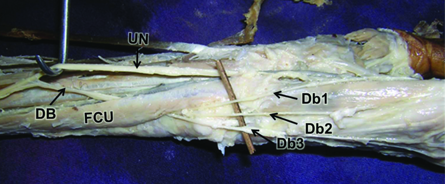
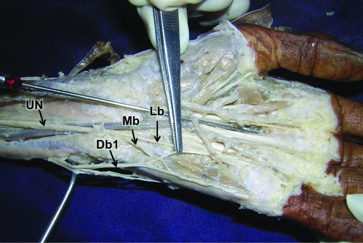
While dissecting the palm, a 70-year-old male cadaver presented with the following variation in the course and distribution of dorsal cutaneous branch 1 of ulnar nerve. The observed variation was unilateral and it was observed in the right hand. In the forearm, ulnar nerve coursed downwards and gave a Db1 in the lower part of the forearm. Then the ulnar nerve coursed into the palm where it divided into superficial and deep terminal branches. The superficial branch further divided into medial and lateral branches (Lb). The Db1 coursed underneath the flexor carpi ulnaris muscle to reach the dorsum of hand. At the wrist joint, dorsal cutaneous branch divided into three branches (Db1, Db2, Db3 in [Table/Fig-1&2]) which supplied the skin of dorsum of the hand. The Mb of superficial branch of ulnar nerve joined one of the branches of the Db1 and supplied the skin of the medial aspect of little finger. Before this connection, the Db1 gave another branch that passed superficial to the hypothenar muscles finally joined the Lb of superficial branch of ulnar nerve. To summarize we have observed a communications between the digital branches of the superficial and Db1 of the ulnar nerve. On the contralateral side, course and distribution of ulnar nerve was found to be normal.
Dissection of the right forearm and hand showing the origin and course of dorsal cutaneous branches (DB). Note the three branches (Db1, Db2, Db3) of DB, UN: ulnar nerve, FCU: flexor carpi ulnaris
Dissection of right hand showing the communications between the medial branch (Mb) and lateral branch (Lb) of the superficial branch of the ulnar nerve with one of the branches of the dorsal cutaneous branch (Db1) of the ulnar nerve. (UN: ulnar nerve)
Discussion
Ulnar nerve arises from medial cord of the brachial plexus. In the axilla it lies between the axillary artery and axillary vein. In the arm, it lies medial to the brachial artery. It gives off a dorsal branch about 5 cm above the wrist. It passes in front of the flexor retinaculum and enters the palm. There it ends by terminating into superficial and deep branches [1].
Ulnar nerve variations are usually found during cadaveric dissections and surgical practice. Earlier published reports described dorsal branch of ulnar nerve dividing into two or three branches as in our case [2]. Kaplan has described the division of Db1 joining the volar branch of the ulnar nerve. Kaplan`s anastomosis has been termed as the communication between the dorsal branch of ulnar nerve and ulnar digital nerves of the fingers [3]. It has also been reported in a study about the higher origin of the dorsal branch of the ulnar nerve in the upper part of the forearm [4]; however in the present case Db1 arose at a normal level. It has been reported that above the level of pisiform bone the dorsal branch of the ulnar nerve divided into three branches and skin of the dorsum of the hand was supplied by the lateral two of it, the most Mb crossed the pisiform bone and established three communications after looping through the belly of the deep head of abductor digiti mini [5]. In another study it was observed that dorsal branch of the ulnar nerve originated approximately at the junction of upper one fourth and lower three-fourth of the forearm. It passed on the dorsal aspect of the wrist lying between flexor carpi ulnaris and ulna and further divided into Mb and Lb.The lateral branch supplied the medial half of dorsum of hand and medial two and half digits. The medial branch again divided to supply the muscles of the hypothenar eminence [2].
The knowledge of present finding may be useful in surgeries and electrophysiological examination. The innervation pattern of dorsal branch plays an important role in designing superficial skin flaps that are harvested from dorsal aspect of the hand [2]. It is essential to know the various branching patterns of the dorsal branch of ulnar nerve for the correct placement of electrodes during any electrophysiological studies, for a successful regional nerve block, forearm flap, carpal tunnel release, and other surgical interventions of forearm and hand [4].
[1]. Standring S, Gray’s Anatomy. The Anatomical basis of clinical practice 2005 39th online EditionNewyorkElsevier Churchill Livingstone:932 [Google Scholar]
[2]. Paul S, Das S, Chaudhary S, Higher origin of the dorsal branch of ulnar nerve and connection between it and deep branch throughout the hypothenar muscles: a case reportEur J Anat 2006 10(1):37-40. [Google Scholar]
[3]. Paraskevas G, Ch Gekas C, Tzaveas A, Spyridakis I, Stoltidou A, Ph Tsitsopoulos P, Kaplan anastomosis of the ulnar nerve: a case reportJ Med Case Reports 2008 2:107 [Google Scholar]
[4]. Lama P, Potu BK, Bhat KM, High origin of dorsal branch of the ulnar nerve and variations in its branching pattern and distribution: a case reportCases Journal 2009 2:9130 [Google Scholar]
[5]. Ghabriel MN, Makar PH, Anatomical variations in the ulnar nerve and hypothenar musclesIJAV 2011 4:131-33. [Google Scholar]