A Rare Case of Isolated Partial Anomalous Pulmonary Venous Connection to the Inferior Vena Cava
Arunthathy Thangarajah1
1 Assistant Professor, Department of Radiology and Imaging, Sri Muthukumaran Medical College and Research Institute, Chikkarayapuram, Near Mangadu, Chennai, India.
NAME, ADDRESS, E-MAIL ID OF THE CORRESPONDING AUTHOR: Dr. Arunthathy Thangarajah, 9, Venkateswara Street, Dhanalakshmi Colony, Vadapalani, Chennai-600026, India. Email : gnaneshwarenator@gmail.com
Atrial septal defect, Inferior vena cava, Partial anomalous pulmonary venous connection
Images in Medicine
A 34-year-old male came to our hospital for a routine master health check up. He was apparently normal and had no significant history. His chest X-ray postero anterior (PA) view showed a curvilinear radio-opaque shadow in the right, mid and lower paracardiac location, coursing towards the diaphragm [Table/Fig-1]. A preliminary diagnosis of scimitar sign was made and a CT pulmonary angiogram was performed to confirm the diagnosis and to identify the extent of the vascular anomaly and any associated anomaly. It revealed an anomalous right superior pulmonary vein draining into the inferior vena cava [Table/Fig-2]. All the other pulmonary veins were draining normally into the left atrium. The pulmonary arteries were normal. There was no associated lung hypoplasia, sequestration or any bronchial anomaly. A 2D Echocardiogram ruled out associated cardiac anomalies such as atrial septal defect (ASD). A diagnosis of isolated partial anomalous pulmonary venous connection was made. Though the patient was asymptomatic, he was advised follow up in the cardiac outpatient department due to the possibility of developing pulmonary venous hypertension in the future.
Plain chest X-ray PA view showing curvilinear radio-opaque shadow ( arrows) in the right mid and lower paracardiac region coursing towards the right cardiophrenic angle.

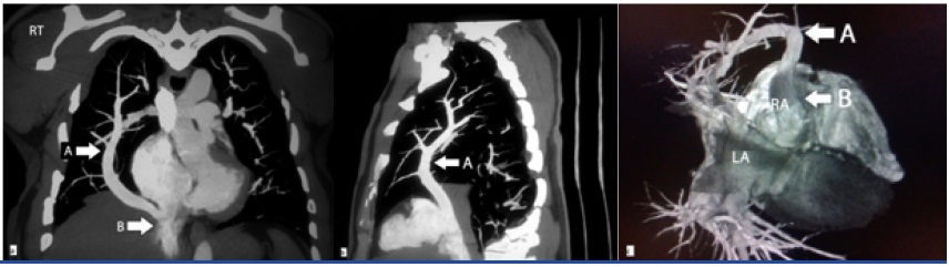
Reconstructed maximum intensity projection (MIP) coronal (a) and sagittal (b) images alongwith volume rendered images (c) of computed tomography pulmonary angiogram showing the right upper lobe pulmonary vein ( arrow A) draining into inferior vena cava ( arrow B ). The left atrium (LA) and the right atrium (RA) have been marked in (c) to provide a better understanding of the anatomy.
Discussion
Winslow in 1739 was the first to describe partial anomalous pulmonary venous connection (PAPVC). Embryonic anastomosis, between the systemic and pulmonary venous plexus, persists and, results in either a single or multiple anomalous pulmonary veins. An uncommon anomaly, it is found in 0.4 to 0.7 % of autopsies [1].
PAPVC can be classified into, supra cardiac, draining to superior vena cava (SVC), cardiac, draining to right atrium or innominate vein and infracardiac, draining to the inferior vena cava (IVC ) or portal vein. PAPVC to the IVC is rare [2] and results in a radiographic finding called the scimitar sign where there is a curvilinear radio-opaque shadow in the right paracardiac region due to the pulmonary vein draining into the IVC. The combination of a scimitar vein anomaly, pulmonary hypoplasia, dextrocardia and lung sequestration is called the scimitar syndrome. Isolated partial anomalous connection to the IVC without associated anomalies is very rare and only a few cases have been reported [3]. Amongst all the variants of PAPVC, the most common variant is the right upper lobe pulmonary vein draining into the SVC or right atrium, which is almost always associated with an ASD [4]. Isolated PAPVC without ASD is extremely uncommon [5] and when present, it drains into the SVC on the right and the innominate vein on the left. In our patient, surprisingly, the right upper lobe pulmonary vein drained into the IVC and there was no ASD or any other associated anomaly.
The complications arising due to the PAPVC and its severity are determined by the magnitude of the shunt. A lone anomalous vessel does not produce a significant shunt and therefore precludes any symptoms, while patients with large shunts present with dyspnoea, palpitations and chest pain. This patient was asymptomatic due to the presence of isolated single anomalous pulmonary vein.
[1]. Healy JE, An anatomic survey of anomalous pulmonary veins. Their clinical significanceJ. Thoracic Surg 1952 23(5):433-44. [Google Scholar]
[2]. Hijii T, Fukushige J, Hara T, Diagnosis and management of partial anomalous pulmonary venous connection. A review of 28 pediatric casesCardiology 1998 89(2):148-51. [Google Scholar]
[3]. Idris MT, Diagnostic aid of transoesophageal echocardiography in an adult case of scimitar syndrome: confirmation of the findings at surgery and review of the literatureJ Am Soc Echocardiogr 1998 11:387-92. [Google Scholar]
[4]. Alsoufi B, Cai S, Van Arsdell GS, Williams WG, Caldarone CA, Coles JG, Outcomes after surgical treatment of children with partial anomalous pulmonary venous connectionAnn Thorac Surg 2007 84(6):2020-26. [Google Scholar]
[5]. Masiello P, Panza A, Morena E, Total anomalous left pulmonary venous connection with intact atrial septum: surgical treatment of a rare caseEur J Cardiothorac Surg 1995 9:102-03. [Google Scholar]