Study of Morphometry of Carotid Canal in Skulls of South Indian origin
Vidya C S1, Shamasundar N M2
1Associate Professor, Department of Anatomy,JSS University, Mysore, India.
2Professor and Head, Department of Anatomy,JSS University, Mysore, India.
NAME, ADDRESS, E-MAIL ID OF THE CORRESPONDING AUTHOR: Dr. Vidya CS, Associate Professor, Department of Anatomy, JSS University, Mysore, India.
E-mail: Vidyasatish78@rediffmail.com
Background: The internal carotid artery supplies the anterior part of the brain, the eye and its appendages and sends branches to the forehead and nose.Carotid canal is a curved channel in the petrous temporal bone through which internal carotid artery enters the skull.
Aim: In the present study 20 skulls of unknown sex were considered. The morphometric study of carotid canal was studied by direct bone method and by silicone cast method. The present study aimed to show the differences of measurements by two methods and the silicon cast method is of its kind to measure the length of the carotid canal.
Materials and Methods: Carotid canal were measured at three different levels by both methods. Length of carotid canal to bend, diameter at the bend, length from the bend to foramen lacerum. The data was analysed for all the measurements of two methods and comparison of mean and SD at three different levels was done.Significant difference between two methods at three different levels was observed. Independent sample t-test was applied for total length of carotid canal.
Results: By observation there is a mean significant difference between two methods in the measurements of carotid canal from lower opening till the angulation on left side of the skull.The study showed that there is a bilateral significant difference in measurements of diameter between two methods. There was no significant difference observed in total length of the canal in both left and right sides.
Conclusion: Morphometrical details of the canal are of great significance to neurosurgeons and otologists. Silicon cast method was cost effective and much easier method to study the length and measurements of carotid canal as it exactly depicts the curvature of carotid canal.
Carotid canal, Curvature, Dry skulls, Length, Siliconcast, Vernier calipers
Introduction
The carotid canal was described as a bony canal directed antero-inferiorly transmitting the internal carotid artery within its internal curvature. The length of the canal is 2cms approximately. It is separated from the semilunar ganglion by a thin plate of bone which forms floor of the fossa for the ganglion [1]. It is remarkable for the number of curvatures that it presents in different parts of its course. It occasionally has one or two flexures near the base of the skull, while in its passage through the carotid canal and along the side of the body of the sphenoid bone it describes a double curvature and resembles the italic letter S [1].
Calguner studied the carotid canal as a landmark for neurosurgeons, who considered the canal the most vital and easily visualized structures on MRI angiography and DSA [2]. The position, dimensions and extensions of the carotid canal are of vital importance in cases of skull base surgery as in the identification and isolation of the internal carotid artery throughout its petrous course [3].The present study aimed to show the differences in measurements of the canal by direct bone and cast method and also on the right and left sides of the skull through individual method.
Materials and Methods
Twenty dry adult skulls were obtained from Department of Anatomy, JSS Medical College in the year 2013 and duration of the study was three months. Carotid canal dimensions were measured by direct bone and silicon cast method as described below.
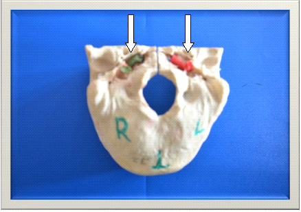
Cast method: The canal was smeared with glycerin, the foramen lacerum was plugged with wax to prevent leakage. Silicon gel was pushed into the external opening of the canal with the help of syringe. Red and green colour was mixed with silicon gel to differentiate left and right side of the cast respectively [Table/Fig-1]. After 24 h the floor of the canal was opened by using electric saw with finest blade to remove the solidified cast. The cast obtained was S-shaped with double curvatures. The measurements of the cast were taken by using digital sliding calipers [Table/Fig-2,Table/Fig-3,Table/Fig-4,Table/Fig-5]. Carotid canal were measured at three different levels by cast method (Length of cast from the lower opening to the angulation – C1, Diameter at the bend - C2, Length from the bend to foramen lacerum–C3).
Direct method: The measurements of the canal from dry skull were taken after dehiscence of floor of carotid canal as shown in [Table/Fig-5] (Length of carotid canal from the lower opening to the angulation– B1, Diameter at the bend - B2, Length from the bend to foramen lacerum – B3).
Statistical Analysis
The obtained values were collected and statistically analysed. Mean and SD at all the levels for both methods and also for both left and right sides was estimated. The data was subjected to statistical analysis with SPSS Version 14. Paired sample t-test was applied for both the methods in left and right sides for all the three levels of measurements. p <0.05 was considered significant. Independent sample t-test was done for total length of carotid canal by summation of individualvalues at first and third levels for both methods (C1+C3, B1+B3).
Results
The data is tabulated as shown in [Table/Fig-6,7].The mean dimensions were higher in all the three levels (C1,C2,C3) on the right side by cast method [Table/Fig-6].
It was observed that there is a mean significant difference between two methods in the measurements of carotid canal from lower opening till the angulation on left side of the skull.
The study showed that there is a bilateral significant difference in measurements of diameter between two methods. There was no significant difference observed in total length of the canal in both left and right sides.The mean total length of canal by bony method was 22.64 and 22.40 on left and right sides respectively. The mean total length of canal by cast method was 21.26 and 22.09 on left and right sides. There was no significant difference observed in total length of carotid canal on left and right sides [Table/Fig-7].
Discussion
Mohamed Abo Aoun et al., studied the carotid canal on 150 skulls considering their shape, direction, length and diameter both in male and female skulls by vernier calipers only. The internal length of the canal measured 22.56±2.87 mm and 24.4±2.5 mm in male skulls and 22.5±1.99 mm and 21.5±1.62 mm in female skulls on right and left sides respectively [4]. In the present study the diameter of the canal studied at the bend is important to note any kinking of the artery which in turn can lead to vascular complications and there was a bilateral significant difference in measurements of diameter between direct bone method and cast method. Namita sharma studied maximum transverse and antero–postero diameters of foramen lacerum and carotid canal in 50 dry skulls of Indian population by using vernier calipers. The mean transverse diameter and antero-postero diameter of carotid canal was 7.01 and 5.42 respectively. The mean dimensions of both diameters were higher on right side [5].
Absence of the floor of the carotid canal was described by Quint et al., [6] and Sharma et al., [7] as being attributed to developmental defects. This defect was considered to make exposure of the internal carotid artery to be vulnerable during surgical approaches and may give rise to complications during skull base surgeries. In the present study dehiscence of floor of the canal was not found in any of the skull.
Somesh Shiva prasad and others studied the morphometry of the External Aperture of carotid canal (AECC) in the base of 82 dry human skulls of mainly Indian population. The length and width of the AECC of both sides were determined using verniercalipers and area (A) was calculated and analysed.The values for the right side were 8.12 ± 0.99 mm, 6.31 ± 0.64mm and 40.61 ± 7.79 mm2 and for the left side the values were 8.15 ± 1.00 mm, 6.19 ± 0.80 mm and 40.032 ± 8.10 mm2 respectively, for the mean length, width and area of the AECC [8].
It is observed that the accuracy by cast method is beyond, even minor surfaces, depressions, projections and recesses can be perfectly cast by this method, which cannot be delineated by CT. Since luminal cast is a direct method, it has more value than the 3D CT analysis. The luminal cast plastination method can aid determiningvolume of air cavities of skull which has importance in forensicto identify gender [9].
Silicon cast in carotid canal, Left side–red colour, right side–green colour
Length of carotid canal from the opening to bend(C1)

Diameter at the bend (C2)

Length of the canal from the angulation to foramen lacerum(C3)

Measurement of bony canal, B1 Length from the opening of carotid canal to the angulation,B2 Diameter at the bend, B3 Length from the angulation to foramen lacerum
Mean and SD of measurements of carotid canal by two methods in both left and right sides of the skullsCast method C 1 B1-Length from the opening of carotid canal to the angulation C2 B2- Diameter at the bend, C3 B3- Length from the angulation to foramen lacerum
| Method | Left | Right |
|---|
| Mean+SD | Significance | Mean+SD | Significance |
|---|
| C1 | 5.99+1.45 | 0.001 | 6.67+0.95 | 0.688 |
| B1 | 6.79+1.59 | 6.58+1.32 |
| C2 | 3.77+0.63 | 0.001 | 3.89+0.57 | 0.014 |
| B2 | 4.61+0.84 | 4.33+0.64 |
| C3 | 11.49+1.48 | 0.298 | 11.52+1.27 | 0.907 |
| B3 | 11.22+1.43 | 11.48+1.46 |
Mean and SD of total length of carotid canal by two methods
| Method | Left Mean+SD | Right Mean+SD | p-value of total length of cast and bone method |
|---|
| Direct bone method – B1+B3 | 22.64+3.22 | 22.40+3.15 | Left 0.172 |
| Cast method – C1+C3 | 21.26+ 3.04 | 22.09+1.57 | Right 0.697 |
Conclusion
Knowledge of dimensions of the carotid canal would guide clinicians towards a correct interpretation of radiographs and would be of help in surgical approaches to this complicated region.The rubber silicone produces an excellent, soft, flexible cast showing an unimaginable 3-dimensional orientation of the canal including the abnormal extension if any. In the present study silicon cast method was first of its kind, to study the dimensions of the carotid canal.
Acknowledgements
I sincerely thank Head of the Institution for constant support to perform this study
Recommendations
The sample of the study is less for exact conclusion; silicon cast can also be measured by metallic wire for the diameter at the bend of the canal. The study can be extended in same population and compared with CT scan analysis.
[1]. S Standring, The Anatomical Basis of Clinical Practice 2008 40th EditionGray’s Anatomy, Elsevier limited:734 [Google Scholar]
[2]. E Calguner, HB Turgut, R Gozil, E Tunc, A Sevim, S Kekil, Measurements of the carotid canal in skullsActa Anat 1997 158:130-32. [Google Scholar]
[3]. JP Leonitti, PG Mith, FH Linthicu, The petrous carotid artery. An anatomic relationship in skull base surgeryOtolaryngol Head Neck Surg 1990 102(1):3-12. [Google Scholar]
[4]. Aoun Mohamed Abo, Y Ashraf, Nasr Adel, M Abdel, Morphometric Study of the Carotid CanalLife Science Journal 2013 10(10):2559-62. [Google Scholar]
[5]. AS Namita, SG Rajendra, Morphometric evaluation and a report on the aberrations of the foramina in the intermediate region of the human cranial base: A study of an Indian populationEur J Anat 2011 15(3):140-49. [Google Scholar]
[6]. DJ Quint, R Silpergtleit, WC Young, Absence of the carotid canals at skull baseCT Radiology 1992 182:477-81. [Google Scholar]
[7]. PK Sharma, PK Lakhtakia, KK Bisaria, A variant of the floor of the carotid canalAnat Anz 1993 175(2):199 [Google Scholar]
[8]. Shivaprasad Somesh, HB Sridevi, BV Murlimanju, Pai Shakunthala, Morphological and Morphometric study of carotid canal in Indian PopulationInternational Journal of Biomedical Research 2014 5(7) [Google Scholar]
[9]. CS Vidya, NM Shamasundar, B Manjunatha, Raichurkar Keshav, Plastination: An attempt to estimate size and volume of maxillary sinus of dry crania to determine genderMedico legal journal 2013 13(2):147-50. [Google Scholar]