The term “aesthetic” is used as a connotation that is pleasing to the eyes. Missing anterior teeth often create an unpleasing appearance to an individual, which affects their confidence, especially in younger individuals [1]. In the natural dentition, incident light is reflected, scattered, and transmitted; which is influenced by the surface texture and thickness of the tooth and affects the translucent properties of a tooth [2–4]. To attain a perfect match of the tooth being replaced, translucency should also be considered while proper selection of shade, without which the shade selection is not completed.
With the advent of All-ceramic restorations, the metal copings are eliminated. The improved aesthetics and the translucency of the all-ceramic crowns are attributed to the characteristics of the core material [4,8]. Translucency of material depends on the transmission of light but not the scattering. Intensely scattered and diffusely reflected materials will appear more opaque [9]. Core translucency therefore becomes one of the primary factors in controlling aesthetics and is a critical consideration in selection of materials [10] but the drawback of the All-Ceramic restorations was lack of strength [11].
In recent years the ceramics have reached a pinnacle with modification of the basic constituent materials like Alumina reinforced Lithium disilicate and Zirconia which is being used as the core material to enhance the strength of ceramic restorations and overcome the darkening effect of the metal substructure [11]. These core materials improved the strength of the All-ceramic crowns but effect of the translucency needs to be evaluated.
In modern era of dentistry lot of materials are available in market selection depends on material properties but, due to advertisements and techniques involved puts clinician in dilemma so that the proper selection of material for successful restoration without proper material knowledge makes it impossible. Therefore, this study was planned to evaluate and compare the translucency of crowns fabricated with three different All – Ceramic materials with the Null hypothesis that there is no significant difference in the translucency among these three materials.
Materials and Methods
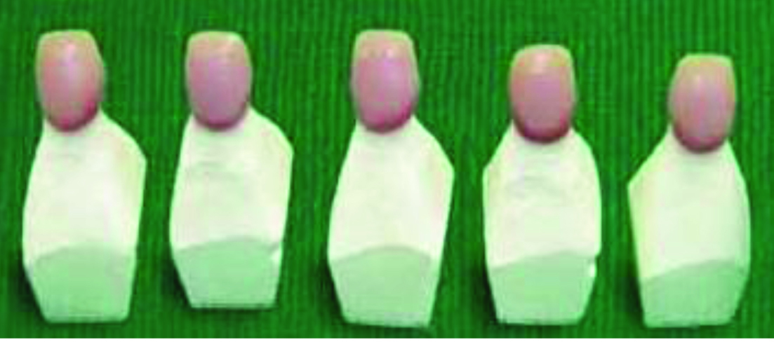
Extracted maxillary central incisor which was caries free, with all surfaces intact, and having good anatomy was selected and mounted in dental plaster leaving the Cemento enamel junction and crown portion exposed [Table/Fig-1]. Since it was an invitro study, extracted teeth were collected from private clinics where patient underwent extraction because of periodontal reasons. Tooth having good anatomy were stored in hydrogen peroxide for removal of debris and stored in formalin. Since the patients were not involved directly in this study and study was invitro so ethical committee clearance was not obtained. An index [12] of the mounted tooth was made in silicone putty (Aquasil, Dentsply, New Delhi, India) and was used as a guide for even reduction of tooth and the even thickness of the crowns [Table/Fig-2]. The tooth preparation was done according to biomechanical principles; with overall preparation of 2mm incisally,1.5mm labially & lingually with a rounded shoulder finish margin and checked with the silicone index for even reduction. Five crowns of each material consisting total of 15 samples, were fabricated on the prepared tooth and grouped as follows (Group-I)Alumina crowns-CAD-CAM Procera, (Nobel Biocare, Goteberg, Sweden). (Group II)Lithium Di-silicate crowns-Pressable IPS e.max Press (Ivoclar Vivadent Mumbai, India) Group III: - Zirconia crowns-CAD-CAM Lava (3M ESPE, Lava, Mumbai, India)
Selected Maxillary Central incisor

Silicone index of the master tooth
Fabrication of Alumina Crowns-CAD-CAM Procera Group I
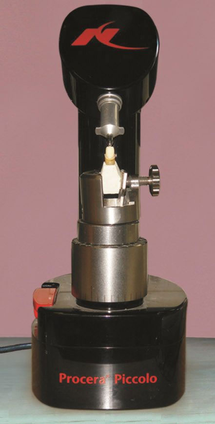
The prepared tooth was mounted on a rotating platform in a digital scanning device (Procera scanner) that was attached to a computer. After scanning of the tooth, two-dimensional image was displayed on the computer screen. The thickness of the coping was kept at 0.4mm as advised by the company for anterior restorations. A file was sent via e-mail to the production station in Sweden after design of the coping [Table/Fig-3]. Five copings of CAD-CAM Procera all ceramic system were then evaluated over the prepared tooth for their fit objectively by visual examination, i.e., the crown Placed on the prepared tooth and focused light on it to see that no gap exists between the crown and the margin and a sharp probe passed from the tooth to the crown. The probe runned smoothly from the tooth to the crown without any catch [13].Thickness of 0.4 mm was verified with gauge. Later, ceramic build up was carried out using Nobel Rondo Alumina ceramic material of B2 shade using the silicone index for the standardization of the form and thickness of the crown. The total thickness of the crowns was kept at 1.5mm.
Scanning of the prepared tooth With Procera scanner
Fabrication of Lithium Di-silicate crowns – Pressable IPS e.max Press: Group II
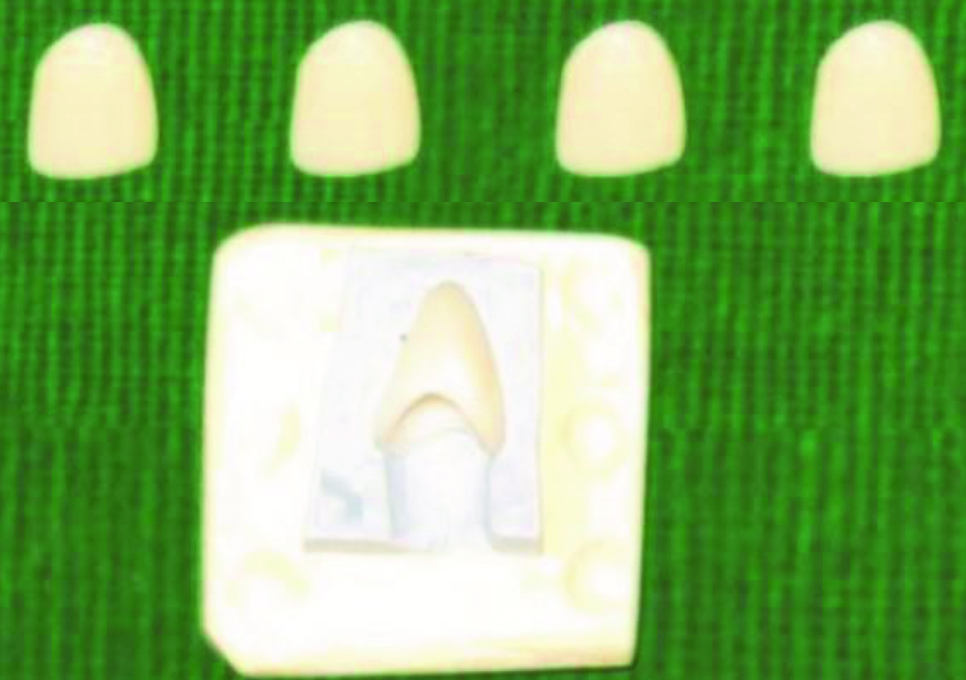
Pressable lithium-disilicate copings were fabricated with the lost wax technique. Five special trays were fabricated with self cure clear acrylic resin (Dental products of India, New Delhi, India), using modeling wax (DPI, New Delhi, India) as spacer of 1.5mm thickness. Impression of the master tooth was made in Elastomeric impression material (Aquasil, Dentsply, New Delhi, India) to fabricate five dies in die stone (Kalabhai, Dentsply, New Delhi, India). Wax patterns (Yeti, New Delhi, India) of 0.9 mm thickness were prepared and measured with the wax gauge to verify their thickness [Table/Fig-4]. The copings were recovered by sandblasting and further trimming was done with diamond finishing burs to finally maintain thickness of 0.8mm as per the manufacturer’s recommendation for this group and evaluated over the prepared tooth for their fit objectively [Table/Fig-5]. Ceramic build up was carried out with the IPS e.max ceramic material using the silicone index. The thickness of the finished crowns was 1.5mm.
Wax Patterns for copings of Group II crowns
Iwansons gauge measuring the copings of Group II crowns

Fabrication of Zirconia crowns – CAD-CAM Lava: Group III
The master tooth was mounted on a rotating platform in an optical scanning device Lava Scan that was attached to a computer [Table/Fig-6]. After scanning the tooth, the three dimensional image got displayed on the computer screen. The thickness of the coping was kept at 0.4mm. After design of the coping, the 3D shape was milled from a pre-sintered Zirconia oxide blank using hard metal tools. The copings were then evaluated for their fit objectively & thickness of 0.4mm was verified with gauge. Later on ceramic build up was carried out using Lava Ceram ceramic material with the help of silicone index. The thickness of the finished crowns was kept at 1.5mm [Table/Fig-7].
Scanning of the prepared tooth in Lava scanner

Group III- Zirconia crowns (CAD-CAM Lava)
The tooth preparation was done and the crowns were fabricated. The thickness of the copings for Group I, Group III was kept as 0.4mm and for Group II as 0.8mm as recommended by the manufacturers for the required strength. The veneering was done using the silicone index as a guide. The final thickness of all the crowns was kept at 1.5mm, accordingly the thickness of the veneering material was 1.1mm for Group I and Group III crowns and 0.7 mm for Group II crowns.
Evaluation of the translucency of all the groups (CR = Yb/Yw)
The translucency was measured quantitatively by comparing reflectance of light through the test specimen over a backing with a high reflectance (white background) to that of low reflectance or high absorbance (black background). A contrast ratio was produced (CR) in which CR =Yb/Yw, the light reflectance of the material on a black surface (Yb) to the reflectance on a white surface (Yw).
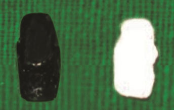
Impression of the master tooth was made in Elastomeric impression and two resin dies were prepared using DPI self-cure clear acrylic resin. One resin die was painted with black acrylic paint and the other resin die was painted with white acrylic paint [Table/Fig-8]. The resin die with the black paint was placed on black modeling clay, so that a black background was formed and the resin die with white paint was placed on white modeling clay, so that a white background was formed.
Black and White resin dies
Reflectance spectrophotometer having light source, Monochro-mator, Detector (i1-XRite, AGS infotech, Mumbai, India) was used. Light source used in the reflectance spectrophotometer is pulsed xenon light source. The xenon flash provides light exactly the same as normal day light and viewing condition is 450/00 means 450 illuminations and 00 viewing. This geometry does not require integrating sphere; it is suitable for textured surface or high glossy surface. Diffraction grating monochromator is used which disperses the reflected light from the sample and Silicone photodiodes are used as detector. The light source emits a beam of light, which strikes on the sample and then reflected to the diffraction grating monochromator, which disperses the reflected light to the silicone photodiodes detector. Information is converted and processed by the Optiview software. The luminous reflectance of the black background was measured with the spectrophotometer and it was found to be 18.16 and that of white background was 80.21; which gave the contrast ratio as 0.22. The range of translucency falls within the range of Contrast Ratio from 0-1. If the Contrast Ratio of an object is about 0.8 or 0.8-1, it is considered as opaque. If the contrast ratio of the object lies between 0.0- 0.8, it is considered as translucent [4,10]. The more the contrast ratio is towards the 0.0 value the more it is translucent. Thus, contrast ratio is not a direct measure of translucency, but is a surrogate measure to approximate the translucency.
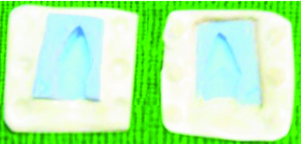
The crowns of each group were placed on both black (Yb) & white (Yw) background and the measurements were made on the Incisal, Middle, Cervical, Mesial and Distal areas of the crown [Table/Fig-9,10]. The mean CR of these areas gave the Contrast ratio of the crown. The mean CR of the five crowns in a group was considered as the CR of that group.
Fabricated crowns over Black and White backgrounds

Measuring the reflectance of the Crowns

Statistical Analysis
Paired t-test was done to find any significant difference in the contrast ratios of the Incisal, Middle, Cervical, Mesial and Distal regions of a crown (p<0.05). Paired t-test was used to find significant differences within the groups. One-way analysis of variance was used to determine which groups differed significantly (p<0.05).
Results
The contrast ratios were obtained for each crown at the Incisal, Middle, Cervical, Mesial and Distal third. The mean contrast ratio of each group was estimated [Table/Fig-11]. The mean contrast ratios for Group I -0.7, Group II -0.54 and Group III-0.75. One-way analysis of variance, showed a significant difference in the contrast ratios of the three groups (p<0.001-[Table/Fig-12]). Paired t-test performed for the variables of Group I-Alumina crowns-CAD-CAM Procera, Group-II - Lithium disilicate crowns-Pressable IPS e.max Press, and Group-III-Zirconia crowns - CAD CAM Lava which showed a significant difference in the translucency of Incisal, Middle, Cervical and Proximal regions of a crown, but there is no significant difference between the Mesial and Distal regions of a crown [Table/Fig-13]. The overall translucency of the three groups- Group II showed the maximum translucency, Group III showed the least translucency and Group I intermediate to Group II and Group III. [Table/Fig-14].
The Contrast ratios of Group I: Alumina crowns -CAD CAM Procera, Group II: Lithium disilicate crowns- Pressable IPS e-max Press, Group III: zirconia crowns - CAD CAM Lava.
| Procera | CR | IPS e-MAX | CR | Lava | CR |
|---|
| P1 | 0.65 | E1 | 0.52 | L1 | 0.71 |
| P2 | 0.69 | E2 | 0.55 | L2 | 0.78 |
| P3 | 0.75 | E3 | 0.57 | L3 | 0.80 |
| P4 | 0.70 | E4 | 0.53 | L4 | 0.70 |
| P5 | 0.72 | E5 | 0.54 | L5 | 0.76 |
| Mean | 0.70 | Mean | 0.54 | Mean | 0.75 |
The Analysis of variance done for the Groups - Group I, Group II and Group III, showed significant difference in their translucency.
| Variables | d.f | t- value | p-value | Result |
|---|
| Procera-IPS e.max | 4 | 93.216 | 0.001 | Significant |
| IPS e.max-Lava | 4 | 87.533 | 0.002 | Significant |
| Procera-Lava | 4 | 83.766 | 0.001 | Significant |
d.f = degrees of freedom, P < 0.05 = Significant, P > 0.05 = Not Significant
Tabulated Value = 2.02
Table showing significant difference in the translucency of Incisal, Middle, Cervical and Proximal regions of a crown, but there is no significant difference between the Mesial and Distal regions of a crown
| Variables | Df | t-Value | p-Value | Result |
|---|
| Incisal-middle | 4 | 2.828 | 0.047 | Significant |
| Incisal-cervical | 4 | 12.649 | 0.000 | Significant |
| Incisal-mesial | 4 | 0.590 | 0.587 | Not Significant |
| Incisal-distal | 4 | 2.236 | 0.089 | Not Significant |
| Middle-cervical | 4 | 3.651 | 0.022 | Significant |
| Middle-mesial | 4 | 4.000 | 0.016 | Significant |
| Middle-distal | 4 | 1.414 | 0.230 | Not Significant |
| Cervical-mesial | 4 | 5.308 | 0.006 | Significant |
| Cervical-distal | 4 | 4.743 | 0.009 | Significant |
| Mesial-distal | 4 | 1.000 | 0.374 | Not Significant |
Graph showing the mean contrast ratios of the three groups

Discussion
A successful dental prosthesis must fulfill the aesthetic, masticatory and phonetic requirements. The aesthetics of the prosthesis depends on translucency and its shade [5]. The shade is determined not only by the color of the porcelain, but also by the thickness of the porcelain, the thickness and the color of the luting agent, and the color of the underlying tooth structure. The ceramics are translucent at clinically relevant thicknesses and with different core materials, the translucencies vary within the ceramics [9,14]. Since natural enamel has inherent translucency, it is important that the restorations reproduce the translucency along with the shade of the natural teeth.
In clinical practice, restoring a single maxillary central incisor is a greatest restorative challenge [3,15]. Several decades of clinical experience has proved metal-ceramics to be the most widely used restorations [4,16,17] but while considering the availability of different materials, ceramics found its rightful place for aesthetics [18]. The ceramo-metal restorations though stronger than ceramic, but were not superior in aesthetics because of more reflectivity of the light due to underlying metal coping. This led to research in ceramic materials and emerges with the new generation of ceramic materials with superior aesthetics and strength [8].
In this study, the translucency was analyzed with the help of a Reflectance spectrophotometer (1) which captures the amount of light reflected from an object. A maxillary central incisor was chosen as the master tooth for preparation, because the test was carried out for translucency and mechanical factors were not considered. Anterior restorations are more in demand for aesthetics, so we had selected the maxillary central incisor [15]. It was important to get the even thickness of the ceramic crowns; so silicone index of the master tooth was prepared. After the fabrication of the crowns, they were subjected to the reflectance spectro-photometer on the white and black back-grounds at the Incisal, Middle, Cervical, Mesial and Distal regions as stated by Aki Yoshida et al., [19]. The null hypothesis that there is no difference in the translucency between the groups was rejected and is in accordance with other studies [8,20]. According to the Contrast ratios obtained in this study, the translucency of Pressable Lithium Di-silicate crowns-IPS e.max Press (0.54) was greater than Alumina crowns-CAD-CAM Procera,(0.7)and Zirconia crowns-CAD-CAM Lava(0.75). In each crown the incisal third showed maximum translucency over the middle and proximal thirds with cervical third showing the least translucency.
Clinical Implications
The tooth that transmits more light is translucent and is considered to be less conspicuous, the tooth which absorbs and scatters more light and transmits less light is considered to be opaque and is more conspicuous [8]. Based on the results of this study, Pressable Lithium Disilicate crowns – IPS e.max Press can be used for matching highly translucent natural teeth. For moderately translucent teeth Alumina crowns – CAD-CAM Procera can be used. For opaque teeth Zirconia crowns - CAD-CAM Lava can be used. This study was limited by the fact that it was an in-vitro study and the heterogeneity in the application of the veneering ceramic and luting agent was not considered which could bring about a change in the overall translucency of the All-ceramic restorations. Further in vivo studies warranted to consider other factors which were not involved in this study.
Conclusion
The study showed the significant difference in the translucency among three groups. The range of translucency between the three Groups were in the following increasing order as Zirconia crowns – CAD CAM Lava, Alumina crowns – CAD-CAM Procera, and Lithium disilicate crowns – Pressable IPS e.max Press. Within each group the entire crowns showed overall maximum translucency in the Incisal area, and least in the cervical area with Proximal surfaces of the crown showing intermediate translucency between the Incisal and Cervical area. The Group II crowns even at 0.8mm thickness of the coping showed higher translucency over Group I and Group III crowns which have 0.4mm thickness of the coping. This is because of the more translucent character of the All-Ceramic material Lithium Di-silicate, over the Alumina and Zirconia All-Ceramic materials.
d.f = degrees of freedom, P < 0.05 = Significant, P > 0.05 = Not Significant
Tabulated Value = 2.02