Mucoid Cyst of the Penis: A Case Report
Mohinder Kumar Malhotra1, Karan Bakhshish Singh2, Prakhar Garg3, Pratibha Sharma4, Karamjot Singh5
1Associate Professor, Department of Surgery,MMISR, Mullana, Ambala, India.
2PG Resident Third Year, Department of Surgery,MMISR, Mullana, Ambala, India.
3PG Resident Third Year, Department of Surgery,MMISR, Mullana, Ambala, India.
4PG Resident Third Year, Department of Surgery,MMISR, Mullana, Ambala, India.
5PG Resident Third Year, Department of Surgery,MMISR, Mullana, Ambala, India.
NAME, ADDRESS, E-MAIL ID OF THE CORRESPONDING AUTHOR: Dr. Mohinder Kumar, Associate Professor, Department of Surgery, MMISR, Mullana, Ambala, Haryana-133207, India.
E-mail: malhotramsfrcs@yahoo.co.in
Penile cysts are rare benign lesions. Their clinical or pathological diagnosis is extremely difficult. Less than 200 cases have been reported in literature. Most of them are asymptomatic and present since birth, but usually they are only detectable in adolescence or adulthood. These lesions rarely interfere with sexual function. These cysts most likely arise from ectopic urethral mucosa sequestered in the penile skin during embryologic development. We report a rare case of a 55-year-old man with a 4.5 × 4 cm nodule on the prepuce (ventral surface), which appeared two years ago and interfered with his sexual function. The nodule was excised by circumcision and a histopathological examination revealed a mucoid cyst of penis with squamous metaplasia. No recurrence was observed at a one year follow-up and the patient’s sexual function returned to normal.
Fungal, Allergic, Charcot, Charcot, Charcot
Case Report
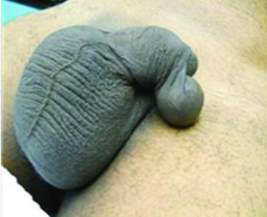
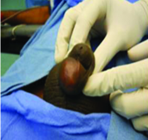
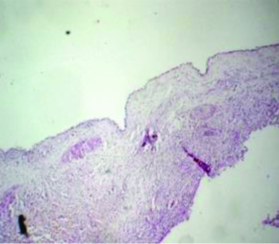
A 55-year-old man developed a progressively increasing nodule on fore skin of his penis since two years, which started to interfere with his sexual function for last three months [Table/Fig-1]. There was no history of trauma, infection or previous surgery. On physical examination, we observed a 4.5 × 4 cm soft, tender and cystic nodule on the prepuce (ventral surface) [Table/Fig-1,Table/Fig-2]. All routine investigations were within normal limits. The lesion was excised by circumcision under local anaesthesia and sent for histopathologic study. Histopathologic examination revealed cyst wall showing single layer of cuboidal to columnar epithelium [Table/Fig-3]. There were no postoperative complications and the patient was discharged on the same day. Patient was followed up in surgery outpatient department every three months and no recurrence was observed and patient had no sexual discomfort.
Discussion
Mucus penile cyst is an uncommon benign lesion affecting mainly young men on ventral surface of glans penis but this case presented in sixth decade. An extensive literature search has revealed only less than 200 reported cases. Among these, only less than 10 have been reported from the Indian subcontinent [1]. The clinical diagnosis is difficult and differential diagnoses include epidermal cyst, lipomas, esteatocystomas, dermoid cysts, pilonidal cysts, tyson glands cysts and urethral diverticulums [2,3]. Several synonymous terms, including mucus cyst of the penis, genitoperineal cyst of the median raphe, parameatal cyst, hydrocystoma and apocrine cystadenoma of the penile shaft are also used in past [1,4]. Although most of them are present since birth, but usually they are only detectable on adolescence or adulthood [5]. Only one case of a spontaneous onset after an intense sexual intercourse has been documented [1,6].They are midline-developmental cysts in the perineum that can be found anywhere from the anus to the urinary meatus. The cysts commonly present around the glans mainly on the penile ventral surface, around near the glans. Mucoid cyst also needs to be differentiated from median raphe cyst of the penis, which generally occurs along the median raphe on the ventral surface of the penis. Median raphe cysts which are usually asymptomatic are lined by urothelium or squamous epithelium [7,8]. Mucoid cysts usually, they are asymptomatic but can be complicated by infection, trauma or can make coitus difficult, as with the patient in this report [9,10]. Surgical excision is required. They are benign cysts and no case of malignancy has been reported till date.
Penile cyst (lateral view)
Penile cyst (ventral view)
Cyst wall showing single layer of cuboidal to columnar epithelium (x100 H &E)
Conclusion
Penile cysts are rare entity and are present since birth but detected in adolescence or adulthood. They are excised by circumcision
[1]. A Amaranathan, SP Sinhasan, SD Dasiah, Median Raphe Cysts of the Prepucial Skin, with Triple Histological Linings: A Case Report and Review of the LiteratureJ Clin Diagn Res 2013 7(1):1466-68. [Google Scholar]
[2]. RR Mendonca, JL Silva, ML Wroclawski, Mucoid cyst of the penis: Case report and literature reviewCan Urol Assoc J 2010 4:155-57. [Google Scholar]
[3]. LA Cole, EB Helwig, Mucoid cysts of the penile skinJ Urol 1976 115:397-400. [Google Scholar]
[4]. E Velasco, Mucoid cyst of penis skin [in Spanish] Urol Colomb 1993 3:99-104. [Google Scholar]
[5]. IH Shao, TD Chen, HT Shao, HW Chen, Male median raphe cysts: serial retrospective analysis and histopathological classificationDiagn Pathol 2012 7:12 [Google Scholar]
[6]. S Giannakopoulos, NE Makris, C Kalaitzis, AG Papatsoris, T Pantazis, S Touloupidis, Two unusual cases of median raphe penile cystsEur J Dermatol 2007 17(4):342-43. [Google Scholar]
[7]. T Otsuka, Y Ueda, M Terauchi, Median raphe (parameatal) cysts of the penisJ Urol 1998 159:1918-20. [Google Scholar]
[8]. R Cardoso, JD Freitas, JP Reis, Median raphe cyst of the penisDermatol Online J 2005 11:37 [Google Scholar]
[9]. P Saini, MN Mansoor, S Jalali, A Sharma, Penile epidermal inclusion cystIndian J.Pediatr 2010 77(7):815-16. [Google Scholar]
[10]. T Yalta, Kazındır G., Mucoid Cyst of The Penile Skin: A Rare EntityAfrican J. Urol 2010 16(4):132-33. [Google Scholar]