Posterior Reversible Encephalopathy Syndrome with Involvement of the Cervical Cord and Medulla: A Case Report
Xuan Hou1, Jinfeng Xu2, Zao Chen3, Gguoliang Li4, Hong Jiang5
1Faculty, Department of Neurology, Xiangya Hospital, Central South University, Changsha, Hunan, P. R. China.
2Faculty, Department of Neurology, Xinjiang Uygur Autonomous Region Chinese Medicine Hospital,Xinjiang Medical University, Urumqi, Xinjiang, P. R. China.
3Faculty, Department of Neurology, Xiangya Hospital, Central South University, Changsha, Hunan, P. R. China.
4Faculty, Department of Neurology, Xiangya Hospital, Central South University, Changsha, Hunan, P. R. China.
5Faculty, Department of Neurology, Xiangya Hospital, Key Laboratory of Hunan Province in Neurodegenerative Disorders, State Key Lab of Medical Genetics,Central South University, Changsha, Hunan, P. R. China.
NAME, ADDRESS, E-MAIL ID OF THE CORRESPONDING AUTHOR: Dr. Hong Jiang, Faculty, Department of Neurology, Xiangya Hospital, Central South University, Changsha, Hunan-410008, P. R. China.
E-mail: huangteng2017@163.com
Posterior reversible encephalopathy syndrome (PRES) is a neurotoxic state, which is associated with symmetrical subcortical areas of vasogenic oedema that are preferentially parieto-occipital, and it typically resolves within a few weeks after appropriate treatment, We hereby report a case of a female with adrenal tumour presenting with PRES, who was featured by a very rare neuroimaging manifestation, the involvement of cervical cord and medulla.
Adrenal tumour, Clinical features, Neuroimaging manifestations, Posterior reversible encephalopathy syndrome (PRES)
Case Report
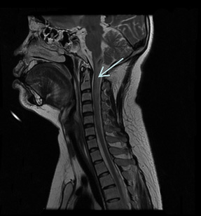
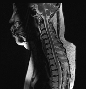
A 26-year-old women experiencing an epileptic seizure, abrupt headache, generalized body weakness, was admitted in the emergency, she was under excessive dieting followed by 15 kg increase of body weight since 3 months. On general medical examination, she was characterized by moon face, papulopustular rashes in the face and anterior of the chest, purple lines on her inner thighs, as well as high blood pressure ranging from 200/100 to 220/110 mmHg. On neurological examination, she was sober, could not move her right limb in the bed, but could lift her left limb. She had no past history of hypertension and did not take any medication before. Non-enhanced computed tomography (CT) showed no evidence of cerebral hemorrhage or subarachnoid hemorrhage (SAH). Magnetic Resonance Imaging (MRI) of brain and spinal cord was thus performed, which should hyperintense lesions involving the medulla and cervical spinal cord in T2- weighted images [Table/Fig-1]. The 24 h urinary vanillyl mandelic acid (VMA) was 11.2μmol/24 hrs, urinary 17-hydroxy corticosteroids was 39.4 μmol/24 h, urinary 17-ketone corticosteroids was 88.7 μmol/24 h, blood cortisol was 34.3 μg/dl, blood cortisol (F-4pm) was 31.55 μg/dl, blood adrenocorticotropic hormone (ACTH) was 0.22pmol/l indicating ACTH-independent Cushing’s syndrome. Abdomen CT of the patient showed a significantly enhancing mass in the left adrenal gland(size about 3.2cm x 2.6 cm) [Table/Fig-2A,B]. The patient was also subjected to adrenal lumpectomy, indicating the tumour of 2cm x 2cm x 3cm in size. The pathological findings showed adrenal adenoma. She was treated with levoamlodipine and prazosin for the hypertension, and 2 weeks later her muscle strength of all four limbs recovered to normal. four weeks follow-up MRI of the spinal cord showed disappearance of previous imaging finding in T2-weighted image [Table/Fig-3] . Considering the characteristics of clinical and imaging performance, an atypical variant of PRES was the most plausible diagnosis. One year after being discharged from hospital, the patient was followed up and remained asymptomatic.
Discussion
Recently, posterior reversible encephalopathy syndrome (PRES) has become increasingly recognized. Hypertensive encephalopathy, toxemia of pregnancy, cyclosporin A toxicity and ureamic encephalopathy are the most common causes of PRES [1]. The mechanism of PRES is controversial. The present more popular theory suggests that severe hypertension exceeds the limits of autoregulation, leading to breakthrough brain oedema [2]. The patient whom we reported here, also suffered from severe hypertension because of adrenal tumour, which further confirmed the above theory. Another theory has also emerged after reports of PRES combined with regions of vasospasm, in which hypoperfusion is assumed to cause injury and subsequent oedema [3]. PRES is a clinic-radiologic syndrome with diverse symptoms. Clinically, headache, confusion, seizure and visual disturbance are common symptoms of PRES, yet muscle weakness of limbs, ataxia are rare [4,5]. The MRI characteristics of PRES are vasogenic oedema involving parieto-occipital lobes (cortical/subcortical white matter). The frontal and temporal lobes were common involvedregions in PRES. Involvement of the basal ganglia, brain stem and deep white matter are the less common but were confidently recognized as part of the PRES process [6]. In recent years, focal hemorrhage, subtle subarachnoid hemorrhage was reported as uncommon imaging features of PRES [7-9]. Involvement of the cervical cord and medulla oblongata was indicated in our patient, which is a very rare neuroimaging manifestation of PRES and was reported in few cases [10-12]. Also from MRI of cervical cord, a high probability for infarction can be considered. But patients with massive brainstem infarction would have a sudden onset and more severe neurodeficit, as well as being comatose at presentation [13].
The cervical cord MRI of the case showed T2-weighted hyperintense involving the cervical cord and medulla
A: unenhanced CT of abdomen showed a mass (size about 3.2cm x 2.6 cm) in the left adrenal gland ,the value of the CT was 21HU. B: enhanced CT of abdomen showed the mass was significantly enhanced, the value of the CT was 56HU

4 weeks follow-up MRI of the spinal cord showed disappearance of previous imaging finding in T2-weighted image
Conclusion
Base on the patient's clinical manifestation, MRI features, and the benign prognosis, diagnosis of PRES seems most reasonable. It needs to be considered whether adrenal tumours or any particular etiology predisposes to cord involvement in PRES. Furthermore, In order to prevent misdiagnosis, the neurologists should recognize this atypical imaging of RPES.
Acknowledgement
Xuan Hou, Jinfeng Xu contributed equally to this work.
[1]. VL Stott, MA Hurrell, TJ Anderson, Reversible posterior leukoencephalopathy syndrome: a misnomer reviewedIntern Med J 2005 35(2):83-90. [Google Scholar]
[2]. RB Schwartz, SM Bravo, RA Klufas, L Hsu, PD Barnes, CD Robson, Cyclosporine neurotoxicity and its relationship to hypertensive encephalopathy: CT and MR findings in 16 casesAJR Am J Roentgenol 1995 165(3):627-31. [Google Scholar]
[3]. WS Bartynski, Posterior reversible encephalopathy syndrome, part 2: controversies surrounding pathophysiology of vasogenic oedemaAJNR Am J Neuroradiol 2008 29(6):1043-49. [Google Scholar]
[4]. J Hinchey, C Chaves, B Appignani, J Breen, L Pao, A Wang, A reversible posterior leukoencephalopathy syndromeN Engl J Med 1996 334(8):494-500. [Google Scholar]
[5]. VH Lee, EF Wijdicks, EM Manno, AA Rabinstein, Clinical spectrum of reversible posterior leukoencephalopathy syndromeArch Neurol 2008 65(2):205-10. [Google Scholar]
[6]. WS Bartynski, JF Boardman, Distinct imaging patterns and lesion distribution in posterior reversible encephalopathy syndromeAm J Neuroradiol 2007 28(7):1320-27. [Google Scholar]
[7]. WS Bartynski, Posterior reversible encephalopathy syndrome, part 1: fundamental imaging and clinical features Am J Neuroradiol 2008 29(6):1036-42. [Google Scholar]
[8]. JA Hinchey, Reversible posterior leukoencephalopathy syndrome: what have we learned in the last 10 years?Arch Neurol 2008 65(2):175-76. [Google Scholar]
[9]. AM McKinney, J Short, CL Truwit, ZJ McKinney, OS Kozak, KS SantaCruz, Posterior reversible encephalopathy syndrome: incidence of atypical regions of involvement and imaging findingsAm J Roentgenol 2007 189(4):904-12. [Google Scholar]
[10]. M Matiello, SM Magana, BG Weinshenker, Asymptomatic spinal cord involvement in posterior reversible encephalopathy syndromeNeurology 2010 74(18):1478-79. [Google Scholar]
[11]. NA Choh, M Jehangir, M Rasheed, T Mira, I Ahmad, S Choh, Involvement of the cervical cord and medulla in posterior reversible encephalopathy syndromeAnn Saudi Med 2011 31(1):90-92. [Google Scholar]
[12]. B Lapuyade, I Sibon, S Jeanin, V Dousset, Neurological picture: Spinal cord involvement in posterior reversible encephalopathy syndromeJ Neurol Neurosurg Psychiatry 2009 80:35 [Google Scholar]
[13]. TY Chen, HJ Lee, TC Wu, YK Tsui, MR imaging findings of medulla oblongata involvement in posterior reversible encephalopathy syndrome secondary to hypertensionAm J Neuroradiol 2009 30(4):755-57. [Google Scholar]