Inexplicable Co- Existence of Eccrine Spiradenoma and Lichen Planus in an old Case of Basal Cell Carcinoma
A. L Hemalatha1, M.S Lavanya2, K Anoosha3, K. P Ashok4, M Rajani Deepa5
1 Professor, Department of Pathology, Affiliated to Rajiv Gandhi University of Health Sciences, Adichunchanagiri Institute of Medical Sciences, B. G. Nagar, Mandya, Karnataka, India.
2 Assistant Professor, Department of Dermatology, Affiliated to Rajiv Gandhi University of Health Sciences, Adichunchanagiri Institute of Medical Sciences, B. G. Nagar, Mandya, Karnataka, India.
3 Post Graduate, Department of Pathology, Affiliated to Rajiv Gandhi University of Health Sciences, Adichunchanagiri Institute of Medical Sciences, B. G. Nagar, Mandya, Karnataka, India.
4 Post Graduate, Department of Pathology, Affiliated to Rajiv Gandhi University of Health Sciences, Adichunchanagiri Institute of Medical Sciences, B. G. Nagar, Mandya, Karnataka, India.
5 Post Graduate, Department of Pathology, Affiliated to Rajiv Gandhi University of Health Sciences, Adichunchanagiri Institute of Medical Sciences, B. G. Nagar, Mandya, Karnataka, India.
NAME, ADDRESS, E-MAIL ID OF THE CORRESPONDING AUTHOR: Dr. Hemalatha. A.L, No: 156, 12th Cross, 2nd Main, Jayanagar, Mysore- 570 014, India. E-mail : halingappa@gmail.com
Eccrine spiradenoma is a rare benign adnexal tumour usually seen in the head, neck or upper trunk in young adults. It is rare in the lower extremities and in the elderly. Co- existence of eccrine spiradenoma with other lesions like cylindroma, trichoepithelioma, hidradenoma and chondroid syringoma has been documented in literature. But, concomitant occurrence of eccrine spiradenoma with a non- neoplastic lesion like lichen planus in an old and treated case of basal cell carcinoma has not been documented in literature till date.
We present a rare case of eccrine spiradenoma occurring on the right thigh in an elderly female along with lichen planus over the left breast. The lady had been diagnosed with basal cell carcinoma 10 years ago and treated with wide local excision. A brief review of literature is also included.
Benign adnexal tumour, Concomitant, Non- neoplastic lesion
Case Report
An 80-year-old woman presented with history of a painless raised blue to blackish lesion over the right upper part of the thigh measuring 2 x 1 cms. On local examination, the nodule was non- tender, with a smooth surface. The overlying skin was pinchable. She also complained of another raised swelling over the medial side of the left breast which was present since 10 years which on examination was a plaque- like lesion measuring 1 x 0.5 cms [Table/Fig-1]. General examination did not reveal any significant findings. Her past history and clinical records revealed that she had been treated for basal cell carcinoma 10 years ago.
Clinical picture of lesion over the breast
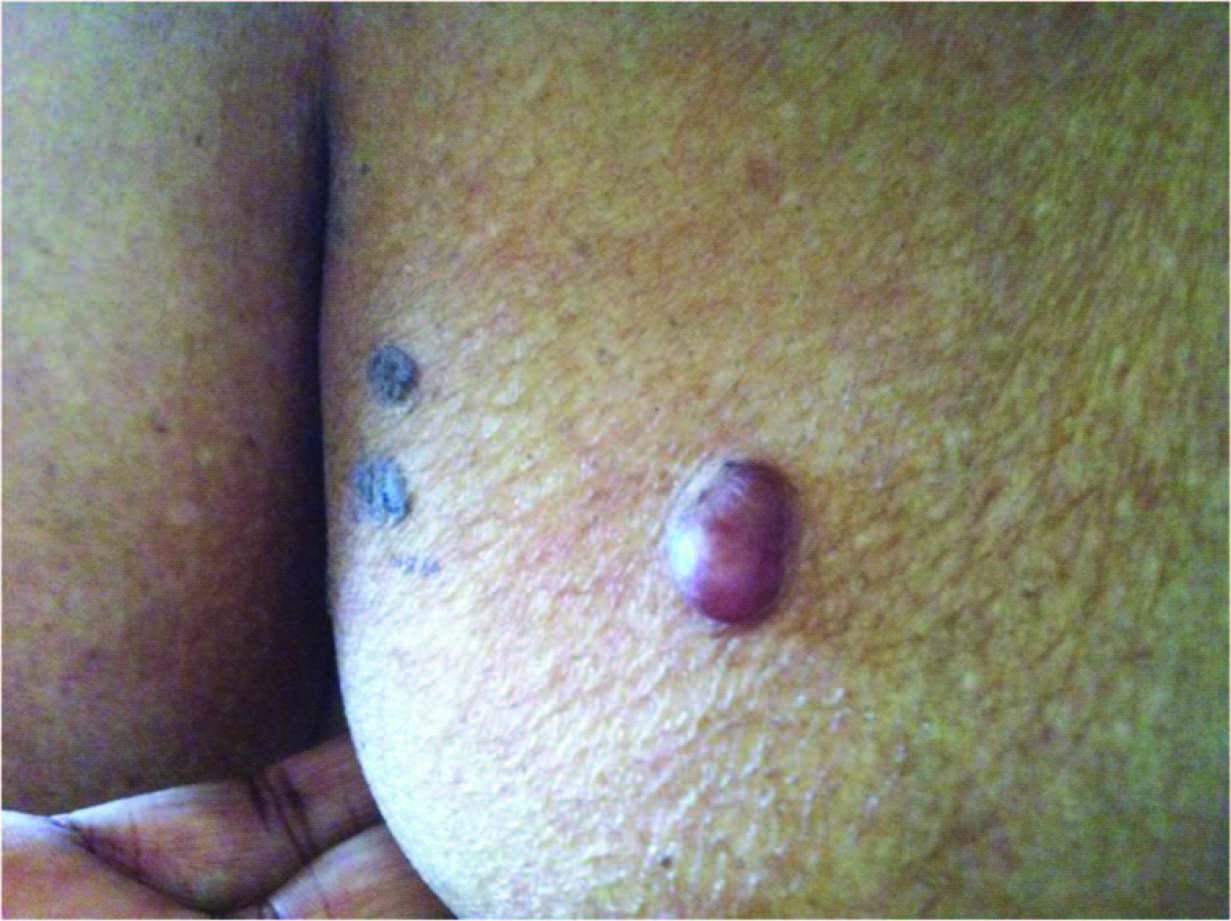
The provisional clinical diagnosis offered for the nodule over the thigh was “Benign Adnexal tumour” and that for the lesion over the breast was “Seborrheic Keratosis.” Both the lesions were excised and submitted for histopathological examination.
Histopathological Findings
Gross examination- Biopsy from the breast showed a skin covered grey- white tissue mass measuring 0.75 x 0.4 cms. Biopsy from the thigh showed a skin covered grey- white tissue mass measuring 1.75 x 0.75 cms. Cut section showed a well encapsulated grey- white, skin covered nodule.
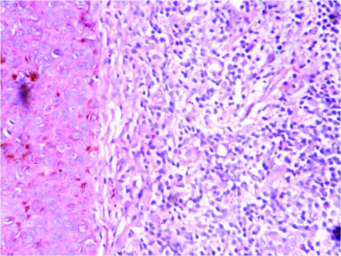
Excisional biopsy from the lesion over the breast showed focal hyperkeratosis, acanthosis, focal spongiosis and melanin incontinence. Papillary dermis showed diffuse and dense band- like lymphocytic infiltrates [Table/Fig-2].
Papillary dermis with diffuse and dense band- like lymphocytic infiltrates in lichen planus, (H & E, ×400)
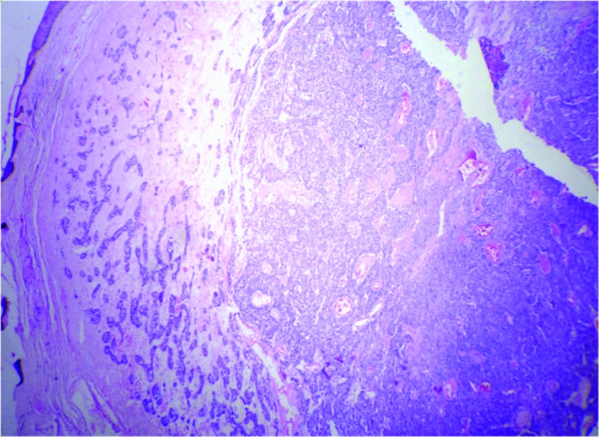
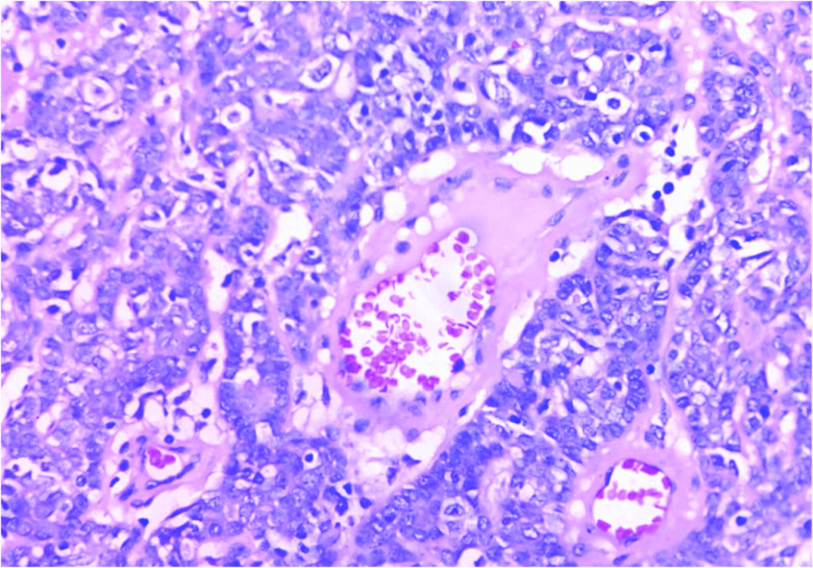
Biopsy from the nodule over the thigh showed thinned out epidermis and a well- encapsulated, lobulated appendageal tumour in the dermis composed of two types of epithelial cells arranged in cords and sheets with evidence of ductal differentiation at places [Table/Fig-3]. The cells in the center were large and basaloid with large pale nuclei. The cells in the periphery were smaller with hypechromatic nuclei and scanty cytoplasm. Some blood vessels showed perivascular palisading by tumour cells [Table/Fig-4].
Scanner view depicting the topography of eccrine spiradenoma, (H & E, ×40)
Perivascular palisading by tumour cells. Two types of tumour cells- 1)Large basaloid cells with pale nuclei 2)Small cells with hyperchromatic nuclei, (H & E, ×400)
Final diagnosis- Eccrine spiradenoma over the thigh co- existent with lichen planus over the breast.
Discussion
Eccrine spiradenoma, an uncommon benign tumour of sweat gland origin with debatable histogenesis occurs mostly in young adults aged between 15 to 35 years [1,2]. Most spiradenomas are benign. Malignant spiradenoma was described in the year 1972 by Dakske for the first time [3]. About 50 cases of malignant spiradenomas have been described in literature. Spiradenomas have been known to occur in Brooke Spiegler syndrome which manifests with combined lesions like cylindromas, spiradenomas and trichoepitheliomas [4,5]. But an eccrine spiradenoma co- existing with lichen planus in an old and treated case of basal cell carcinoma, as in the present case is an extremely uncommon occurrence. Extensive literature search did not reveal any other case with similar co- existence. On a cytogenetic study Dijkhuizen et al., [6] observed similar karyotypes for a spiradenoma and two metastatic lymph nodes. Whether a genetic causal relationship exists between eccrine spiradenoma, lichen planus and basal cell carcinoma is an integral issue which needs to be resolved by advanced research and cytogenetic studies in this regard.
Eccrine spiradenomas are known to arise from the coiled duct and secretory portion of the eccrine apparatus [7]. They present as blue to red dermal and subcutaneous nodules and hence referred to as “blue balls in the dermis”. The tumour in the present case presented as a blue to blackish colored nodule. They occur predominantly in young adults with a peak incidence in the second, third and fourth decades of life. The present case is an exception with the lesion presenting at 80 years [7]. Majority of these lesions are solitary [8] as in our case. Rarely, they may be associated with cylindroma [8], eccrine hidradenoma [9], trichoepithelioma [7] and pilar components [9].
Nath AK et al., have reported a co- existent chondroid syringoma in their case of eccrine spiradenoma [10]. The present case seems to be unique in the co- existence of eccrine spiradenoma with a non- neoplastic condition like lichen planus in a patient with previously treated basal cell carcinoma.
Conclusion
The present case is an unusual combination of pathological conditions which bear no causal relationships with each other as per the current knowledge available regarding these entities. Advanced research in future may help unravel this interesting mystery shrouding the co- existence of multiple pathological lesions like eccrine spiradenoma and lichen planus in a previously treated case of basal cell carcinoma.
[1]. Ekmekci TR, Koslu A, Sakiz D, Congenital blaschkoideccrine spiradenoma on the faceEur J Dermatol 2005 15:73-74. [Google Scholar]
[2]. Scheinfeld NS, Tarlow MM, Burgin S, Blaschkoideccrine spiradenomaCutis 2002 70:73-75. [Google Scholar]
[3]. Dabska M, Malignant transformation of eccrine spiradenomaPol Med J 1972 11:388-96. [Google Scholar]
[4]. Bumgardner AC, Hsu S, Nunez- Gussman JK, Schwartz MR, Trichoepitheliomas and eccrine spiradenomas with spiradenoma/ cylindroma overlapInt J Dermatol 2005 44:415-17. [Google Scholar]
[5]. Clarke J, Ioffreda M, Helm KF, Multiple familial trichoepitheliomas: a folliculosebaceous- apocrine genodermatosisAm J Dermatopathol 2002 24:402-05. [Google Scholar]
[6]. Dijkhuizen T, van den Berg E, Nikkels PG, Hoekstra HJ, de Jong B, Cytogenetics of a case of ecrine spiradenomaHum Pathol 1992 23:1085-87. [Google Scholar]
[7]. Gupta S, Jain VK, Singh U, Gupta S, Multiple eccrine spiradenomas in zosteriform distribution in a childPediatr Dermatol 2000 17:384-86. [Google Scholar]
[8]. Bedlow AJ, Cook MG, Kurwa A, Extensive naevoid eccrine spiradenomaBr J Dermatol 1999 140:154-57. [Google Scholar]
[9]. Gupta S, Radotra BD, Kaur I, Handa S, Kumar B, Multople linear eccrine spiradenomas with eyelid involvementJ Eur Acad Dermatol Venereol 2001 15:163-66. [Google Scholar]
[10]. Nath Ak, Kumari R, Thappa DM, Eccrine spiradenoma with chondroid syringoma in Blaschkoid distributionIndian J Dermatol Venereol Leprol 2009 75:600-02. [Google Scholar]