Case Report
A 34-year-old female patient presented to Department of Oral and Maxillofacial Surgery of Government Dental College and Hospital, Ahmedabad, India, seeking treatment for a slowly enlarging, asymptomatic mass in posterior left part of maxilla on palatal aspect that appeared three years earlier. Patient was psychologically obsessed about the growth in the maxilla and was irritated with the constant involuntary movement of the tongue over the growing mass. There was no history of trauma in that region. Medical history was non contributory.
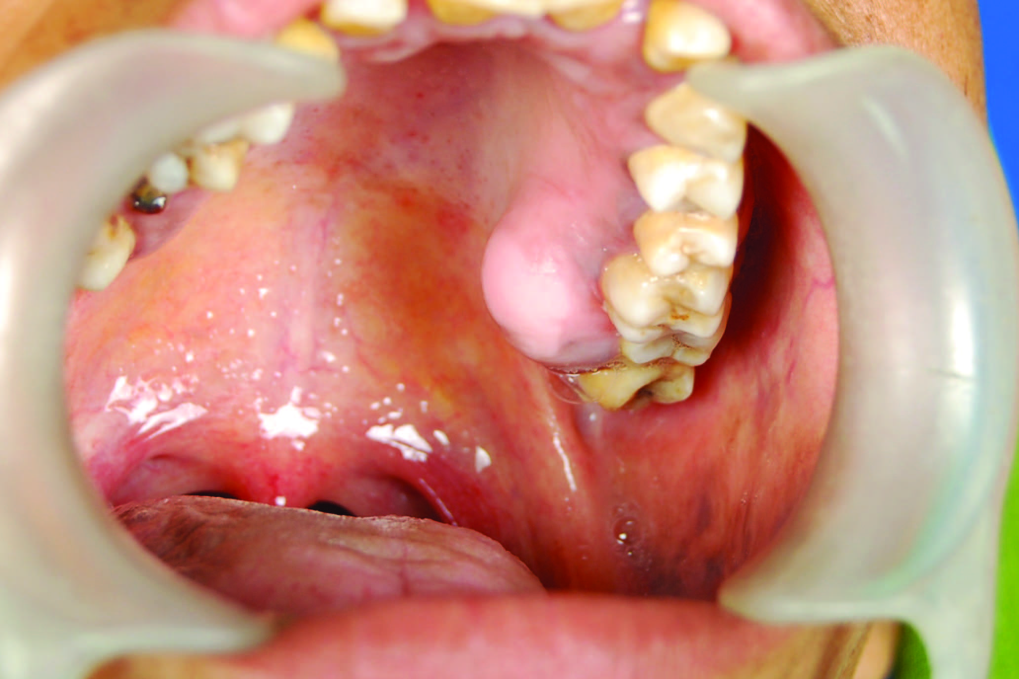
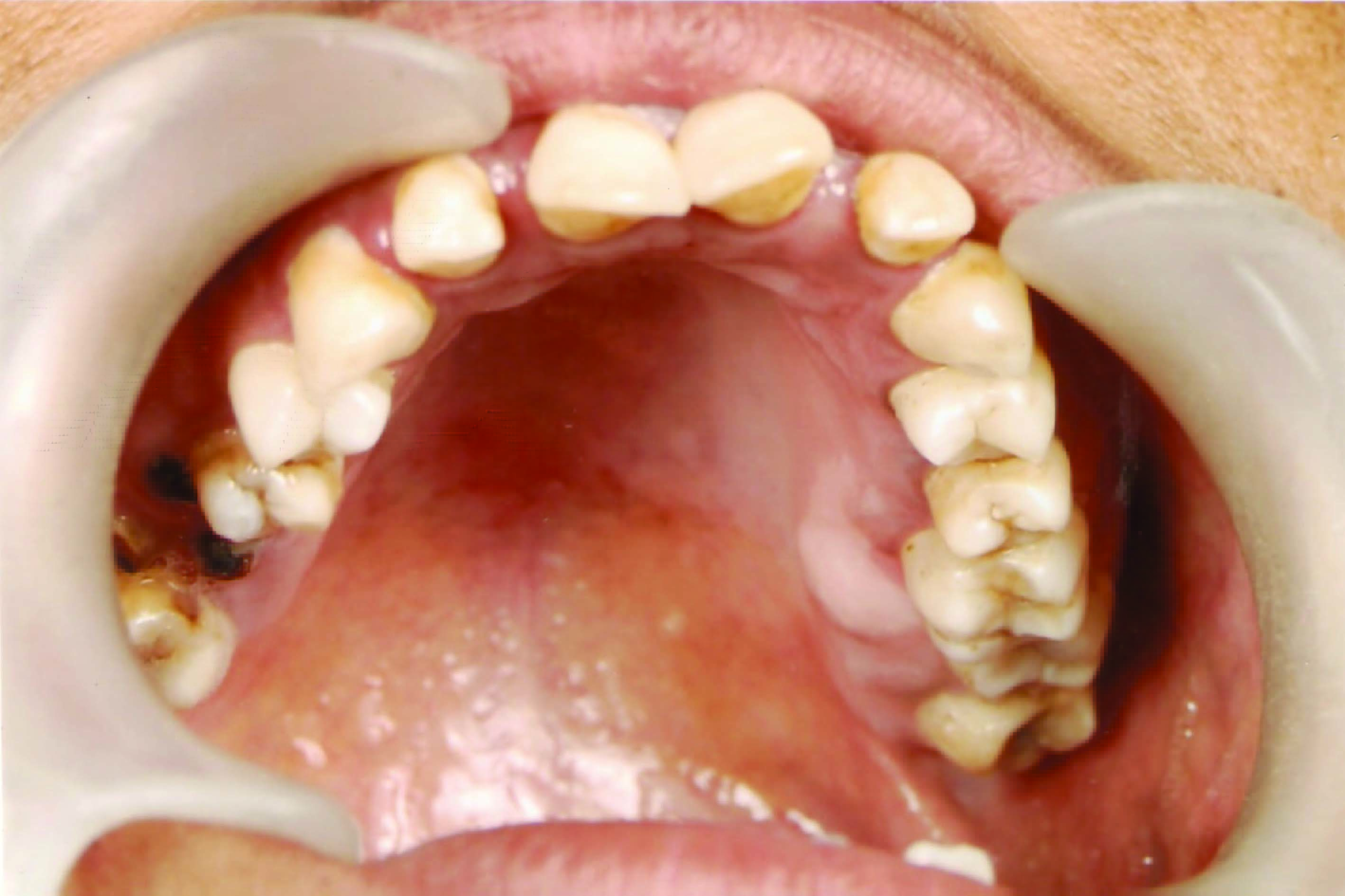
Intraoral examination revealed a well defined sub-periosteal mass on the palatal aspect of left maxilla in posterior region, which extended from lingual aspect of middle of upper left first molar to distal end of upper left second molar [Table/Fig-1]. The lesion was hard on palpation, sessile, and measured 20 x 15 mm in diameter. Lesion was non-tender on palpation with blanching of overlying mucosa. All teeth in upper left quadrant responded normally to electric pulp testing. No regional lymphadenopathy was present.
Intra Oral view with slowly enlarging, asymptomatic mass in posterior left part of palatal
Intraoral radiographs including maxillary occlusal radiographs were taken to rule out any radiolucent cystic lesion. Maxillary occlusal radiograph showed no abnormalities. CT Scan in coronal section through upper left first molar region demonstrated well defined bony lesion arising from alveolus with pedunculated base without involvement of any tooth [Table/Fig-2]. A provisional clinical diagnosis of peripheral osteoma was made.
C.T. Scan in coronal section through upper left first molar demonstrates well defined bony lesion arising from alveolus with pedunculated base without involvement of any tooth

The mass was approached intra-orally under local anaesthesia. The mass appeared well circumscribed and attached with the palatal alveolar bone in upper left first and second molar region [Table/Fig-3]. A decision to surgically excise the mass was made. Superficial cut made with fissure bur and lesion removed with the help of chisel and mallet [Table/Fig-4]. Residual bed of lesion was debrided, smoothened and flap was closed with absorbable sutures [Table/Fig-5]. Palatal splint was given to reduce dead space. Patient tolerated the procedure well.
Surgical excision of the bony mass

Complete excision with smoothening of the palatal bone

Post operative view of surgical site

Grossly, the specimen consisted of a homogenous mass of cancellous bone [Table/Fig-6]. Postoperative CT scan showed complete excision of the lesion [Table/Fig-7]. Histologically, the diagnosis of cancellous type of osteoma was confirmed [Table/Fig-8]. Patient was scheduled for regular follow up. Intraoral examination at one year follow up revealed normal mucosal tissue without any recurrence of lesion [Table/Fig-9].


Histopathological slide shows irregular bone with empty lacunae and hemorrhagic area

Post operative 1 year follow up
Discussion
Osteomas are benign osteogenic tumors of the bone [1–7] arising from the proliferation of compact or cancellous bone. They may be peripheral, central, or extraskeletal. Peripheral osteomas arise from the periosteum and central osteomas arise from the endosteum, whereas extra skeletal osteomas develop from the soft tissues, usually within a muscle [2].
Osteomas are found mainly in the cranio maxillofacial bones. Peripheral osteomas (PO) are uncommon. Clinically, the PO is usually an asymptomatic slow growing lesion which can produce swelling and asymmetry. The pathogenesis of PO is unclear. Some investigators consider it a true neoplasm, while others classify it as a developmental anomaly [8]. The possibility of a reactive mechanism, triggered by trauma or infection has also been suggested [3,9]. In the present case no history of trauma or infection was elicited. The association between maxillofacial osteomas, cutaneous sebaceous cysts, multiple supernumerary teeth and colorectal polyposis is known as Gardner’s syndrome [10].
Osteomas, usually asymptomatic, and appear to have a very slow growth rate. They often remain undetected unless incidentally found on a routine radiographic survey or until they cause facial asymmetry or functional impairment [5,11]. They produce symptoms by compression, rather than by invasion or destruction [5,11]. Depending on the location, they might cause headaches, facial pain or swelling, exophthalmos and/or limited mandibular movements or deviation of the mandible on opening [11].
Peripheral osteoma occurs most frequently in the para nasal sinuses in the maxillofacial area. The most common site is the frontal sinus, followed by the ethmoidal and maxillary sinuses. PO has also been described in the external auditory canal, and rarely in the temporal bone and pterygoid plates [10]. PO of the jaw-bones is uncommon [3]. These lesions usually appear as unilateral, pedunculated mushroom-like masses.
In the mandible, the most common sites are the angle and lower border of the body, locations that are more susceptible to trauma [9], also, the location of PO of the jaws is usually in close proximity to areas of muscle attachment, suggesting that muscle traction may play a role in its development [3,8].
In the maxilla, osteomas arise from the maxillary sinus [4,12], from the buccal plate in molar region and the tuberosity of the maxilla, and from the anterior [13] or posterior [11] part of the maxilla. The exact aetiology and pathogenesis of PO is still unclear, traumatic, congenital, inflammatory and endocrine causes have been considered as possible etiologic factors [14].
It has also been considered that osteomas arise either from embryological cartilaginous rests or from persistent embryological periosteum [12]. Therefore, osteomas originate from the suture between bones with different embryological derivation.
Osteomas can also arise from the proliferation of the periosteum related osteogenic cells stimulated by the chronic infection of the paranasal sinuses [13].
Some consider osteomas as a reactive condition triggered by trauma. This contention is strengthened by the fact that peripheral osteomas are generally located on the lower border or buccal aspect of the mandible, which are areas usually susceptible to trauma [2]. It has been suggested that muscle traction along with trauma may play a role in development of osteomas [3]. Our patient did not report any history of trauma in the area.
Osteomas have no definite sex predilection [2,6]. However, Kaplan et al.,[3], in their case series found a male to female ratio of 2:1. Osteomas can arise at any age though it seems to be more common among young adults [2,3,5–7].
Peripheral osteomas should be distinguished from exostoses, osteoblastomas and osteoid osteomas. Exostoses are bony excrescences of reactive or developmental origin that occur on the buccal aspect of alveolar bone. They are not thought to be true neoplasm’s [15]. Osteoblastomas and osteoid osteomas are frequently painful and may exhibit a more rapid rate of growth than osteomas.
Peripheral osteoma can be distinguished into two different types [15]. The compact or “ivory” osteoma, usually has a sessile base with normal-appearing dense bone with minimal marrow spaces, and occasional haversian canals. The size may range from several millimeters to several centimeters; however, part of the lesion may be in bone, masking the true size. The cancellous osteoma is usually corticated and has a smooth or irregular surface. It is pedunculated in nature and contains bony trabeculae and bone marrow. Overall it fairly resembles the bone of origin.
Patients with osteomas should be evaluated for Gardner’s syndrome [10]. Symptoms consistent with this syndrome include rectal bleeding, diarrhea, and abdominal pain. Patients with Gardner’s syndrome usually suffer from colorectal polyposis, skeletal abnormalities, and multiple impacted or supernumerary teeth. Onset occurs in the second decade, with malignant transformation of the colorectal polyps approaching 100% by the age 40. The skeletal involvement includes peripheral and endosteal osteomas in the skull, ethmoid sinuses, mandible and maxilla. The mandibular osteomas are usually lobulated and located at the angle of the mandible [16].
The patient in this case report underwent complete oral and physical examination for ruling out Gardner syndrome. Radiograpic evaluation including chest X-ray and PA skull was done to rule out multiple impacted and supernumerary teeth and multiple osteomas and odontomes of the jaws. CBC and LFT and Thyroid function tests were within normal limits. Patient also gave no history of any gastrointestinal complaints.
Imaging of PO can be achieved by traditional radiography (i.e.: panoramic radiograph, Water’s view) or by CT scan. The use of CT scanning with 3-D reconstruction makes it possible to achieve a better resolution and more precise localization [17].
An individualized approach for management of osteomas is recommended considering the size and location of the tumour [11]. Partial removal of the neoplasm can be considered in order to preserve the bone tissue indispensable for prosthetic rehabilitation [18]. Patients with known asymptomatic osteomas should be evaluated every 1 to 2 y to assess growth and to monitor the development of complications.
Definite treatment of the osteoma consists of complete surgical removal at the base where it unites with the cortical bone [3]. Surgery is indicated if there is painful or active lesion growth [19], or in order to correct asymmetry or other secondary problems, such as blockage of cavities, nerve foramina and vital organ compression, desire of definite histopathological diagnosis, or when there are symptoms or complications secondary to the osteoma that have failed to improve despite appropriate medical therapy [11].
The surgical approach should be case specific. For the mandible there are intraoral or extraoral approaches. The intraoral approach is preferable when possible, mainly for cosmetic reasons. For the maxillary antrum, the sub-labial gingivo-buccal (Caldwell-Luc) approach is convenient [20]. Endoscopic nasal approach for the resection of ethmoidal and frontal osteomas has been reported [11]. For the temporal, frontal and fronto-orbito-ethmoidal lesions the coronal or bi-coronal approaches have been classically used. However, these require an extensive amount of dissection, and carry the potential for significant morbidity, especially considering that the lesion to be excised is benign [20].
Recurrence of PO after surgical excision is extremely rare [21]. There are no reports of malignant transformation of PO in the literature [5,6].
Conclusion
Peripheral osteomas are relatively uncommon in the palate. Though the pathogenesis of peripheral osteomas is still controversial, the definite treatment is surgical excision. The present case was also treated with surgical excision. Ruling out the presence of Gardener syndrome is extremely important. Periodic clinical and radiological follow up should be done though recurrence of peripheral osteomas after excision is extremely rare.
[1]. World Health OrganizationApplication of the International Classification of Diseases to dentistry and stomatology 1995 3rd edGenevaWorld Health Organization:139 [Google Scholar]
[2]. Bodner L, Gatot A, Sion-Vardy N, Fliss DM, Peripheral osteoma of the mandibular ascending ramusJ Oral Maxillofac Surg 1998 56:1446-49. [Google Scholar]
[3]. Kaplan I, Calderon S, Buchner A, Peripheral osteoma of the mandible: a study of 10 new cases and analysis of the literatureJ Oral Maxillofac Surg 1994 52:467-70. [Google Scholar]
[4]. Richards HE, Strider JW, Short SG, Theisen FC, Larson WJ, Large peripheral osteoma arising from the genial tubercle areaOral Surg Oral Med Oral Pathol 1986 61:268-71. [Google Scholar]
[5]. Aghabeigi B, Evans AW, Crean SJ, Hopper C, Simultaneous repair of an orbital floor fracture and removal of an ethmoid osteoma: case report and review of the literatureInt J Oral Maxillofac Surg 2003 32:94-96. [Google Scholar]
[6]. Swanson KS, Guttu RL, Miller ME, Gigantic osteoma of the mandible: report of a caseJ Oral Maxillofac Surg 1992 50:635-38. [Google Scholar]
[7]. Shafer WG, Hine MK, Levy BM, A textbook of oral pathology 1974 3rd edPhiladelphiaWB Saunders:151-52. [Google Scholar]
[8]. Sayan NB, Ucok C, Karasu HA, Gunhau O, Peripheral osteoma of the oral and maxillofacial region: a study of 35 new casesJ. Oral Maxillofac Surg 2002 60:1299-301. [Google Scholar]
[9]. Rodriguez R, Rizzo S, Fiandrino G, Lupi S, Galiotio S, Mandibular traumatic peripheral osteoma: a case reportOral Surg Oral Med Oral Pathol 2011 112:44-48. [Google Scholar]
[10]. Lew D, DeWitt A, Hicks RJ, Cavalcanti MG, Osteomas of the condyle associated with Gardner’s syndrome causing limited mandibular movementJ Oral Maxillofac Surg 1999 57:1004-09. [Google Scholar]
[11]. De Chalain T, Tan B, Ivory osteoma of the craniofacial skeletonJ Craniofac Surg 2003 14:729-35. [Google Scholar]
[12]. Varboncoeur P, Vanbelois HJ, Bowen LL, Osteoma of the maxillary sinusJ Oral Maxillofac Surg 1990 48:882-83. [Google Scholar]
[13]. Seward MHE, An osteoma of the maxillaBr Dent J 1965 5:27-30. [Google Scholar]
[14]. Lucas RB, Pathology of tumors of the Oral Tissues 1984 Edinburgh, ScotlandChurchill Livingstone:191-94. [Google Scholar]
[15]. Regezi JA, Sciubba J, Oral Pathology 1993 ed 2Philadelphia, PASaunders:407 [Google Scholar]
[16]. Jones K, Korzcak P, The diagnostic significance and management of Gardner's syndromeBr J Oral Maxillofac Surg 1990 28:80-84. [Google Scholar]
[17]. Bodner L, Bar-Ziv J, Kaffe I, CT of cystic jaw lesionsJ Comp Assist Tomog 1994 18:22-26. [Google Scholar]
[18]. Navotti M, Pignanelli M, Banfi L, Caronni EP, Osteomas of the jaws. A clinical and rehabilitative problemMinerva Stomatol 1991 40:591-97. [Google Scholar]
[19]. Woldenberg Y, Nash M, Bodner L, Peripheral osteoma of the maxillofacial region. Diagnosis and management: a study of 14 casesMed Oral Patol Oral Cir Bucal 2005 10(Suppl. 2):E139-42. [Google Scholar]
[20]. Brodish BN, Morgan CE, Sillers MJ, Endoscopic resection of fibro- osseous lesions of the paranasal sinusesAm J Rhinol 1999 13(11):1-6. [Google Scholar]
[21]. Bosshardt L, Gordon RC, Westerberg M, Morgan A, Recurrent peripheral osteoma of the mandible: report of a caseJ Oral Surg 1971 29:446-50. [Google Scholar]