The primary aim of chemo mechanical preparation is to completely remove the microorganisms, pulp tissue and debris and enlarging the canal diameter to receive an obturating material [1]. At times, in the zeal of biomechanical preparation of the canal, we inevitably end up damaging the root dentin which becomes a gateway to dentinal cracks and minute intricate fractures or even vertical root fractures, thereby failure of treatment [2]. Complexities in canal preparation may be attributed to variation in the design of the cutting instrument, taper and composition of the material from which it is made [3].
In the last decades, the emergence of NiTi rotary instrumentation has transfigured the root canal treatment by reducing the operator fatigue, time required to complete the preparation and minimized the procedural errors as compared with hand instrumentation [4]. However, rotary files with large tapers may cause significantly more complete and incomplete dentinal cracks [5].
Recently, single file nickel titanium (NiTi) reciprocating systems has been introduced which completes the canal preparation with only one instrument, which is claimed to relieve stress on the instrument because of its peculiar counter clockwise (cutting action) and clock wise (release of instrument) requiring even lesser time than rotary full-sequence systems [6]. It is assumed that this movement reduces the risk of cyclic fatigue caused by tension and compression [7–9].
Debate continues regarding the best motion of action for NiTi rotary files. Till date no studies have compared the incidence of dentinal micro cracks of Protaper, Protaper Next, Reciproc, One shape files using two different motions {Rotary (Rot) and reciprocation (Rec)} with that of hand files during root canal preparation using stereomicroscopy.
Materials and Methods
This study was conducted in August, 2014 for a period of 3 weeks, In Mamata Dental College. Khammam, India. One hundred human extracted mandibular central incisors were selected and kept in distilled water. Radiographs were taken from buccolingual and mesiodistal angles. Specimens with single root and single patent canal were included in the study. Root fractures, cracks, open apices,curved canals,multiple roots,caries or restorations, severe anatomic variations, calcified canal were excluded. To ensure standardization, decoronization was done under water cooling with a low-speed saw (Isomet; Buehler Ltd, Lake Bluff, IL) maintaining 16 mm from the apex.
During this study specimens were wrapped in 4x4 gauze and kept moist. A silicon impression material (Oranwash; Zhermack SpA, Rovigo, Italy) was used for coating the surface of roots to simulate periodontal ligament space.
Cleaning and shaping
The working length of the canals was determined by inserting a size #10 K file (Dentsply Maillefer, Ballaigues, Switzerland) into the root canal terminus and subtracting 1 mm from this measurement. A glide path was performed via a size #15 Kfile (Dentsply Maillefer, Ballaigues, Switzerland). The root canals were irrigated with 1% sodium hypochlorite solution after each instrument change. Each instrument was changed after preparing four canals. A total of 12 mL 1% sodium hypochlorite was used in each canal. After preparation, the specimens from the prepared groups were rinsed with 5 mL distilled water.
The specimens were divided into 10 groups (n=10). All instruments were set in rotation and reciprocating motion through X-Smart Plus (Dentsply Maillefer, Ballaigues, Switzerland) and the speed and torque was programmed according to manufacturer instructions and the flutes of every instrument were cleaned after three pecking motions.
Group 1 (Positive Control, n = 10): No preparation.Group 2 (NiTi hand K file, Negative Control, n = 10): Canals were enlarged to #40 size K file using the balanced force technique.
Group 3 (Protaper – rot, n=10): Canals were prepared using Protaper rotary files (Dentsply Maillefer, Ballaigues, Switzerland) mounted in a 6:1 reducing hand piece,and X-Smart Plus motor (Dentsply Maillefer, Ballaigues, Switzerland) and was set in rotary speed program (300 rpm). The Protaper shaping SX will be used in coronal Enlargement, then S1, S2, F1, F2, F3 files will be sequentially used to the working length. Group 4 (Protaper– rec, n=10): Protaper files (Dentsply Maillefer, Ballaigues, Switzerland) were mounted in a 6:1 reducing hand piece, and the X-smart Plus motor (Dentsply Maillefer, Ballaigues, Switzerland) was set at the reciproc program.Group 5 (Protaper Next - rot, n=10): Canals were prepared using Protaper Next (Dentsply Maillefer, Ballaigues, Switzerland) mounted in a 6:1 reducing hand piece and X-SmartPlus motor and was set at speed program (300 rpm, 200 g/cm torque). The Protaper Next files (Dentsply Maillefer, Ballaigues, Switzerland) were used in the sequence Protaper Universal SX and then Protaper Next X1, X2, and X3. Group 6 (Protaper Next – rec, n=10): Protaper Next files (Dentsply Maillefer, Ballaigues, Switzerland) were mounted in a 6:1 reducing hand piece, and the X-smart Plus motor (Dentsply Maillefer, Ballaigues, Switzerland) was set at the reciproc program. Group 7 (One shape – Rot, n=10): The canals were first prepared with NiTiflex files k-files (Dentsply Maillefer, Ballaigues, Switzerland)to # 15.Canal preparation was then performed to the apical foramen with Oneshape rotary file (Micro-Mega, Besancon Cedex, France) #25,0.06 taper at a constant speed of 400 rpm in pecking motion. Group 8 (One shape – Rec, n=10): One shape files were mounted in a 6:1 reducing hand piece, and the X-smart Plus motor (Dentsply Maillefer, Ballaigues, Switzerland) was set at the reciproc program. Group 9 (Reciproc - Rot, n=10): The canals were first prepared with NiTiflex files k-files (Dentsply Maillefer, Ballaigues, Switzerland) (to # 15. A Reciproc file with size #25 .08 taper was then used in continuous motion at a constant speed of 300 rpm. Group 10 (Reciproc – Rec, n=10): The canals were first prepared with NiTiflex files k files to # 15. A Reciproc file (VDW, Munich, Germany), with size #25 .08 taper was then used in a reciprocating motion to the apical foramen using the “reciprocal” mode.
Sectioning and Microscopic Examination: All the specimens were sectioned perpendicular to the long axis at 3, 6, and 9 mm from the apex using a low-speed saw (Isomet; Buehler Ltd, Lake Bluff, IL) under water cooling. Slices were observed under a digital stereomicroscope (Expert DN) at X25 magnification and pictures were taken (Olympus BX43).
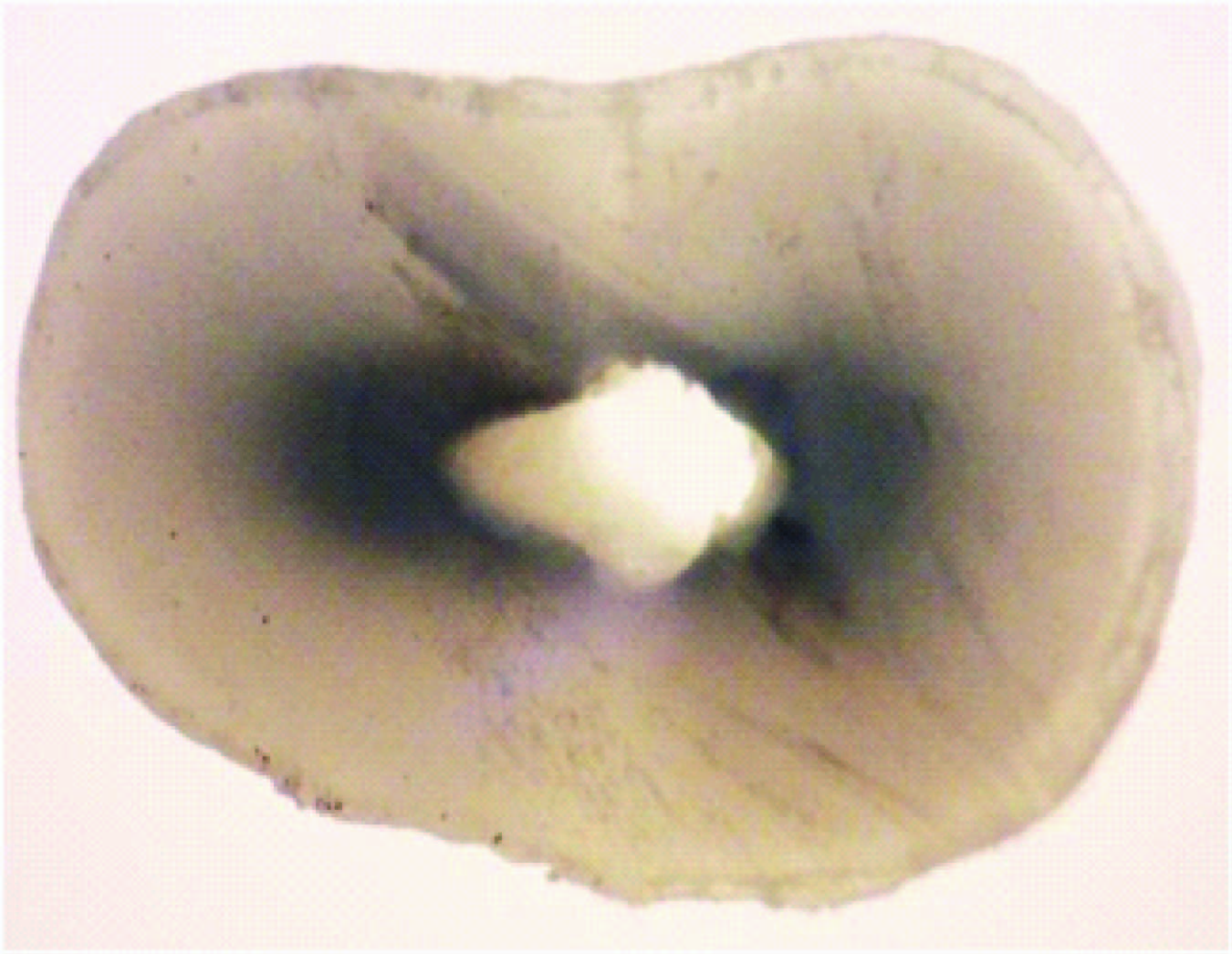
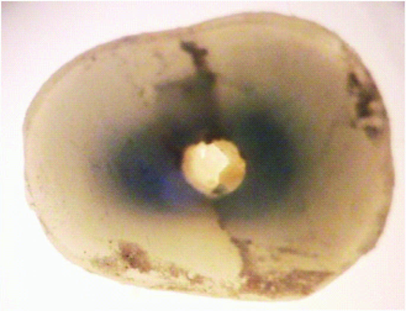
Definition of dentinal microcracks: To define crack formation, 2 different categories were made (“no crack” and “crack”) [Table/Fig-1a,b]. To avoid the confusing description of root cracks they were divided in to two categories:
Cross sectional image showing no crack
Cross sectional image showing crack
No crack- No crack was defined as root dentin without cracks or craze lines either at the internal surface of the root canal wall or the external surface of the root.
Crack- Crack was defined as all lines observed on the slice that either extended from the root canal lumen to the outer surface or from the outer root surface into the dentin [10].
Statistical Analysis
The results were expressed as the number and percentage of cracked roots in each group. The data were analysed with a chi-squaretest and Kruskal – Wallis test. All statistical analyses were performed using SPSS software (SPSS Inc, Chicago, IL).
Results
No cracks were observed in the negative control group (unprepared) and in the hand files group.Vertical root fractures were not observed in any group. There was a statistically significant difference between the groups (p<0.05). There were no significant differences in crack formation between the groups (Protaper Next - Rot, Protaper Next - Rec, Reciproc – Rec); (ProTaper - Rot, ProTaper- Rec, Oneshape - Rot); (Oneshape - Rot,Reciproc - Rot); (One shape - Rec, Reciproc – Rec); (p > .05) represented in [Table/Fig-2].
Graph showing % of defects in all groups

Discussion
In the present study, dentinal cracks were observed in all groups except group 1 which implies that the sectioning method did not induce damage, so it may be concluded that the cracks were a result of the preparation procedures and currently no method is able to avoid completely such cracks.
This study is in accordance with the previous studies done by Bier CAS et al., and Shemesh H et al., who compared the incidence of dentinal defects of manual Flexo files with different rotary files systems: ProTaper (Dentsply-Maillefer, Ballaigues, Switzerland), ProFile (Dentsply-Maillefer), SystemGT (Dentsply-Maillefer), or S-ApeX (FKG Dentaire, La Chaux-de-Fonds, Switzerland) and concluded that no defects were found in the unprepared roots and those prepared with hand files and S-ApeX. ProTaper, ProFile, and GT preparations resulted in dentinal defects in 16%, 8%,and 4% of teeth, respectively [5,10].
Hand instrumentation did not cause much damage to the root canal wall which could be because of its less aggressive movements of the hand files in the canal compared with engine operated files [11]. In the present study, although cracks were observed in all groups, cracks in the coronal region are more compared to cracks in apical region which is in accordance with the previous studies done by Adorno CG et al., and Liu R et al., respectively [12,13]. Least amount of cracks were observed in the canals instrumented with Protaper Next files either in rotary or reciprocating motion and more cracks were observed in canals instrumented with Protaper in rotary or reciprocating motion.It might be because of high levels of stress concentrations in root canal walls that may result in crack formation and also the taper (F1,F2,F3 0.07, 0.08, and 0.09, respectively) which is greater than Protaper Next (X1, X2, and X3; 0.04, 0.06, and 0.07, respectively) which could explain the incidence of cracks observed [14]. The reason for less cracks in Protaper Next files is due to its off-centered rectangular design which generates a swaggering motion, which decreases the screw effect, dangerous taper lock and torque on any given file by minimizing the contact betweenthe file and the dentin [15]. In addition, Protaper Next files are made of M-wire alloy which shows more flexibility than those made from conventional NiTi wire [16–18].
There was no significant difference found when we compared the cracks of One Shape and Reciproc single file systems when set in rotary motion, whereas the same files when set in reciprocating motion showed less cracks comparatively and among these two single file systems Reciproc files showed less cracks than one shape files. This might be because of the reciprocating movement that minimizes torsional and flexural stresses, reduces canal transportation and also due to its cross sectional design [19,20]. Furthermore, the reciprocating motion showed significantly higher resistance to cyclic fatigue [21,22].
In the present study it is found that the canals instrumented with full sequence systems showed less cracks than single file systems where only, one instrument causes more stress generation leading to crack formation. Full sequence systems showed less cracks both in rotary and reciprocating motion.
Sectioning method was used which allowed the evaluation of the effect of root canal treatment procedures on the root dentin by direct inspection of the roots [23].
Conclusion
Least defects were seen in canals with hand instrumentation.
Among engine driven instrumentation Pro Taper Next files showed least cracks when set in rotary or reciprocating motion.
Full sequence systems showed less cracks than single file systems and reciprocating motion was found to be better for both full sequence and single file systems.