Brachial artery is the continuation of the axillary artery beyond the lower boarder of the teres major muscle, opposite the neck of the radius in the anterior cubital region it divides in to radial and ulnar arteries. Variations in upper limb arteries have been frequently observed majority of these variations occur in radial artery followed by ulnar artery [1], however brachial artery variations are less common [2]. Accurate knowledge of muscular and neurovascular variations is important for both surgeons and radiologists, which may prevent diagnostic errors [3].
The term accessory brachial artery was first established by McCormack and embryologically it referred to as the superficial brachial artery which is based on the persistence of more than one intersegmental cervical artery which does not deteriorate but persists and can even enlarge its diameter [4,5]. Tohno Y et al., reported a case of double brachial arteries in which superficial brachial artery descended in the arm superficial to the median nerve and deep brachial artery with its normal course descended behind the median nerve [6]. A detailed knowledge of variations of branching pattern of vessels is essential for providing accuracy during vascular diagnosis and re-constructive surgery and also in evaluation of angiographic images.
Accordingly, the present study was designed to evaluate the anatomical variations of the brachial artery and its morphology, embryogenesis and clinical implications.
Materials and Methods
In this study a total of 140 upper limb specimens of 70 embalmed cadavers were examined, which were dissected as the part of routine dissection for teaching undergraduate students in the Department of Anatomy- Mayo Institute of Medical Sciences-, Barabanki, Department of Anatomy- KMCT Medical College, Manassery, Calicut, Kerala and Department of Anatomy-Melaka Manipal Medical College-Manipal University. Out of 70 cadavers there were 35 males and 35 females, and the age of death ranged from 50 to 70 yrs.
The flexor (anterior) compartment of arm, cubital fossa and forearm were dissected according to the instructions given in the standard dissection manual. The skin, superficial fascia, deep fascia and muscles were separated using a scalpel and forceps and the anatomical variations of the brachial artery and its terminal branches with their relation to the surrounding structures were examined and representative anatomy was photographed for the proper documentation. Length of the accessory brachial artery is measured by 2 points (a) the midpoint of the width of the artery where it begins (b) point of termination.
Results
Accessory brachial artery was arising from the axillary artery at the lower border of teres major along with main brachial artery was noted in eight female cadavers (11.43%). Accessory brachial artery was placed superficially and medially, compared to main brachial artery, which was placed deeply and laterally.
Out of eight cadavers the following variable course of accessory brachial artery was noted
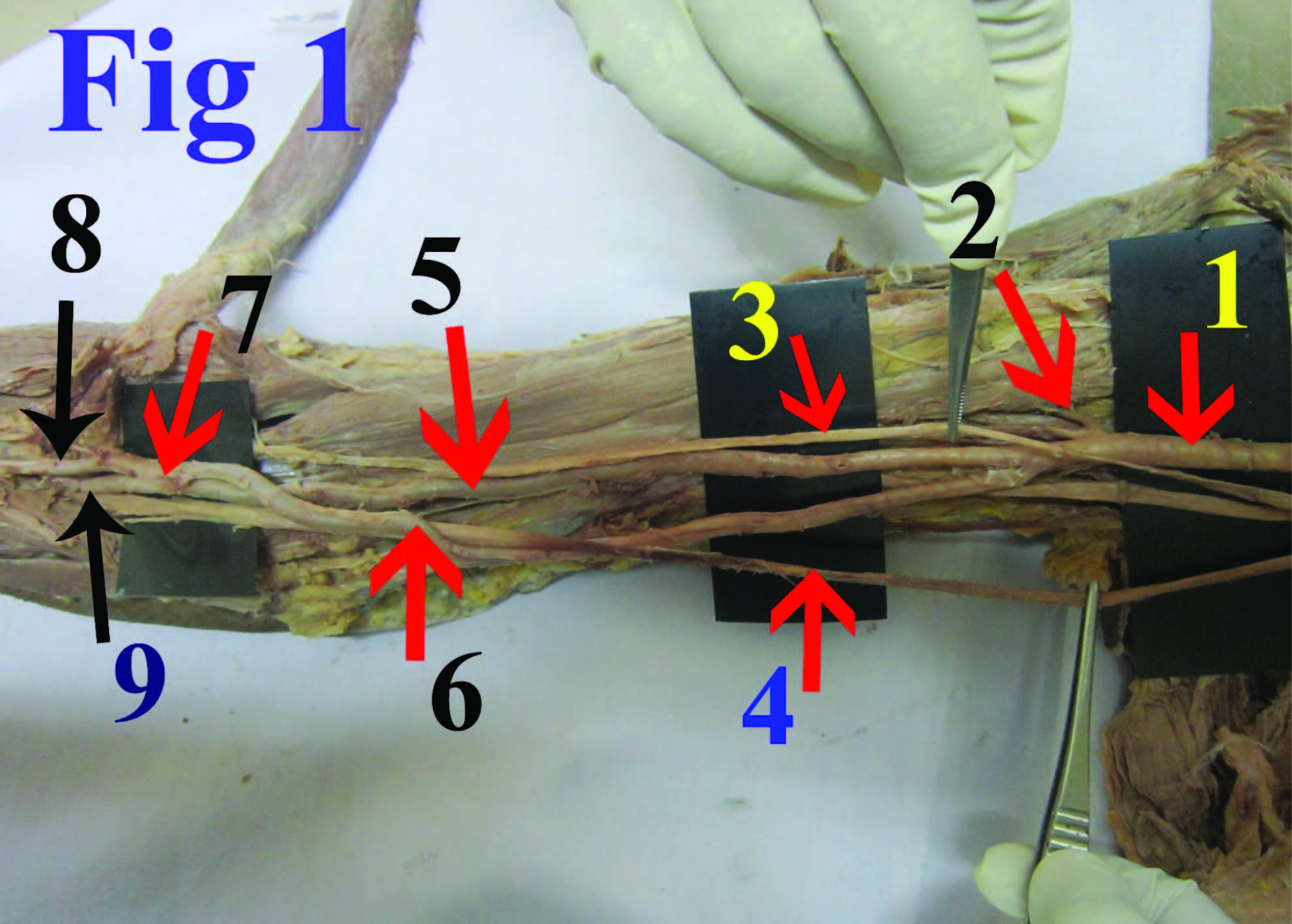
1. Rare unusual unilateral accessory brachial artery and its reunion in the cubital fossa with the main brachial artery in relation to the musculocutaneous nerve and median nerve were noted in five cadavers (Four left upper limbs and in one right upper limb) (7.14%). [Table/Fig-1,2].
Right upper limb showing unusual accessory brachial artery and its reunion in the cubital fossa with the main brachial artery in relation to median and musculo cutaneous nerve, 1- axillary artery; 2- profunda brachii artery arising from the lower part of the axillary artery; 3- musculo cutaneous nerve; 4-median nerve; 5-main brachial artery; 6- accessory brachial artery; 7- accessory brachial artery reunion in the cubital fossa; 8-radial artery; 9-ulnar artery
Left upper limb showing accessory brachial artery and its reunion in the cubital fossa with the main brachial artery in relation to Median Nerve 1- Axillary Artery; 2- Accessory brachial artery; 3-Main brachial artery; 4-Median Nerve; 5- Accessory brachial artery reunion in the cubital fossa; 6-Radial Artery; 7-Ulnar Artery

In relation to median nerve and musculocutaneous nerve:
In five cadavers unilateral accessory brachial artery in the upper part the flexor compartment of the arm is related medial to the medial nerve, where as in the lower part of the arm it crossed the median nerve ventrally from medial to lateral. At base of the cubital fossa the accessory brachial artery united with the main brachial artery where it is more tortuous across the median nerve [Table/Fig-1].
In two cadavers accessory brachial artery descends downwards parallel to the main brachial artery separated by median nerve. At base of the cubital fossa the accessory brachial artery united with the main brachial artery where it is crossed ventrally by the median nerve and after a short course the united trunk of accessory and main brachial artery divided into radial and ulnar arteries [Table/Fig-2].
In three cadavers at the middle of the arm musculocutaneous nerve was plastered to the deep brachial artery and at the base of the cubital fossa it passed behind the arterial trunk (formed by the union of accessory and main brachial arteries) [Table/Fig-1].
2. An unusual bilateral accessory brachial artery arising from axillary artery and is continuing in the forearm as superficial accessory ulnar artery were noted in the three cadavers (4.29%), whereas the main brachial artery is dividing into radial and ulnar arteries in the cubital fossa. [Table/Fig-3,4].
Right upper limb showing accessory brachial artery and its continuation in the forearm as superficial accessory ulnar artery, 1- Axillary Artery; 2-Median Nerve; 3-Main brachial artery; 4-Ulnar Artery; 5- Superficial accessory ulnar artery; 6- Accessory brachial artery; 7-Radial Artery

Left upper limb showing accessory brachial artery and its continuation in the forearm as superficial accessory ulnar artery, 1- Axillary Artery; 2 and 3- Accessory brachial artery; 4- Ulnar Artery ;5- Superficial accessory ulnar artery; 6- Main brachial artery; 7-Median Nerve; 8 and 9-Radial Artery

In relation to median nerve: An unusual bilateral accessory brachial artery throughout its course in the arm accompanies the median nerve on its medial side.
3. Accessory brachial artery does not have any branches and origin of profunda brachii artery from the third part of the axillary artery was noted in six cadavers (8.57%) [Table/Fig-1].
4. Accessory brachial artery length varies between 19 cm to 22 cm [Table/Fig-5].
Showing length of accessory brachial artery
| Serial Number | Sex | Side | Length (Cm) |
|---|
| 1 | Female | LEFT | 19 |
| 2 | Female | LEFT | 20 |
| 3 | Female | LEFT | 19 |
| 4 | Female | LEFT | 22 |
| 5 | Female | LEFT | 22 |
| 6 | Female | RIGHT | 19 |
| LEFT | 19 |
| 7 | Female | RIGHT | 20 |
| LEFT | 20 |
| 8 | Female | RIGHT | 19 |
Discussion
It is thought that the main blood supply to the forearm was dependent on the superficial brachial artery. Variations in upper limb arteries have been frequently observed either in routine dissections or in clinical practice. Persistent of superficial brachial artery was observed mostly in the right upper limb [6,7,8] and few cases also reported in the left upper limb [9]. In this study we also reported the left dominance of persistent of accessory brachial artery. Keen suggested that the superficial brachial artery is in fact high origin of the radial artery [10], whereas prevalence of the superficial brachial artery originating from the axillary artery was reported as 3% by Muller [11], 0.24% by Adachi [12], 1.25% by Kachlik et al., [13]. Whereas in this study prevalence of accessory brachial artery was noted as 11.43%. Such superficial course of accessory brachial artery can serve as a route for a catheter during the radial approach to coronary procedures for catheterization. At the same time existence of such superficial brachial artery is more prone to injuries which can lead to bleeding and ischaemia.
Kachlik et al., reported accessory brachial artery emerging from the third part of axillary artery and its reunion with the main brachial artery in the cubital fossa [14]. Yoshinaga et al., reported bifurcation of brachial artery into large superficial and small deep branches at the lower border of teres major muscle [15]. The superficial branch further divided into radial and ulnar arteries in the cubital fossa, while the deep branch mainly supplied the muscles of arm. Baeza et al., noted duplication of brachial artery and reported that superficial brachial artery ended by anastomosing with the radial artery in the cubital fossa and in few cases, it continued as antibrachial artery [16]. Whereas in this study prevalence of accessory brachial artery reunion in the cubital fossa with the main brachial artery was noted in five cadavers (11.43%). We considered that the knowledge of the anastomotic blood vessel, particularly its position in the cubital fossa, are clinically important and may complicate intravenous drug administration venipuncture and percutaneous brachial catheterization.
Embryological Explanation: In the foetal life radial and ulnar arteries appears in the forearm from the axial artery. The axis artery is derived from the lateral branch of the seventh intersegmental artery. Along the axial line the axis artery grows outwards and proximal part of it forms the axillary and brachial artery. Initially the radial artery arises more proximally than the ulnar artery, later it establishes a new connection with main trunk at or near the level of origin of ulnar artery. In the later stages of development the upper portion of radial artery (above the connection with main trunk) usually disappears. The type of anomalies presented in this study is due to origin of radial artery from the axial artery in the arm and persistence of upper portion of radial artery above its connection with axial artery or the abnormal bifurcation of axial artery in the arm and its reunion in the cubital fossa.
Profunda brachii artery is the largest branch of the brachial artery its variations in the origin and termination are rarely described in literature. Prevalence of the profunda brachii artery originating from the axillary artery was reported as 8.7% by Charles et al., [17], 16.6% by Anson [18], 2% by Patnaik [19] and 4% by Chauhan K et al., [20], where as in our study it was noted as 8.57%. Knowledge of this unusual anatomy is important during brachial artery catheterization and harvesting of lateral arm flaps.
In few cases higher origin of radial artery in arm with normal course in forearm have been reported [21,22]. Maruti Ram et al., [23] reported the continuation of the superficial brachial artery as radial artery, Shweta Solan et al., [24] reported the continuation of the superficial brachial artery as ulnar artery and Kodama [25] reported the continuation of the superficial brachial artery as radial artery and deep brachial artery as ulnar artery. In a study of 68 specimens unilateral superficial brachial artery reportedly divided into superficial radial and superficial ulnar arteries [26]. Where as, in our study bilateral accessory brachial artery was continued as a superficial accessory ulnar artery and main brachial artery was dividing into radial and ulnar arteries in the cubital fossa was noted in three cadavers. Such variant superficial accessory ulnar artery may complicate intravenous drug administration, venipuncture, and percutaneous brachial catheterization. Their superficial course can cause misinterpretation of incomplete angiographic images and also makes them more prone to injury, which may result in bleeding.
Embryological Explanation- Arey reported that anomalous blood vessels may be due to unusual paths in primitive vascular plexuses or persistence of vessels normally obliterated or disappearance of vessels normally retained or incomplete development or fusion and absorption of parts usually distinct [27]. Whereas bilateral persistence of superficial accessory ulnar artery noted in this study may be due to the persistence of embryological vessels or abnormal bifurcation of axial artery results in accessory brachial artery in the arm and its continuation in the forearm as a superficial accessory ulnar artery.
Course of variable brachial artery in relation to median and musculocutaneous nerve: Uneven tortuous course of superficial brachial artery in relation to the median nerve noted in this study may lead to compression of the medial nerve in the lower part of the anterior compartment of the arm. Such abnormal superficial tortuous accessory brachial artery course in the lower part of the anterior compartment of arm noted in this study may be mistaken for basilic vein during cannulation [28,29]. Such neurovascular variations are clinically important because symptoms of median nerve compression arising from such variations are often confused with more common causes, such as radiculopathy and carpal tunnel syndrome or pronator teres syndrome [30,31] In the arm musculocutaneous nerve passes obliquely downwards and laterally between the biceps brachii and brachialis then it penetrates the deep fascia slightly above the elbow, where as in this study at the middle of the arm it was plastered to the deep brachial artery and passed behind the arterial trunk (formed by the union of accessory and main brachial arteries) in the cubital fossa such unusual course was not reported in the literature. Such variant course of the musculocutaneous nerve at the middle of the arm and in the cubital fossa may lead to its compression resulting in paraesthesia and weakness of elbow flexion and supination.
There are only a few references in the literature on sex and laterality about accessory brachial artery. Fuss et al., [32], Rodriguez-Niedenfuhr et al., [33] and Musaed et al., [34] reported the incidence of superficial brachial artery was more frequent in males and on the right side. Whereas prevalence of accessory brachial artery noted in this study was more in females (11.43%) and on the left side. Dimensions (length) of the accessory brachial artery from the point of its origin to termination are clinically impartment. The length of accessory brachial artery noted in this study varies between 19 cm-22 cm. Knowledge of dimensions of brachial artery variations may reduce the incidence of iatrogenic injuries in angiographic studies preceding coronary artery bypass surgery.
Conclusion
An accurate knowledge anatomical variation of the brachial artery course, branching, bifurcation/termination, the course of its terminal branches and relationship with the surrounding structures is essential prerequisite during vascular and reconstructive surgeries of arm and forearm. Anatomical variations of brachial artery noted in this study are rare and very important clinically. Accessory brachial artery and superficial accessory ulnar arteries noted in this study may be mistaken for a vein and may complicate intravenous drug administration and venipuncture in general, also percutaneous brachial catheterization. A detailed knowledge of such vascular variations is essential not only to anatomists, but also to radiologists, orthopedists, vascular and plastic surgeons.