Benign Triton Tumour of Upper Lip- A Rare Neoplasm at an Extremely Uncommon Site
Hemalatha A. L1, Sanjay M2, Anoosha K3, Ashok K. P4, Shantha Kumari B.R5
1Professor, Department of Pathology, Affiliated to Rajiv Gandhi University of Health Sciences, Adichunchanagiri Institute of Medical Sciences, B. G. Nagar, Mandya, Karnataka, India.
2Assistant Professor, Department of Pathology, Affiliated to Rajiv Gandhi University of Health Sciences, Adichunchanagiri Institute of Medical Sciences, B. G. Nagar, Mandya, Karnataka, India.
3Post Graduate, Department of Pathology, Affiliated to Rajiv Gandhi University of Health Sciences, Adichunchanagiri Institute of Medical Sciences, B. G. Nagar, Mandya, Karnataka, India.
4Post Graduate, Department of Pathology, Affiliated to Rajiv Gandhi University of Health Sciences, Adichunchanagiri Institute of Medical Sciences, B. G. Nagar, Mandya, Karnataka, India.
5Post Graduate, Department of Pathology, Affiliated to Rajiv Gandhi University of Health Sciences, Adichunchanagiri Institute of Medical Sciences, B. G. Nagar, Mandya, Karnataka, India.
NAME, ADDRESS, E-MAIL ID OF THE CORRESPONDING AUTHOR: Dr. Hemalatha. A.L, No: 156, 12th Cross, 2nd Main, Jayanagar, Mysore- 570 014, India. Phone : 8453399335,
E-mail: halingappa@gmail.com
Benign Triton tumours are exceedingly rare tumours occurring predominantly in young children. Fewer than 20 cases have been reported in literature. The tumours develop as masses in various large nerve trunks, the most common of them being the brachial and the sciatic. They are very rarely encountered in the head and neck region. Neurological symptoms may manifest due to their strategic locations. Various case studies in literature support their benign nature. Early diagnosis and complete excision avoids deformities and other associated complications. We present one such rare case of benign triton tumour of the head and neck region in a 10 year old female child who presented with a diffuse nodular swelling in the upper lip involving the philtrum and extending upto the left ala of the nose along with a brief review of literature.
Choristoma, Head and neck, Neuroskeletal
Case Report
A 10-year-old female child presented to the surgical department with history of deformity and painless diffuse swelling over upper lip present since 3 years. The swelling started on the left side of the upper lip and gradually progressed to involve the left ala of the nose. Local examination revealed a diffuse, non- tender, soft swelling with ill- defined borders on the left side of the upper lip extending from the philtrum to the angle of the mouth on the left side upto the left ala of the nose [Table/Fig-1]. Ear, nose and throat examination and systemic examination were within normal limits. The provisional clinical diagnosis offered was “Neurofibroma”. A biopsy was performed and the specimen submitted to histopathologic examination for a definitive diagnosis.
Histopathological Findings
Gross examination- Specimen consisted of multiple, irregular, grey- white to grey- brown tissue bits with firm consistency.
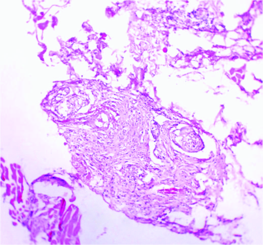
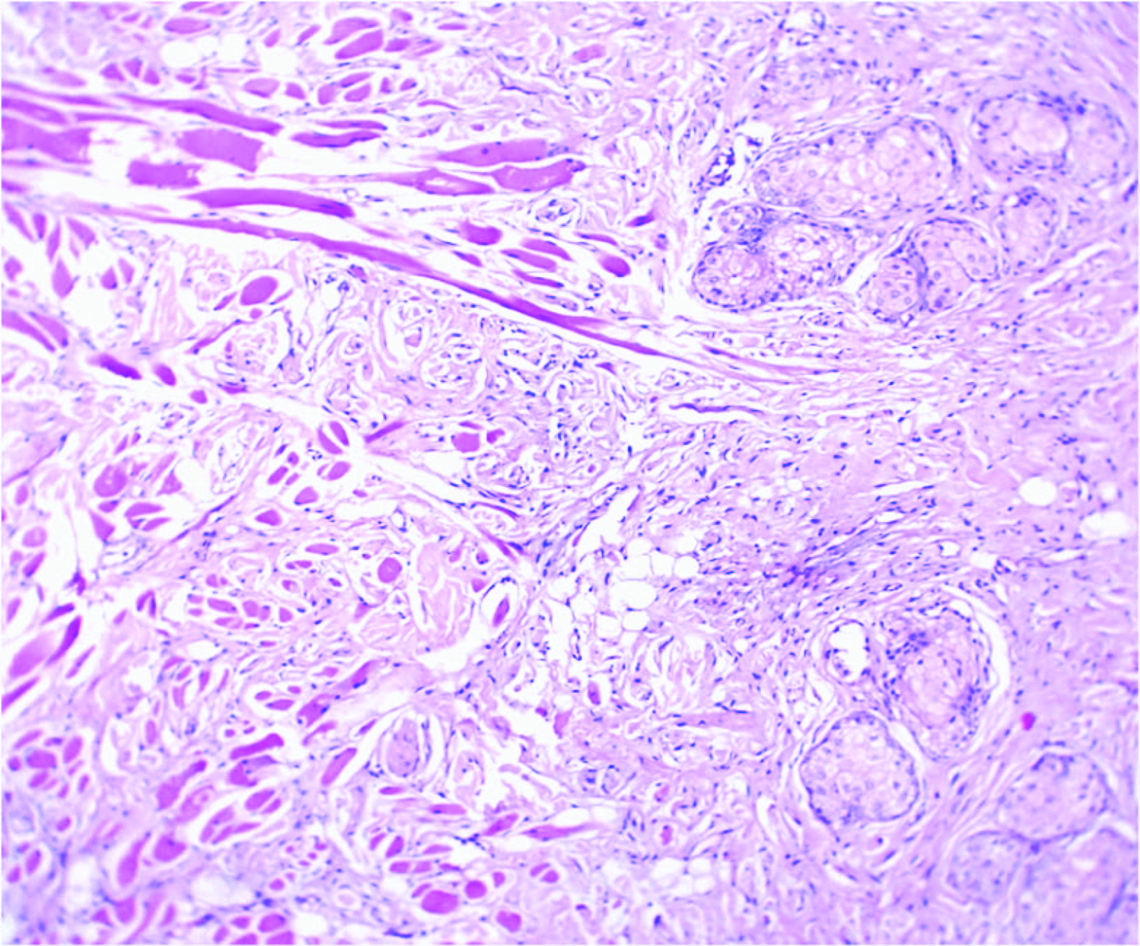
Microscopy- Showed nodules and fascicles composed of well differentiated skeletal muscle fibres of varying sizes separated by fibrous tissue bands admixed with nerve bundles, some of which were enclosed within the perimysium of the skeletal muscles [Table/Fig-2,3].
Discussion
Benign triton tumours, also referred to as neuromuscular hamartomas or neuromuscular choristomas are extremely rare neoplasms with a variable combination of peripheral nerve and skeletal muscle differentiation in an organized fashion in a manner somewhat akin to neuroskeletal development.They are believed to be hamartomatous lesions of neuroectodermal- mesenchymal origin which could have resulted due to incorporation of mesenchymal tissue into nerve sheaths during embryogenesis or aberrant differentiation of neuroectodermal elements into mesenchymal components or hamartomatous growth of muscle spindles [1-4].
Majority of the reported cases in literature have been seen in infants and children in association with large nerve trunks in the sciatic and brachial regions [1]. Overall,15 [5] to 20 [6]cases have been reported .Very few cases have been reported in the head and neck region [2,3]. A careful literature search revealed five cases reported in head and neck region till date [2-4]. Our case will be an additional contribution to this figure.
The name “Triton tumour” is derived from the amphibian called Triton salamander in which the skeletal muscle regeneration was believed to be induced by nerves [7]. Mitchell et al., described this entity as neural neoplasm exhibiting focal skeletal muscle differentiation [8].
Very occasionally benign triton tumours have been reported in the head and neck region as in the present case where it involved the facial nerve extending from left side of the upper lip, along the philtrum and the angle of the mouth upto the left ala [2-4].
The tumours which occur in the cranial nerve trunks like the fifth, seventh and the ninth cranial nerves are more aggressive and are often seen in infants and children. They need extensive surgical treatment [2,3,5,6], whereas, the tumours which arise from the peripheral nerves as in the present case are usually non- aggressive and may be seen in infants, children or adults [4,9,10].
These tumours respond well to surgical excision. However, Demir et al., have reported a case of benign triton tumour arising from the mental region which recurred after excision [11]. Daley et al., propose an alternative theory of histogenesis for peripheral lesions. They suggest that there could be an alteration either epigenetic in the infants or acquired in the adults involving a motor end plate or synaptic junction defect. The motor end plate defect could be due to protein alterations such as defective and non- functioning nicotinic cholinergic receptors. The synaptic abnormalities could be due to acetylcholine production or secretion. Both of these result in a reactive hyperplasia of both the nervous and the muscular elements due to a compensatory mechanism [10] .
Clinical picture after biopsy- Diffuse swelling involving left part of upper lip, philtrum and left ala

Nervous tissue within the perimysium, (H & E, ×100)
Well differentiated skeletal muscle fibres admixed with nerve bundles, (H & E, ×100)
Conclusion
Diagnosis of a benign triton tumour depends solely on the histopathological examination since the clinical diagnosis is difficult. Sometimes, the fibrous component may predominate, thereby mimicking fibromatosis. After a correct diagnosis, the treatment should be conservative and primarily aimed at maintaining the integrity of the nerve. A complete excision is totally curative.
[1]. SW Weiss, JR Goldblum, Enzinger and Weiss’ soft tissue tumours. 2008 8345th EditionPhiladelphiaMosby Elsevier:917-19. [Google Scholar]
[2]. JC Tiffee, EL Barnes, Neuromuscular hamartomas of the head and neckArch Otolaryngol Head Neck Surg 1998 124:212-16. [Google Scholar]
[3]. S Tobias, CH Kim, B Sade, SM Staugaitis, Jh Lee, Neuromuscular hamartoma of trigeminal nerve in adultActaNeirochir 2006 148:83-87. [Google Scholar]
[4]. JX O’connell, AE Rosenberg, Multiple cutaneous neuromuscular choristomasAm J SurgPathol 1990 14:93-96. [Google Scholar]
[5]. RS Oeppen, SP Harden, JD Argent, Neuromuscular hamartoma: imaging features of a rare pediatric craniofacial tumourPediatrRadiol 2003 33:50-52. [Google Scholar]
[6]. G Vajramani, I Devi, V Santosh, T Hegde, BS Das, Benign triton tumour of trigeminal nerveSpringer- Verlag 1999 15:140-44. [Google Scholar]
[7]. SF Markel, FM Enzinger, Neuromuscular hamartoma: a benign “triton tumour” composed of mature neural and striated muscle elementsdCancer 1982 49:140-44. [Google Scholar]
[8]. A Mitchell, BW Scheithauer, H Ostertag, A Sepehrnia, A Sav, Neuromuscular choristomaAm J ClinPathol 1995 103:460-65. [Google Scholar]
[9]. DE Castro, K Raghuram, CD Phillips, Benign Triton tumour of the trigeminal nerve Am J Neuroradiol 2005 26:967-69. [Google Scholar]
[10]. TD Daley, MR Darling, B Wehrli, Benign triton tumour of the tongueOral Surg Oral Med Oral Pathol Oral RadiolEndod 2008 105:763-66. [Google Scholar]
[11]. Y Demir, O Uluoglu, S Ozmen, ZM Boyacioglu, K Atabay, Neuromuscular hamartoma in the mental region J Oral MaxillofacSurg 2003 61:397-400. [Google Scholar]