Thorough debridement of root canal system is essential for the successful outcome of root canal therapy. The removal of debris is often neglected or overlooked and the influence of smear layer on the outcome of endodontic treatment is still controversial [1].
Mechanically well prepared canals harbored areas that were never contacted by endodontic instruments. These findings prompted the investigators to look at the effect of mechanical preparation under Scanning electron microscope [2]. Use of irrigating solutions are ineffective in completely removing hard and soft tissue debris, especially in apical portion of the canal [3–5].
Many of our currently accepted methods of chemo mechanical preparations being inadequate in producing debris free canal. Therefore, emphasis has been placed on improving the endodontic instruments and developing more effective cleaning and shaping procedures.
A new generation of rotary endodontic instruments developed from Nickel–Titanium alloys has brought a path breaking change in endodontics. They potentially allow shaping of canals, procedure being noticeably easier, faster than hand preparation. They are effective in removing debris and smear layer in apical third of the canal compared to hand instrumentation [6].
Use of rotary nickel- titanium instruments with various tapers lead to good instrumentation of the canal. However, little is known about their cleaning effectiveness. LightSpeed is not “just another root canal instrument”. Its design is different from all other rotary instruments. The design gives the ability to negotiate canal curvature, ‘feel’ canal diameter and instrument to an apical size large enough to clean all walls of a canal [7].
Endowave is the next generation NiTi rotary files, designed by J Morita and developed to increase the safety factor, cutting efficiency with ‘continuous wave’ design of instrument [8].
The purpose of the study was to determine the efficacy of LightSpeed, and Endowave rotary instruments in removing debris and smear layer from the canal surface.
Materials and Methods
Thirty freshly extracted human mandibular premolars with fully formed apices, free of apical root resorption and caries were collected and were stored in 10% formalin.
The collected samples were randomly divided into 2 groups of 15 each. A small piece of modeling compound was placed at the root tip of each tooth to prevent the flow of irrigants through apical foramen.
An ideal access cavity was prepared for each tooth to obtain a straight-line access to the root canal. Teeth were decoronated 2mm above cemento enamel junction using diamond disc to obtain root segment for the preparation. The working length was obtained by measuring the length of initial instrument #10 visible at apical foramen minus 1mm for all the groups.
Root canal preparation for all the teeth was carried out with 2 different types of rotary instruments [Table/Fig-1,2].
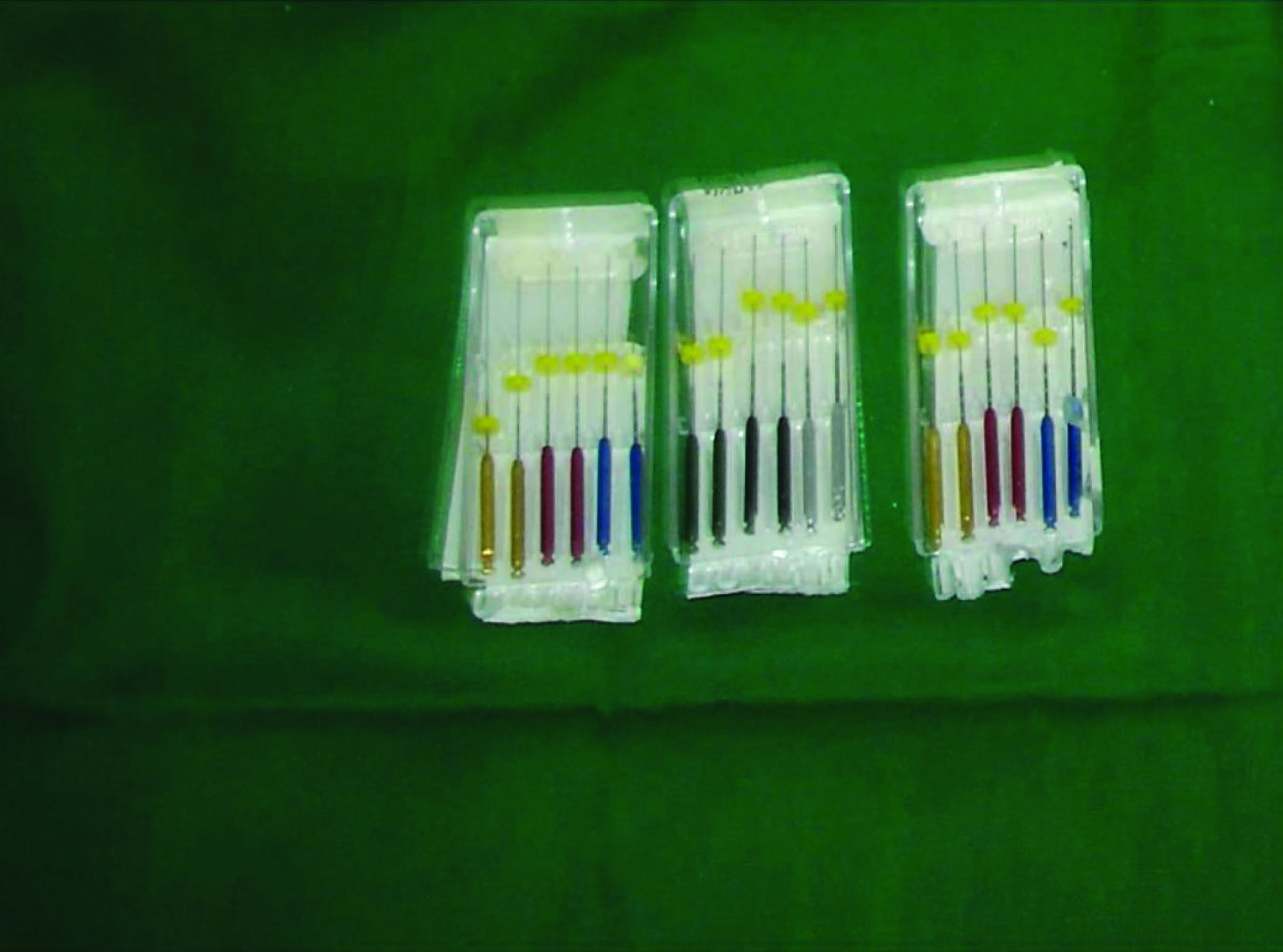

Group I- The canals were prepared with LightSpeed instrument.
Group II-. The canals were prepared with Endowave instrument.
Crown down preparation technique was carried out in all the teeth according to manufacturer’s recommendation, using alternate 5.25% sodium hypochlorite and 17% EDTA (dent wash, Prime Dent) as irrigants.
Procedure For Root Canal Preparation
LightSpeed recommended method: Instrumentation was performed at constant speed of 1300 rpm. Coronal flaring was done with Gates Glidden drill. Preparation involved 5 steps as described below [Table/Fig-3].
LightSpeed instrumentation

Step 1 : Determining the Light Speed size that was used to begin rotary instrumentation (sizing or gauging the apical canal diameter). A LightSpeed instrument can reach working length, if its cutting head is smaller than the canal’s diameter from orifice to working length. Sizing apical 3rd by hand was continued with smaller to sequential larger sizes, until the instrument did not reach the working length. This is known as first LightSpeed size to bind (FLSB). FLSB was chosen to begin instrumentation.
Step 2 : Determining the apical preparation size: Instrumentation with FLSB was started with slow continuous movement until it engaged the canal walls. At this point, the instrument progressed apically in advance and withdrawal motion (pecking). This pecking movement was continued until FLSB reached the working length. Sequential larger instruments were used with pecking movement to enlarge the apical 3rd. The instrument that takes at least 12 pecks to reach working length is known as MAR (Master Apical Rotary). This is called 12 pecks rule.
Step 3 : Completing apical instrumentation: After determining MAR, the next LightSpeed size which is short by 4 mm to working length was used. This enables the 5 mm long simplifill plug to closely match the size and shape of canal preparation.
Step 4 : Mid root instrumentation: The middle 3rd of root canal was prepared with sequential larger instruments with 4 to 8 light pecks, which means stopping after 4 pecks if LightSpeed did not advance, but continuing with 8 pecks if light speed advanced. This was continued until to reach the size of the instrument, which did not advance easily past the apical extend.
Step 5 : Recapitulation: Recapitulation to working length of each canal was done with respective MAR [9].
Endowave recommended method
Crown down preparation technique was employed to enlarge the canal by using file series from large to small size. A speed of 280 50 rpm was maintained. Enlargement by using files from large to small resulted in smooth coronal flaring without creating steps on the canal wall.
No. 35/08 file was used to prepare the coronal half of the canal with back and forth motion. This was followed by # 30/06, then #25/06, which was 2-3 mm short of the estimated working length. # 20/06 instrument was used to prepare the canal to the full working length. If resistance occurred with # 20/06, a smaller instrument # 15/02 was used. Then apical preparation was completed with # 25/06 instrument.
The teeth were embedded in the alginate mold, which was used as the conducting medium for the electronics apex locator and lip clip electrode of the Tri Auto Zx was inserted into the alginate to complete the circuit. This model was used for the preparation of the canal with Endowave instrument because manufacturers recommended Tri Auto Zx hand piece with this system [9,10] [Table/Fig-4].

Preparation for scanning electron microscopic study
After completion of the instrumentation, each canal was flushed with sodium hypochlorite solution and dried with absorbent points. Longitudinal grooves were made on the buccal and lingual root surfaces with diamond disk without penetrating the canal. The chisel and hammer was used to complete the fracture of the specimen. The specimens were stored in 2.0% glutaraldehyde aqueous solution till the SEM was carried out. The specimens were dehydrated using aqueous ethanol solution and were dried in a desiccator for 48 hours. They were mounted on aluminum stubs, sputter coated with gold. Sections were mounted on the scanning electron microscope (JSM-840A Scanning Electron Microscope, JEOL-Japan) to evaluate the presence of debris and smear layer at coronal 1/3rd, middle 1/3rd and apical 1/3rd. Using x200 and x1000 magnification respectively.
SEM photomicrographs for each specimen were taken and cleanliness of the canal was evaluated in three areas by means of numerical evaluation scale.
Hulssman has given 5-step scale rating the debris and smear layer depending upon the amount of clumps present on the canal walls. Debris was defined as dentine chips, pulp remnants and particles loosely attached to the root canal wall.
Score 1: Clean canal wall, only a few small debris particles.
Score 2: A few small agglomeration of debris.
Score 3: Many agglomeration of debris covering less than 50% of root canal wall.
Score 4: More than 50% of root canal wall covered by debris.
Score 5: Complete or near complete root canal wall covered by debris.
Smear layer was defined as a surface film of debris retained on dentine.
Score 1: No smear layer, dentinal tubules open.
Score 2: Small amount of smear layer, some dentinal tubule open.
Score 3: Homogeneous smear layer covering the root canal wall, only few dentinal tubules open.
Score 4: Complete root canal wall covered by homogeneous smear layer, no open dentinal tubules.
Score 5: Heavy, inhomogeneous smear layer covering the complete root canal wall [11].
All measurements and reading were noted and statistically analysed and compared among two groups.
Result
The score for debris and smear layer at coronal, middle and apical third were analysed by One-way-analysis of variance (ANOVA) which indicated that there was significant variation when compared between group I & II (p< 0.05). Mann-Whitney test was performed for group wise comparison [Table/Fig-5,6]. There was significant difference for removal of debris and smear layer at coronal, middle and apical third for LightSpeed and Endowave system (p<0.01). The entire group showed higher removal of debris and smear layer in coronal third followed by middle third and lower scores in apical third [Table/Fig-7&8]. Overall, LightSpeed instrumentation was significantly more efficient in removal of debris and smear layer compared to Endowave instrument.
Comparison of scores for debris removal S=Significant
| Score | Coronal | Middle | Apical |
|---|
| Group I | Group II | Group I | Group II | Group I | Group II |
|---|
| Mean (SD) | 1.7 (1.0) | 3.9 (0.7) | 2.9 (0.8) | 3.9 (0.6) | 3.4 (0.8) | 4.4 (0.8) |
| p valve | p=0.001,S | p=0.001, S | p=0.001 S |
Comparison of scores for smear layer removal
| Score | Coronal | Middle | Apical |
|---|
| Group I | Group II | Group I | Group II | Group I | Group II |
|---|
| Mean (SD) | 1.5 (0.9) | 3.0 (0.5) | 2.5 (0.5) | 3.4 (0.6) | 2.8 (0.6) | 4.2 (0.8) |
| P valve | p=0.001,S | p=0.001, S | p=0.001 |

SEM comparison of smear layer

Discussion
The present study was conducted to evaluate the efficacy of LightSpeed, and Endowave rotary instrumentation to remove debris and smear layer from the root canal.
In the present study, results indicated, statistically significant differences between LightSpeed and Endowave for debris and smear layer removal. This observation was in accordance with previous studies [12,13].
Cleaning efficiency of instruments in coronal and middle third was better because,
Large preparation obtained with LightSpeed, and Endowave was #45-55, #25/06 taper file respectively, allows larger volume of irrigants to be in contact with canal wall.
Use of irrigants such as 5.25% NaOCL and 17%EDTA solution.
File designs such as presence of radial land and U shape may prevent risk of debris jamming in the canal.
Endowave NiTi files employ modified blade design with shaper edge along with the process of electropolshing. Electropolishing will greatly enhance the cutting efficiency of an edge. The variable helical angle helps to remove debris and smear layer [14].
Cleaning ability of all the instruments in the apical third of the canal was less than middle and coronal third regardless of instrument used. This could be due to, use of torque control hand piece reduces the cutting efficiency of instrument and progression of the file into apical third becomes more difficult [15].
In general LightSpeed instrument was more efficient in removing debris and smear layer. This may be because:
Light Speed instrumented canal had larger apical stops, which enabled large volume of irrigating solution to react in apical area and their spade design would allow the movement of debris coronally in an irrigant flooded canal.
Manufacturer has recommended irrigation of canal with 5.25% NaOCL and 17%EDTA.
Instrument was used in advance and withdrawal motion. Cutting occurs with advancement and withdrawal removes debris.
The clinical relevance of the current study indicated that none of the rotary instrumentation produced completely clean canal. But LightSpeed demonstrated better results compared to Endowave systems.
Conclusion
Within the limitations of this in-vitro study the following conclusions can be drawn from the results of this study:
None of the rotary instrumentation rendered the canal completely free of debris and smear layer.
Overall Light Speed instrument was significantly more efficient in removing debris and smear layer from the root canal than compared to Endowave instrument.