Scrotal Hemangioma: A Case Report
Deepak Chavan1, Anita P Javalgi2
1Assistant Professor, Department of Surgery, BLDEU’s Shri B M Patil Medical College, Sholapur Road, Bijapur, India.
2Assistant Professor, Department of Pathology, BLDEU’s Shri B M Patil Medical College, Sholapur Road, Bijapur, India.
NAME, ADDRESS, E-MAIL ID OF THE CORRESPONDING AUTHOR: Dr Deepak Chavan, Assistant Professor, Department of Pathology, BLDEU’s Shri B M Patil Medical College, Sholapur Road, Bijapur-586103, India.
Phone: 09880771234,
E-mail: dipdeepak@yahoo.co.in
We report a case of scrotal-hemangioma in a 14-year-old boy. Subcutaneous scrotal-perineal hemangioma may mimic an inguinal hernia, thus forming a diagnostic and therapeutic challenge. Histopathological study confirms the diagnosis. Definitive treatment by en bloc excision is recommended
Hemangioma, Scrotal swelling
Case Report
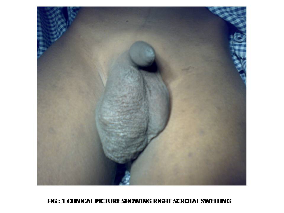
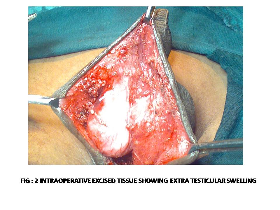
A 14-year-old boy presented with Right sided scrotal swelling since birth, insidious in onset and gradually increased in size. On examination swelling was measuring 5X5cm in size [Table/Fig-1], firm in consistency, non tender and both the testis were freely mobile. There was no significant past history of trauma or any other disease. At posterio-lateral part of the scrotum around 0.5cm was adherent to the swelling. Preoperative impression was HEMANGIOMA of scrotum. Patient was referred for radiological and routine laboratory investigation. Blood routine, coagulation profile was within normal limits and USG showed features of benign vascular lesion suggesting hemangioma, Doppler study revealed vascular malformation, extra testicular within scrotal wall. Patient was posted for surgery. Intra operatively a highly vascular mass measuring around 5X6 cm [Table/Fig-2] was removed along with the redundant skin after ligating the feeding vessels. The excised tissue was send to histopathiological study for confirmation.
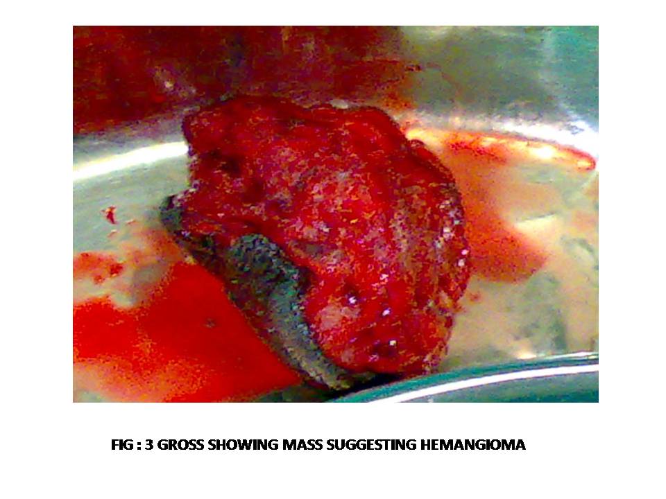
Grossly skin covered mass was noted measuring 5X5X4cms [Table/Fig-3], on cut section it was hemorrhagic and brown in colour. Microscopic examination showed features of hemangioma mixed variant of capillary and cavernous heamngioma.
The diagnosis of scrotal hemangioma was made.
Discussion
Very few articles have been reported in literature describing about penile hemangioma [1]. Giant penile cavernous hemangioma with intrapelvic extension has been reported by Froehner and Tsatalpas [2]. A case of mutiple hemangiomas of the scrotum, perineum and pelvis along with mega penis and associated with agenesis of the corpus spongiosum along with scrotal and pelvic hemangiomas have been reported by Nouira et al., [3]. The findings observed in the cases described by the above authors are similar to that seen in the present case. Cavernous hemangioma of the scrotum primarily presents during childhood. Subcutaneous scrotal-perineal hemangioma may mimic an inguinal hernia, thus forming a diagnostic and therapeutic challenge [4] Large scrotal hemangioma may be associated with testicular damage believed to be due to the heat generated by hemangioma [5].
Imaging can help assessing the extent of the hemangioma, as well as detecting any associated abnormalities. The most typical radiographic finding is a soft-tissue mass or prominence containing phleboliths (small calcifications). Presence of phleboliths is highly suggestive of cavernous hemangioma [6,7] Phleboliths are usually better demonstrated on plain radiographs or CT scans.
In sonographic images cavernous hemangiomas may appear hyperechoic or hypoechoic depending upon the content of the lesion (such as septa, blood containing units). Phleboliths may be seen as echogenic foci with distal acoustic shadowing. Colour Doppler may demonstrate blood flow within these lesion but the absence of flow does not rule out the presence of these lesions [8]. CT and MRI provide a simple, noninvasive method of diagnosing and determining the extent of these lesions in addition to delineating their relationship with adjacent structures. Therefore, these are the imaging techniques of choice for this condition and are considered mandatory before surgical procedures are taken [7]. CT scans reveal a soft tissue mass with phleboliths. In the rectum and sigmoid, they are seen as nodular thickening of the wall with phleboliths. Involvement of the urinary bladder may cause hematuria. A non-homogeneous, subtle to intense enhancement of the lesion is evident on postcontrast image [9]. Selective arteriography may detect the lesions, but it is of little importance for diagnosis. In addition, it reveals normal results in most patients, because of the presence of thrombosis in dilated vascular spaces within the hemangioma. Recently, it has been suggested that radionuclide studies, particularly Tc-99 scans, may play a role in the assessment of the extension of these lesions [7].
Once a diagnosis has been established, eradication of the lesion should be recommended. Treatment options include a sclerosing agent (such as alcohol), surgery or laser therapy. In one study, the success rate of laser treatment was 92.8% with a low immediate complication rate (approximately 3.57%) that included minimal scarring and deformity. No long-term complications of laser therapy were noted. The study concluded that laser treatment of these lesions enables good results with a very low incidence of complications. Surgery and other treatment modalities are not always satisfactory, yield similar or less efficient results, and have a higher complication rate. Laser treatment may be the treatment of choice in some settings [10].
Clinical picture showing right scrotal swelling
Intraoperative excised tissue showing extra testicular swelling
Gross showing mass suggesting hemangioma
[1]. Y Lin, GH Sun, DS Yu, Intrascrotal hemangioma Arch Androl 2002 48(4):259-65. [Google Scholar]
[2]. M Froehner, P Tsatalpas, Giant penile cavernous hemangioma with intrapelvic extension-review of the literatureUrology 1999 53(2):414-15. [Google Scholar]
[3]. Y Nouira, I Kbaier, F Attyaoui, E Menif, A Horchani, Megapenis associated to corpus spongiosum agenesis with Scrotal and Pelvic HemangiomasEur Urol 2001 40(5):571-74. [Google Scholar]
[4]. FA Ferrer, PH McKenna, Cavernous hemangioma of the scrotum: a rare benign genital tumor of childhoodJ Urol 1995 153(4):1262-64. [Google Scholar]
[5]. M Gotoh, S Tsai, T Sugiyama, K Miyake, H Mitsuya, Giant scrotal hemangioma with azospermiaUrology 1983 22(6):637-39. [Google Scholar]
[6]. H Djouhri, L Arrive, T Bouras, Diffuse cavernous hemangioma of the rectosigmoid colon: imaging findingsJ Comput Assist Tomography 1998 22(6):851-55. [Google Scholar]
[7]. D Hervias, JP Turrion, M Herrera, Diffuse cavernous hemangioma of the rectum: an atypical cause of rectal bleedingRev Esp Enferm Dig 2004 96(5):346-52. [Google Scholar]
[8]. LJ Yeoman, D Shaw, Computerized tomography appearances of pelvic hemangioma involving the large bowel in childhoodPediatr Radiol 1989 19(6- 7):414-16. [Google Scholar]
[9]. C Perez, J Andreu, J Llauger, J Valls, Hemangioma of the rectum: CT appearanceAbdom Imaging 1987 12(1):347-49. [Google Scholar]
[10]. O Sarig, S Kimel, A Orenstein, Laser treatment of venous malformationsAnn Plast Surg 2006 57(1):20-24. [Google Scholar]