Urinary stones have afflicted humankind since antiquity, with the earliest recorded example being bladder and kidney stones detected in Egyptian mummies dated to 4800 BC [1]. Obstructive uropathy can be defined as an interruption of normal urine flow at some point along the urinary tract from the renal tubule to the urethra. Obstruction results in an increase in pressure within the urinary tract, causing structural and physiologic changes. Without intervention, obstructive nephropathy eventually leads to an irreversible loss of renal function. The various imaging techniques differ substantially in their ability to provide anatomical detail and physiological information.
Conventional gray scale ultrasonography retains its pivotal role in screening for subacute and chronic urinary obstruction, especially in utero, children and in patients with renal failure. In acute obstruction, pyelocaliectasis is minimal or even absent. The introduction of duplex Doppler ultrasound with Resistivity Index measurement [2] takes ultrasound into the functional arena and plays a role in cases of acute obstruction.
This study aimed i) to evaluate the intra renal Resistivity Index (RI) of acutely obstructed kidney, ii) to determine the significance of Resistivity Index Ratio (RIR) and difference in Resistivity Index of obstructed and contralateral non obstructed kidney and iii) to compare the Resistivity Index, Resistivity Index Ratio and difference in Resistivity Index following relief of obstruction with those of obstructed kidney.
Materials and Methods
This was a prospective study, with study period of one year (01.01.2006 –31.12.2006). Eighty three patients between the age group 20-45 y presenting to the Department of Radiodiagnosis, with acute unilateral ureteric obstruction were initially included in the study. Of the 83 patients, 2 with bilateral hydronephrosis, one with renal artery stenosis, two with renal parenchymal disease, three each with diabetes mellitus and systemic hypertension were excluded from the study, with the final sample size being 72. Ethical clearance obtained from the college ethical committee.
After obtaining informed consent Duplex sonography was performed using Philips Envisor unit, 3.5MHz transducer. The kidneys were evaluated for their size, pelvicalyceal system dilatation and presence of calculus. Ureter if dilated was measured. The Doppler parameters were measured at the corticomedullary junction (arcuate arteries) or along the border of medullary pyramids (interlobar arteries)
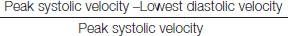
Resistivity index was calculated using the formula
Resistivity index of the obstructed kidney was compared with that of the non obstructed contralateral kidney. These patients were followed up with Resistivity index following relief of obstruction
Statistical Analysis
Statistical analysis was done using SPSS Software V13 and p-value <0.05 was considered as statistically significant.
Results
A. Demographic data
27.78% of the study population was in the age group of 20-29 y, 43.05% within 30-39 y and 29.17% within the 40-49 y age group. Male subjects constituted 79.17 % of the study population and 20.83% were females.
B. Clinical features
Of the subjects who presented with pain, 22.2% had history of accompanying dysuria. 15 % had history of haematuria.
C. Sonographic features
Gray scale: The size of the obstructing calculus was <0.5 cm in 14 subjects (19.4%), 0.51-1.0 cm in 51 subjects (70 .8%) and >1.1 cm in 7 (9.7%). Maximum size of the calculus in our study was 1.4 cm. Hydroureteronephrosis was mild in 42 subjects (58.33%), moderate in 28 (38.9%) and severe in 2 (2.77%)
Doppler
Association of Resistivity index (RI) with degree of hydronephrosis [Table/Fig-1]
Association of Resistivity index with size of calculus [Table/Fig-2]
Distribution of Resistivity Index in ureteric obstruction [Table/Fig-3]
Distribution of delta Resistivity Index (Delta RI)
Association of Resistivity index (RI) with degree of hydronephrosis, Abbreviations: RI- Resistivity Index
| S. No. | Degree of hydronephrosis | RI |
|---|
| ≤ 0.7 | > 0.71 |
|---|
| 1 | Mild | 38 | 4 |
| 2 | Moderate | 10 | 18 |
| 3 | Severe | 1 | 1 |
Association of Resistivity index with size of calculus, Abbreviations: RI- Resistivity Index
| S. No. | Size of calculus (cm) | RI |
|---|
| ≤ 0.7 | > 0.71 |
| 1 | <0.5 | 13 | 1 |
| 2 | 0.5-1.0 | 32 | 19 |
| 3 | >1.1 | 4 | 3 |
Distribution of Resistivity Index in ureteric obstruction, Abbreviations: RI- Resistivity Index
| S. No. | RI | Frequency | Percentage |
|---|
| 1 | ≤0.7 | 49 | 68.1 |
| 2 | ≥0.71 | 23 | 31.9 |
| Total | | 72 | 100 |
Forty Seven of the obstructed kidneys showed delta RI value less than 0.1 and 25 of those kidneys showed delta RI value more than 0.11.
After the relief of obstruction, 70 of the kidneys showed delta RI value less than 0.1 and only 2 of those kidneys showed delta RI value more than 0.11.
Relief of obstruction
The calculus passed spontaneously in 81.94% (59) of the subjects, with intervention (Ureteroscopic removal) in 18.06% (13). The average size of calculus in cases with spontaneous passage was 0.64±0.03 cm and that in cases of intervention was 1.02±0.02 cm
a) Mean Resistivity index
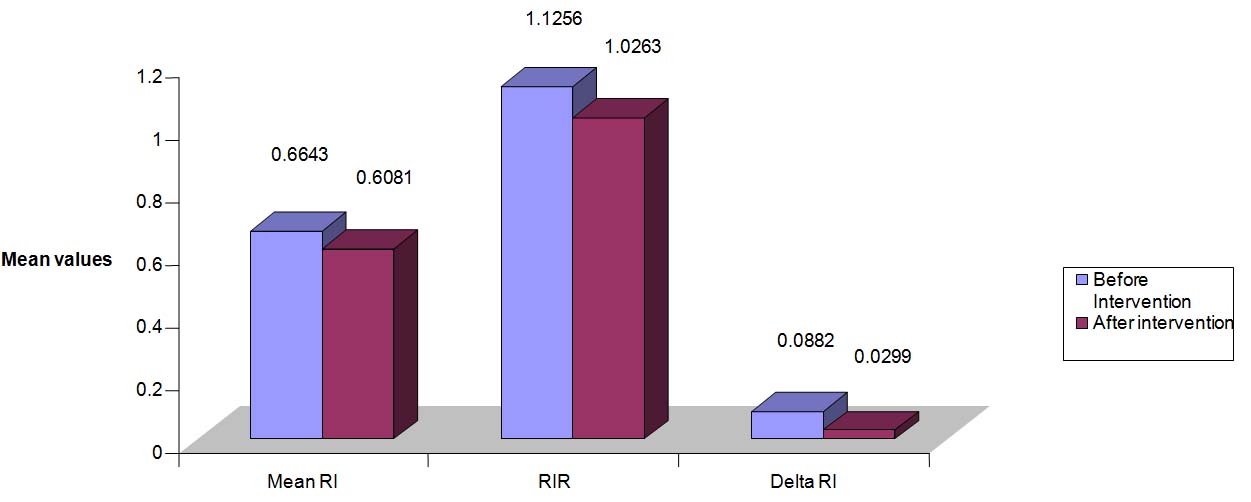
The mean resistivity index of the obstructed kidneys was 0.66 ±0.08, while that after relief of obstruction was 0.60±0.49, with p-value of 0.00
b) Mean delta Resistivity index
The mean delta resistivity index of the obstructed kidneys was 0.09 ±0.14, while that after relief of obstruction was 0.03±0.12, with p-value of 0.01
c) Mean Resistivity Index Ratio (RIR)
Forty two kidneys showed elevated RIR. After relief of obstruction, two kidneys showed persistent elevation, while rest of the kidneys showed RIR within normal limits. Mean RIR (with obstruction) 1.12 ± 0.14. Mean RIR (relief of obstruction) 1.03 ± 0.00. p-value = 0.001
Summary of distribution of RI, RIR and Delta RI values before and after intervention illustrated in [Table/Fig-4].
Summary of distribution of RI, RIR and Delta RI values before and after intervention Abbreviations: RI- Resistivity Index, RIR-Resistivity Index Ratio, Delta RI-Delta Resistivity Index
Sensitivity and specificity of RI, Delta RI and RIR illustrated in [Table/Fig-5].
Sensitivity and specificity of RI, Delta RI and RIR Abbreviations: RI- Resistivity Index, RIR-Resistivity Index Ratio, Delta RI-Delta Resistivity Index
| S. No. | Parameter | Sensitivity (%) | Specificity (%) |
|---|
| 1 | RI | 32 | 98.6 |
| 2 | Delta RI | 34.72 | 97.2 |
| 3 | RIR | 41.67 | 97.2 |
Discussion
Hydronephrosis
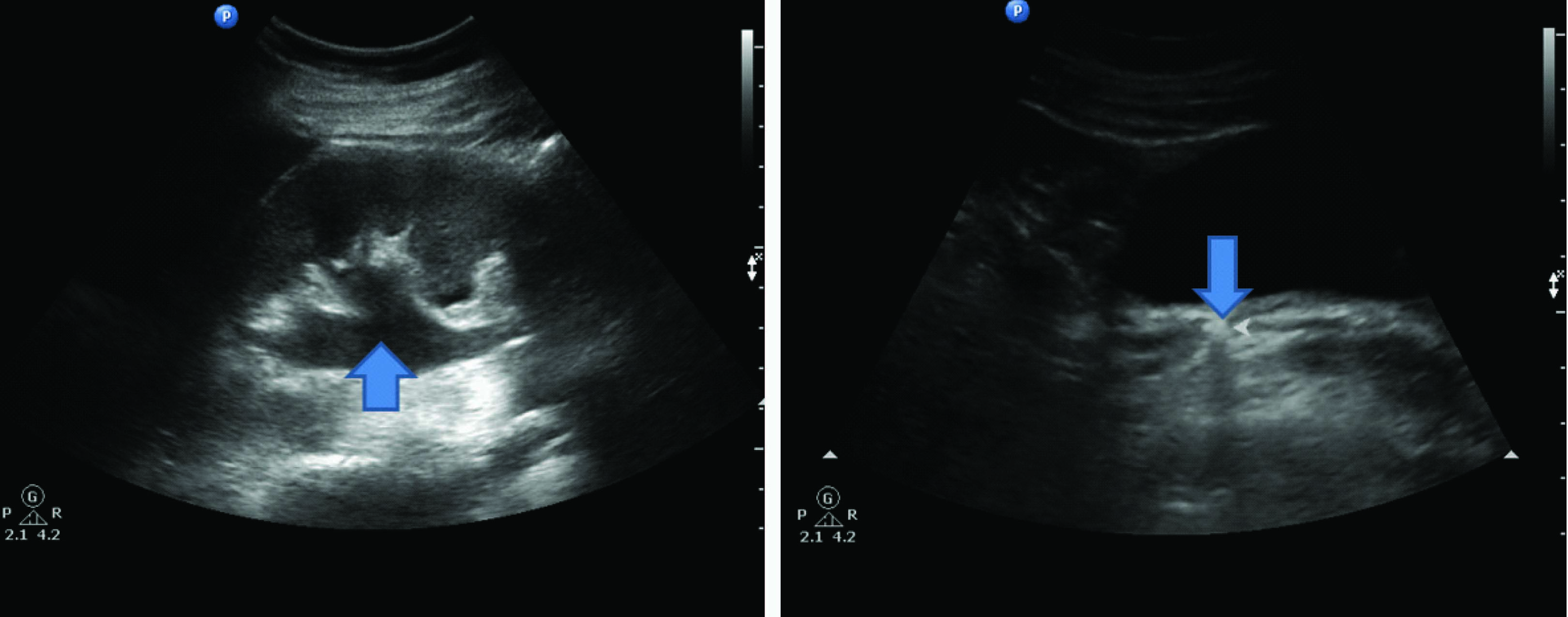
Pyelocaliectasis is the main finding that suggests the presence of obstruction at examination with conventional sonography [Table/Fig-6a,b]. Forty two of our patients (58.33%) had mild, 28 (38.89%) moderate and 2 (2.78%) severe hydronephrosis. None of the patients in this study showed absence of hydronephrosis. This is in contrast to the study of Platt JF et al., [3]; 30% of their patients showed no dilatation of the pelvicalyceal system, in the presence of obstruction. The contrast in our study might be due to fluids administered prior to sonography and the degree of obstruction caused by the calculus.
a): Shows mild Hydroureteronephrosis (as denoted by →) b): Demonstrates distal ureteric calculus of size 0.6 cm (as denoted by →)
Resistivity Index
In present study, the mean RI of the obstructed kidney was 0.66±0.88 and that of contralateral non obstructed kidney was 0.59±0.05 [Table/Fig-7]. Both of these values are within the normal limit of 0.7 but the difference appears to be statistically significant with p-value 0.00. However, only 31.94% of the subjects with obstruction showed elevation of RI. The mean RI after relief of obstruction was 0.61±0.05 and that of the contralateral nonobstructed kidney was 0.59±0.04 with p value of 0.03. The study of Platt et al., [3] had revealed that the mean RI value of 0.77±0.05 in obstructed kidneys. They have included both acute and chronic cases of obstruction and also cases with coexisting renal disease. Their results indicated that obstruction is not the only renal abnormality that can elevate the RI, as over half of the patients with non dilated renal disease had RI values greater than or equal to 0.70. The probable reason for reduced mean RI in our study might be that only 31.94% of patients with obstruction showed elevation of RI above 0.71. The mean RI of the obstructed kidneys which showed elevated RI was 0.75±0.03 (comparable with the previous study [3]) which was also statistically significant (p=0.00) when compared that after relief of obstruction. Results comparable to the present study have been reported by Cronan JJ et al., [4], where only 37% of their patients with any degree of obstruction were diagnosed via the use of Resistivity index. This discrepancy of results in different studies might be due to the varying degrees of obstruction. Moreover the fixed biphasic response to acute obstruction (vasodilatation followed by vasoconstriction) may not occur consistently in clinical practice. The episodic nature of renal colic clearly indicates that in many patients, obstruction is intermittent. In these patients, Resistivity index theoretically might not elevate, even the intermittent obstruction persists for days. The normal contralateral kidneys in unilateral ureteric obstruction had no elevated Resistivity index. This suggests that the hemodynamic changes in obstructed kidneys were local intrarenal rather than systemic events [5]. Dwivedi et al., [6] studied both control and unilateral chronic hydronephrotic cases by Doppler ultrasound. In control group the normal Resistivity index was less than 0.65 in all cases. 88% of cases showed raised Resistivity index more than 0.70. All 22 cases of raised Resistivity index was compared by Whitaker's test and found obstructive (100 percent specificity).
Intrarenal Doppler spectrum for the same case as in [Table/Fig-1], showing PSV: 24.5 cm/s, EDV: 8.65 cm/s ,RI: 0.64, Abbreviations: PSV- Peak Systolic Velocity, EDV-End Diastolic Velocity, RI-Resistivity Index

Haroun A [7] studied 14 kidneys with complete obstruction whose mean Resistivity index was 0.70 ±0.06; 42 kidneys with partial obstruction showed mean Resistivity index of 0.65 ± 0.06. Sayani R et al., [8] studied 161 patients with suspected ureteric colic with duplex Doppler Ultrasonography, followed by intravenous urography and 110 patients were found to have unilateral ureteric obstruction. Mean RI of obstructed kidney was 0.67± 0.04 and that of contralateral non obstructed kidney was 0.59±0.04. Jassim SM [9] reported a mean Resistivity index of 0.72±0.03 in hydronephrotic kidneys while that of 0.63±0.02 in non obstructed contralateral kidneys.
Delta Resistivity Index
In this study, the difference in Resistivity index (delta RI) between the obstructed and the contralateral non obstructed kidney was 0.08±0.03. It reduced to 0.03±0.03 after relief of obstruction which was not statistically significant (p=0.01). Haroun A [7] showed that delta RI in healthy individuals was 0.03 ±0.01, while that in complete and partial obstruction were 0.09±0.02 and 0.05±0.02 respectively. Sayani R et al., [10] reported that mean delta RI of obstructed kidney was 0.07±0.03 and that of non obstructed contralateral kidney was 0.03±0.05. Granata A et al., [11] reported a delta RI of 0.116±0.03 and 0.093±0.05 in cases of functionally excluded and delayed excreting hydronephrotic kidneys.
Resistivity Index Ratio
The mean RIR (Resistivity index ratio) between the obstructed and non obstructed kidney was 1.12±0.04 and the same after relief of obstruction was 1.03±0.06. This was statistically significant with p = 0.01
Sensitivity
The sensitivity of Resistivity Index, Delta Resistivity Index and Resistivity Index Ratio in predicting acute ureteric obstruction was 32%, 34.72% and 41.67% respectively. Platt et al., [8] have reported sensitivity of 92% of Resistivity index in diagnosing obstruction. Haroun A [7] has also reported a sensitivity of 64 % and 100% of mean RI and delta RI respectively in cases of complete ureteric obstruction.
Conclusion
The renal resistivity indices thus are less sensitive in diagnosing ureteric obstruction. This is due to the varying degrees of obstruction and pyelosinus extravasation. The obstructed kidneys which showed elevated Resistivity indices (RI more than 0.71, delta RI more than 0.11 and RIR more than 1.16) showed reduction to baseline values, after the relief of obstruction. However, the indices within the normal range did not rule out obstruction. Hence, this study concludes that the renal resistivity indices should not be interpreted in isolation.