Xanthogranulomatous Appendicitis with a Fulminant Course: Report of a Case
Gaurav Kochhar1, Sudipta Saha2, Manoj Andley3, Ashok Kumar4, Ajay Kumar5
1Senior Resident, Department of Surgery, Lady Hardinge Medical College, Delhi, India.
2Assistant Professor, Department of Surgery, Lady Hardinge Medical College, Delhi, India.
3Professor, Department of Surgery, Lady Hardinge Medical College, Delhi, India.
4Professor, Department of Surgery, Lady Hardinge Medical College, Delhi, India.
5Director Professor, Department of Surgery, Lady Hardinge Medical College, Delhi, India.
NAME, ADDRESS, E-MAIL ID OF THE CORRESPONDING AUTHOR: Dr. Gaurav Kochhar, Room No. 331, House Surgeon Block, LHMC Campus, Shaheed Bhagat Singh Marg, New Delhi, India. Phone : +919582935505,
E-mail: gauravkochhar82@gmail.com
Xanthogranulomatous inflammation is a well-described entity with involvement of various body organs. But the involvement of vermiform appendix in the disease process is quite rare with only few cases are reported in literature. This case report describes a 50-year-old man, who was diagnosed as a case of acute appendicitis with appendicular lump on the basis of clinical history, physical examination, and hematological and radiological investigations. Patient underwent surgical interventions twice. But, he succumbed to the disease. We are reporting this case in view of rarity of the disease and the fulminant course, which has not been described in any other reports.
Appendicectomy, Xanthogranulomatous, Vermiform appendix
Case Report
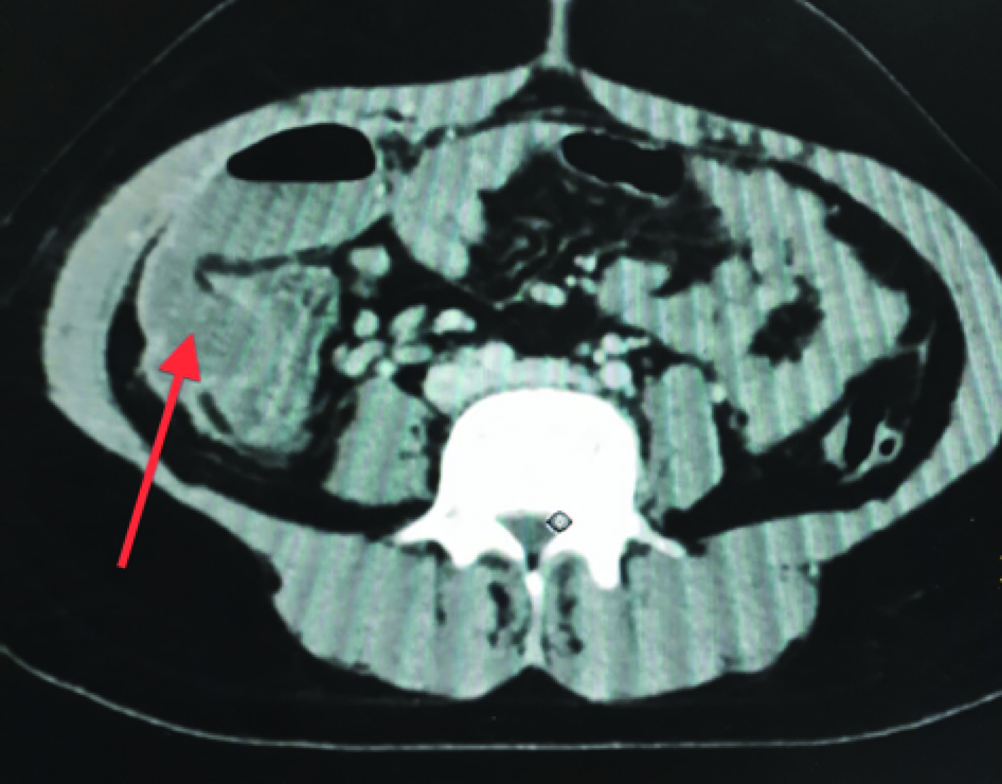
A 50-year-old male presented with the complaints of pain in the right lower abdomen for seven days along with history of fever and anorexia. On examination, there was approximately 8 x 6 cm lump present in right iliac fossa. Total leucocyte count was 24000/ mm3. Ultrasound abdomen showed inflamed appendix with echogenic mesentery. Clinical diagnosis of acute inflammatory appendicular lump was made and patient was started on expectant management. Despite of expectant management his clinical condition did not improve. CECT abdomen was done which suggested abscess formation [Table/Fig-1]. As there was no window for percutaneous aspiration, extra peritoneal drainage of abscess was done on fourth day of admission. Hundred ml of pus was drained. Appendicectomy was not attempted as small intestine, omentum and cecum was forming a mass. But patient developed small intestinal obstruction which did not resolved with conservative management. Patient was then taken up exploratory celiotomy on eighth day of admission. Intra-operatively, there was a larger ileo-cecal phlegmon with dense interloop adhesions and omentum was adhered to the phlegmon with dense interloop adhesions and omentum was adhered to the phlegmon. Small intestine was dilated proximal to the mass. Right hemicolectomy was done with ileostomy and mucous fistula. Postoperatively patient went into septicaemia and later on into multiorgan dysfunction. Despite of aggressive management he patient succumbed to the disease process. The histopathological report was suggestive of transmural inolvement of appendix with infiltration by lymphocytes, histiocytes and gaint cells. Foamy macrophages and plasma cells were also present, and diagnosis of xanthogranulomatous appendicitis was suggested [Table/Fig-2].
Discussion
Xanthogranulomatous inflammation is a rare entity. Any organ of the body can be involved in the disease process with kidney and the gall bladder being the commonest ones [1] . Other than these two predominantly involved sites, other organs where xanthogranulomatous inflammation is reported are endometrium, epididymis, fallopian tubes, bone, skin, appendix, urinary bladder, thyroid and adrenal glands [2,3] . The differential diagnosis includes the conditions showing the presence of xanthomatous cell such as malakoplakia. The disease masquerades as malignancy due to presence of diffuse inflammatory and fibrotic changes [4] .
The exact pathogensis is not known; it probably represents a chronic inflammatory process leading to tissue destruction and localized proliferation of macrophages containing large amounts of lipid which are the characteristic histological features of the disease [5] . Other speculations are defective lipid transport, disorders of neutrophillic chemotaxis, lymphatic obstruction and specific immune response towards Proteus and E. coli [2,6] . The disease has a typical histopathogical appearance. On gross appearance there is presence of golden yellow mass with abscesses. On microscopy the lesion shows recruitment of acute and chronic inflammatory cells, lipid laden macrophages and foam cells [7] .
Xanthogranulomatous inflammation of ileo-cecal region is a rare entity. Around ten cases of involvement of vermiform appendix have been reported in literature [8] . In a histopathological review of intreval and emergency appendicectomies over a period of four years Guo and Greenson found xanthogranulomatous inflammation in 8 out of 22 (36.4 %) in interval appendicectomies group, while no such findings were seen in emergency appendicetomies group [9] . They also derived the conclusion that the xanthogranulomtous is characterstically associated with the chronic inflammatory states, which comprised of granunulomas, xanthomatous cells, mural fibrosis, and transmral chronic inflammatory infiltrates. These changes may mimic as Crohn’s disease.
Munichor et al., reported a case of an adult lady who had acute appendicitis and histopathology of the specimen revealed xanthogranulomatous appendicitis. The electron microscopy showed presence of electron lucent droplets of various sizes in the xanthoma cells and other cell types. They derived inference that various cell types can be damaged by common mechanism which includes obstruction, haemorrhage and hypoxia [7] .
Xanthogranulomatous appendicitis can also masquarede as other diseases. In a case reported by Chuang et al., a 39-year-old man presented with fever, right lower abdominal pain and hard lump. Right hemicolectomy was done in suspicision of cancer. The final histopathological report suggested xanthogranulomatous appendicitis, inferring that this disease may simulate locally invasive right colonic cancer [10] .
We are presenting this case of xanthogranulomatous appendicitis in view of its rarity. Also, the uniqueness in our case is was the fulminant course of the disease and poor prognosis which have not been mentioned in the previous reported case.
CECT showing a large collection in right iliac and lumbar region with collapsed colon, suggestive of appendicular abscess (marked by arrow)
Histopathological slide showing transmural inolvement of appendix with infiltration by lymphocytes, histiocytes and gaint cells, foamy macrophages and plasma cells

[1]. V Franco, F Aragona, G Genova, Xanthogranulomatous cholecystitis. Histopathological study and classificationPathol Res Pract 1990 186:383-90. [Google Scholar]
[2]. YH Oh, SS Seong, KS Jang, Xanthogranulomatous inflammationpresenting as a submucosal mass of the sigmoid colonPathol Int 2005 55:440-44. [Google Scholar]
[3]. Y Gray, NP Libbey, Xanthogranulomatous salpingitis and oophoritis: a case report and review of the literatureArch Pathol Lab Med 2001 125:260-63. [Google Scholar]
[4]. T Maeda, M Shimada, T Matsumata, Xanthogranulomatous cholecystitis masquerading as gallbladder carcinomaAm J Gastroenterol 1994 89:628-30. [Google Scholar]
[5]. RO Peterson, Kidney. In: Peterson RO, ed. Urologic PathologyTetracyclineTeratology 1986 11PhiladelphiaJ.B. Lippincott:40e46 [Google Scholar]
[6]. M Munichor, H Kerner, H Cohen, A Bickel, TC Iancu, Xanthogranulomatous appendicitis—an incidental finding of localized pathologyUltrastructural Pathology 2000 24:33-39. [Google Scholar]
[7]. AZ Anadol, II Gonul, E Tezel, Xanthogranulomatous inflammation of the colon: a rare cause of cecal mass with bleedingSouth Med J 2009 102:196-99. [Google Scholar]
[8]. SM Al-Rawabdeh, V Prasad, DR King, SB Kahwash, Xanthogranulomatous appendicitis in a child: report of a case and review of the literatureCase Rep Med 2013 2013:498191 [Google Scholar]
[9]. G Guo, JK Greenson, Histopathology of interval (delayed) appendectomy specimens: strong association with granulomatous and xanthogranulomatous appendicitisAmerican Journal of Surgical Pathology 2003 27:1147-51. [Google Scholar]
[10]. YF Chuang, TI Cheng, TC Soong, MH Tsou, Xanthogranulomatous appendicitisournal of the Formosan Medical Association 2005 104:752-54. [Google Scholar]