Background: Medial sigmoid depression or medial depression of the mandibular ramus (MDMR) is a known variant of the normal radiographic anatomy. The clinical importance of MDMR has been recognized, however, its prevalence and association with patients with orthodontic needs have been poorly documented.
Aims and Objectives: To estimate the prevalence and characteristics of MDMR on panoramic radiographs of patients with different Angle’s molar relation.
Materials and Methods: Three hundred panoramic radiographs of 100 each belonging to patients with Angle’s class I, II and III molar relationship was recruited from an orthodontic clinic. The radiographs were evaluated for presence of MDMR along with its characteristics such as site and shape. The data was entered in the proforma and subjected for statistical analysis.
Results: The overall prevalence of MDMR in the present study was 23.2%. MDMR was found to be more prevalent in Class II – 28 (9.3%) followed by class III-23 (7.6%) and Class I- 19 (6.3%). There was equal distribution of MDMR with respect to site and the semilunar shape was the most common 36 (34%). All these findings were statistically non-significant.
Conclusion: Although MDMR is considered as a normal radiographic finding, the present study confirms the disparity in the prevalence of MDMR in patients with dentoskeletal deformities. Thus our findings suggest the importance of recognizing this entity prior to orthognathic surgery so as to avoid untoward sequelas.
Introduction
The Medial sigmoid depression or medial depression of the mandibular ramus (MDMR) was first observed on a panoramic radiograph by Steven Bricker and first reported by Langlais and co-authors in 1983 [1]. The MDMR is a normal anatomical depression observed on the medial side of the upper ramus just below and anterior to the greatest depth of the sigmoid notch [1]. On the radiographs, they appear as a radiolucent artefact due to decreased absorption of X-rays. The depression may be unilateral or bilateral and may be misinterpreted as a pathological condition [2,3]. Over the years, studies have shown variable prevalence of MDMR in anatomical specimens, radiograph of normal patients and in patients with skeletal defects. MDMR is considered to increase the potential for complication during orthodontic surgery in patients with dentoskeletal deformities, as this area is thin, an increased difficulty in splitting the ramus can be expected [4,5]. Thus it becomes mandatory to evaluate the existence of MDMR in patients undergoing orthodontic surgery. A very few studies have evaluated MDMR among different facial skeleton patients. Considering this, the present study was done to evaluate the prevalence and characteristics of MDMR among different Angle’s facial skeletal classifications.
Materials and Methods
Three hundred routine panoramic radiographs of patients belonging to both the genders in the age group of 11-30 y were retrieved from an orthodontic clinic. The 300 radiographs obtained belong to three different groups of patients with 100 in each: Group 1- Angles class I molar relation; Group 2- Angles class II molar relation and Group 3 - Angles class III molar relation.
Radiographs of developmental malformations of the face and jaws, pathologies in the maxillofacial region, history of trauma to the maxillofacial region, missing of maxillary/mandibular permanent first molar and patients who had undergone any surgical intervention in the mandibular ramus area were excluded from the study.
Prior to the initiation of the study, a sample of 20 panoramic radiographs was pilot tested by two oral radiologist and two general dentists to establish the presence and shape of the MDMR.The geometric shape considered for interpretation in the present study was based on the types available in the literature: tear-drop, semilunar, circular and triangular. The authors were asked to interpret the MDMR as present/absent and to select a geometric shape which closely represented the above said morphology. After agreement was reached, the prevalence of the anatomical depression was determined in all groups by one radiologist. The radiographs were evaluated for presence of MDMR along with its characteristics such as site and shape. The data was entered in the proforma and subjected for.
Statistical Analysis
The data was analysed by using SPSS (statistical package for social studies) software version 17. Percentages were calculated for each of the categories. Chi-square test was performed to analyse the difference between the groups. Significance for statistical test was predetermined at a probability value of 0.05 or less.
Results
The overall prevalence of MDMR in the present study was 23.2%. It was more common in Angles class II malocclusion samples (9.3%), followed by class III (7.6%) and class I (6.3%) [Table/Fig-1]. MDMR was more prevalent in females (58.57%) and it was more common in class II (64.29%) Whereas in males it was common in class III (47.83)[Table/Fig-2]. These findings were statistically non-significant.
The mean age of the samples with MDMR was 20.96. The mean Age of males samples with MDMR was 21.21 and females were
20.78. The mean age was more with class I (22.53) whereas it was least with class II (19.50) [Table/Fig-3]. There was equal distribution of MDMR samples with respect to site. However, Unilateral MDMR was common with class I (57.89%) and class III
(56.52%). Bilateral MDMR was common with class II (60.71%)(p=0.34) [Table/Fig-4].
In respect to overall MDMR shape, semilunar was the most common(34%), followed by triangular(28.3%), tear drop(20.8%) and circular(16.9%) [Table/Fig-5,6,7,8]. On the right side, semilunar was the most common (25.7%) and circular (11.43%) was the least common (p=0.91) [Table/Fig-9]. The left side too showed similar pattern of predominance i.e., semilunar was the most common (25.71%) and circular (14.29%) was the least common shape (p=0.99) [Table/Fig-10].
The association of the MDMR shape with the type of angle’s classification on the right side showed semilunar as the most pronounced shape and were seen more in class III cases (30.43%) and the least was the circular shape seen with class I cases (5.26%). Similar association on the left side also showed predominately of semilunar shape (31.58%) seen with class I cases and least number of circular shape (10.53%) are seen with class I cases[Table/Fig-9,Table/Fig-10]. The findings were statistically non-significant.
Discussion
MDMR is a normal anatomic radiolucent area in the ramus just below and anterior to the most inferior aspect of mandibular sigmoid notch. It appears as a depression or a foramen-like or notch-like radiolucency on the panoramic radiograph or in a lateral oblique view of the mandible and at times with certain periapical radiographs as well [1]. Although considered normal they can mimic other pathologic entities of the jaws. In such instances, the size, location, appearance of the radiolucent area, and the presence or absence of symptoms can lead to correct diagnosis before redundant surgical exploration [6].
Characteristically, MDMR appear as a small, round, ovoid or triangular well defined radiolucency which usually lacks a cortical margin. More often, they appear less than 5 mm in diameter and can be unilateral or bilateral [1]. The aetiology behind the appearance of MDMR is not known. Some authors considered them as developmental and some as congenital.
Over the years, a very few studies have been undertaken to analyse the existence of MDMR. As early as in 1983, Langlais et al., radiographed the anatomical specimen of dry mandibles and found the prevalence of MDMR as 66% [1]. In the same year, Clarke and Mc Anear performed a similar study on dry mandibles and found the prevalence to be only 5% [2]. Kang BC observed the prevalence as 62% (28% unilateral and 33% bilateral) [6]. Carvalho et al., observed the prevalence as 33.9% (unilateral in 20.8% and bilateral in 13.1%) [7]. Although this varied discrepancy can be correlated with the selection criteria or due to ethnic differences, the non- availability of the intricacies of the study limits us from further discussion.
Another interesting aspect of Langlais et al., study is that the author observed a decreased prevalence of MDMR on patient radiographs. Langlais et al., observed the radiographic appearance as 10% (6% were unilateral and 4% were bilateral) [1]. A Similar study done by Honing showed the prevalence of MDMR as 5.3% [8]. However, other studies done by Kang BC in 1991 and Carvalho et al., showed a high prevalence of MDMR-33% and 20.3% respectively [6,7].
From the above observations it’s understandable that a certain degree of differences exist in the incidence of MDMR among the agemandibular specimen’s radiographs and the patient’s radiographs. The difference may be due to the superimposition of airway shadows, pterygoid plate, soft palate and other soft tissues over the sigmoid notch region [6]. Langlais et al., suggested that the absence of the MDMR in the patient radiographs may be due to the area not being imaged in the focal trough of the panoramic machine. This is possible as the panoramic X-ray machines have a varied focal trough zones and structures which does not fall within the focal trough can be blurred or not visualized [9].
Another interesting observation from the literatures suggests that, few studies have observed a higher prevalence of MDMR in patients with dentofacial deformities. Carvalho et al., performed a study among patients with and without any dentofacial deformities. The authors observed that, the dentoskeletal deformities patients had a higher prevalence of MDMR (31.1%). The prevalence was higher in the class II (32.9%), class III (32.1%) and mandibular asymmetry subgroups (37.0%). Carvalho et al., further quoted from smith et al., and stated that the presence of the MDMR could lead to a much higher incidence of difficulty or unfavourable fracture due
to fusion of the medial and lateral cortical plates. The authors reasoned the inclusion of patients with dentoskeletal deformities in their study was to document the possible surgical difficulty as these patients may be a candidate for orthognathic surgery. As stated earlier, their data also suggested that MDMR is significantly more frequent in patients with dentoskeletal deformities compared with Angle Class I occlusion [7].
Considering the above finding the present study was undertaken to evaluate the prevalence of MDMR among different angles molar relation patients. The findings of the present study was in concordance with the Carvalho et al., study as stated above and also with an another study conducted by Dalizi and co author, who found the prevalence of this depression was more common in Cl II (RT:38.8%, LT:39%) and Cl III patients (RT:23.9%, LT:38.6%) [10]. In the present study too, the prevalence of MDMR was higher in Angles class II molar relation patients (9.3%) followed by Angles class III (7.6%). However, the percentage of prevalence of MDMR was comparatively lower. These differences may be explained by the differences in the methods of patient selection and possibly the ethnicity of the patients.
The findings of the Carvalho et al., study, Dalizi and co author and the present study argues against the possible misconception proposed by Langlais et al., that the absence of MDMR in certain patient populations may be due to the concerns of focal trough. Although positioning errors could be the reason for the absence of the MDMR, this should not be the reason behind the high frequency of MDMR in patients with dentofacial deformities, as they are more prone for positioning errors due to their skeletal disproportion.
Another interesting observation by Carvalho et al., is that the difference in prevalence between the anatomical and patient studies could probably be due to the failure to adequately image the relatively shallow anatomical depressions. It is understandable with the different MDMR shapes it is not always possible to image them clearly as they dependent on the projection angle which is not necessarily a true lateral in the ascending ramus of mandible [7].
Literature suggests that the shape of the MDMR varies and the noted shapes are triangular, semilunar, tear drop and circular. Honing suggests that the shape reflects the functional adaptation that may occur in the ramus, as the medial and posterior attachments of temporal muscle are inserted into this area [8]. Storey suggests that the size and shape of MDMR may be a result of variations in muscle function [11]. Studies evaluating the shapes of MDMR are meagre in the literature. Carvalho et al., observed a highest percentage of triangular shaped MDMR (39.7%), followed by semilunar (31.4%), tear drop (20%) and the least circular shaped MDMR (8.9%) [7]. In the present study, however the semilunar shape was the most common- 36 (34%) and the circular shape was the least common- 18 (16.9%).
The findings of the present study in comparable with the other previous studies suggest that there was no bilateral symmetry in the prevalence or shapes of the MDMR. Although there is a predisposition for vertebrates to form bilateral symmetric structures, nevertheless there are few exceptions to this rule such as the laterality of many organs and motor behaviour [12]. Asymmetric growth in the human craniofacial skeleton and in the orofacial region occurs quite frequently but the basic mechanisms underlying the developmental processes are poorly understood [13]. We believe the MDMR too fall under such category and thus the diagnosis of MDMR as an anatomical variation rather than a pathological entity has to be emphasized.
Prevalence of sigmoid depression in the three groups
| Angles Type of classification | No. of radiographs | No. of MDMR | Percentage |
| Class I | 100 | 19 | 6.3% |
| Class II | 100 | 28 | 9.3% |
| Class III | 100 | 23 | 7.6% |
| Total | 300 | 70 | 23.2% |
Distribution of samples with MDMR by gender
| Gender | Class I | % | Class II | % | Class III | % | Total | % |
| Male | 8 | 42.11 | 10 | 35.71 | 11 | 47.83 | 29 | 41.43 |
| Female | 11 | 57.89 | 18 | 64.29 | 12 | 52.17 | 41 | 58.57 |
| Total | 19 | 100.00 | 28 | 100.00 | 23 | 100.00 | 70 | 100.00 |
Distribution of samples with MDMR by mean age
| Gender | Class I | Class II | Class III | Total |
| Mean age±SD age | Mean age±SD age | Mean age±SD age | Mean age±SD age |
| Male | 22.75±7.98 | 19.50±6.26 | 21.64±6.65 | 21.21±6.79 |
| Female | 22.36±3.53 | 19.50±6.18 | 21.25±4.18 | 20.78±5.06 |
| Total | 22.53±5.63 | 19.50±6.09 | 21.43±5.38 | 20.96±5.80 |
Distribution of samples with MDMR by site
| Type of site | Class I | % | Class II | % | Class III | % | Total | % |
| Bilateral | 8 | 42.11 | 17 | 60.71 | 10 | 43.48 | 35 | 50.00 |
| Unilateral | 11 | 57.89 | 11 | 39.29 | 13 | 56.52 | 35 | 50.00 |
| Total | 19 | 100.00 | 28 | 100.00 | 23 | 100.00 | 70 | 100.00 |
Chi-square= 2.1512 P = 0.3412
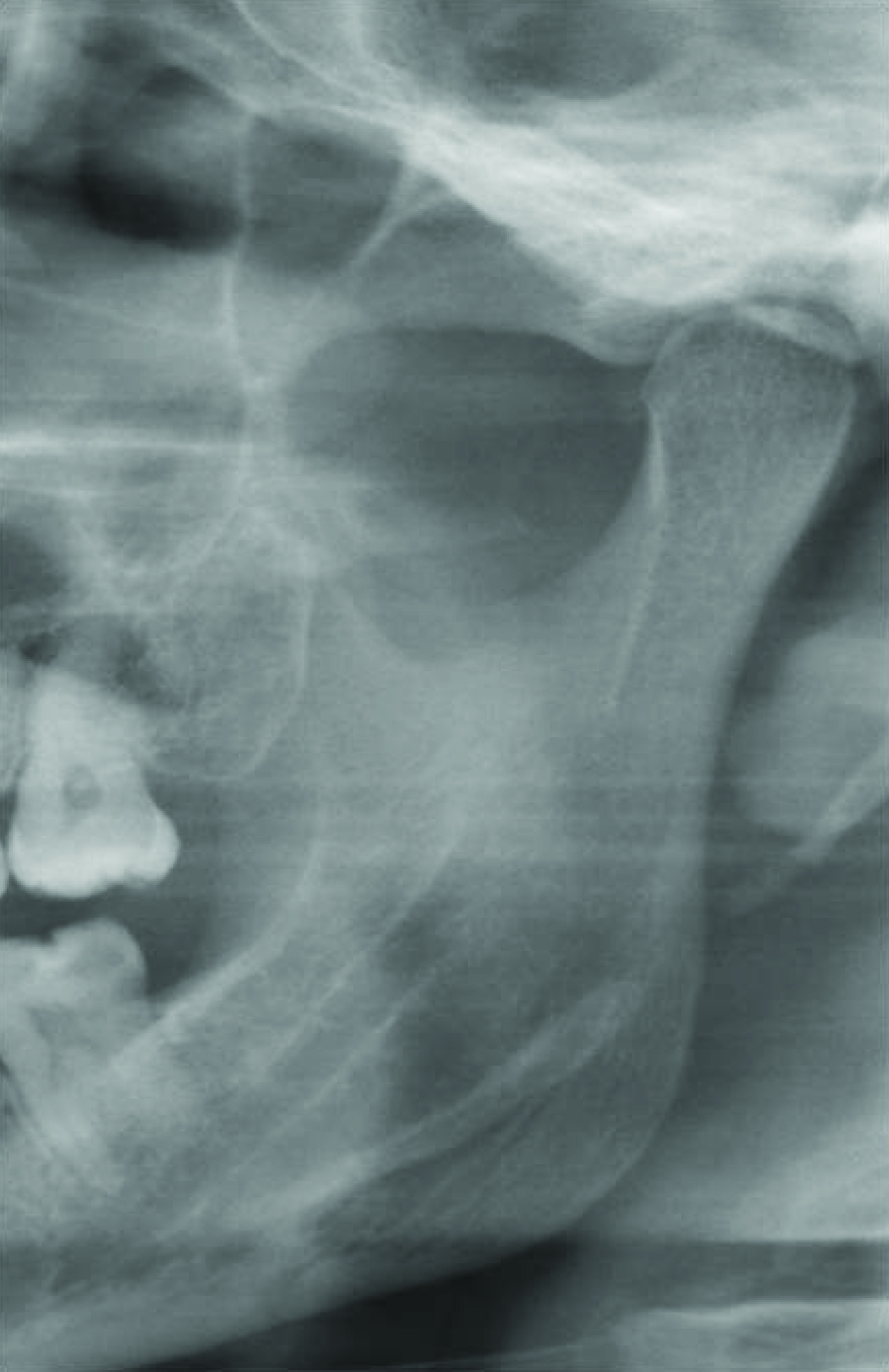
Cropped panoramic radiograph showing semilunar shape MDMR
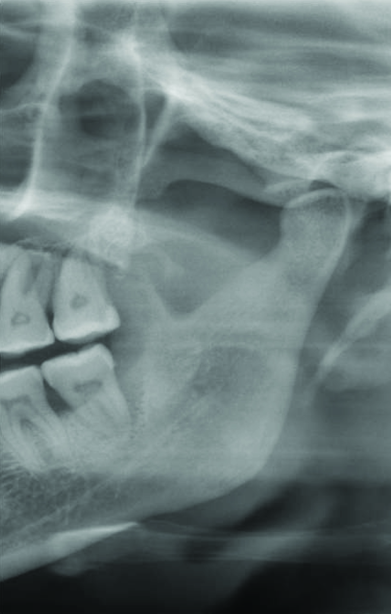
Cropped panoramic radiograph showing triangular shape MDMR

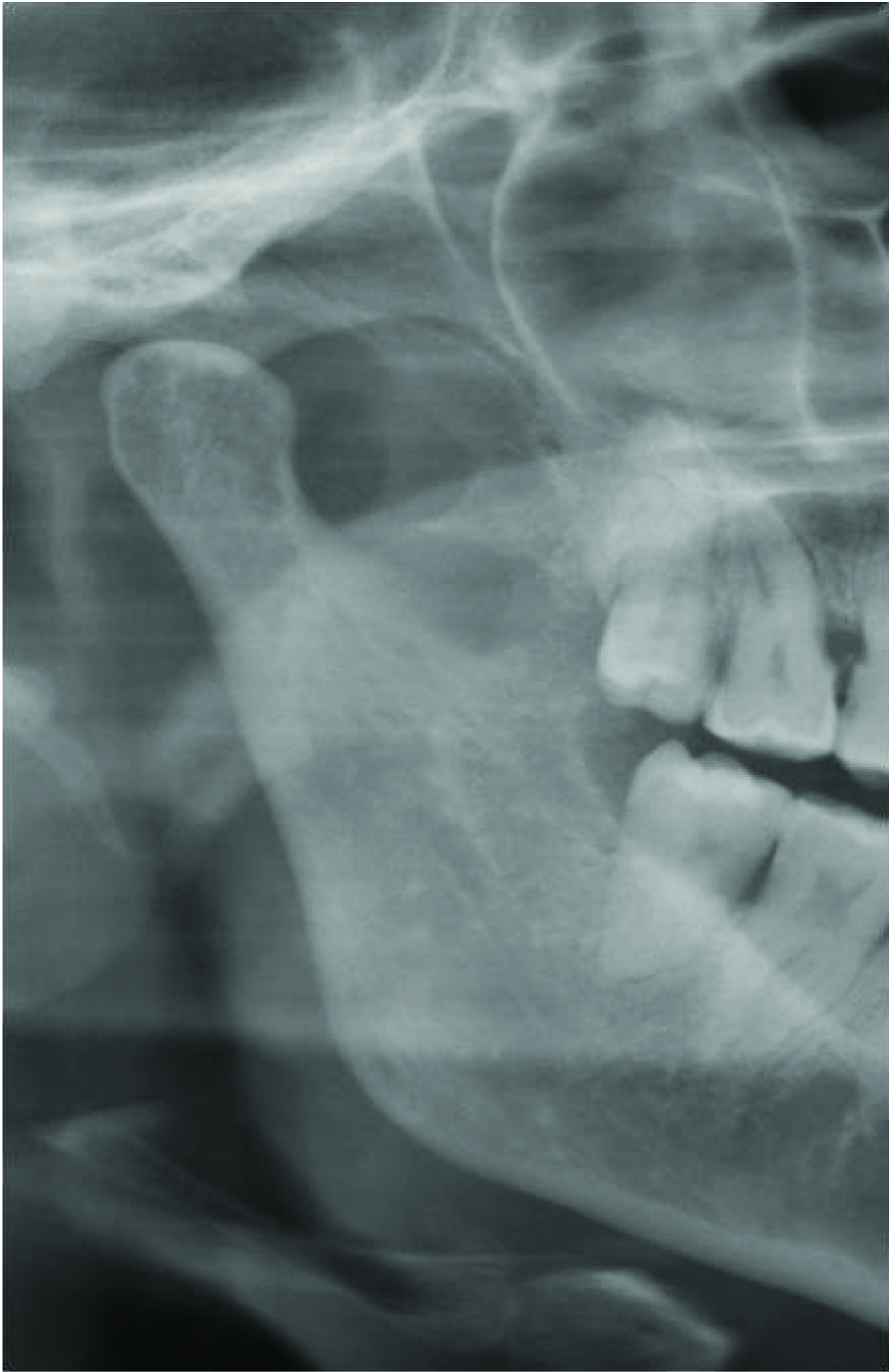
Cropped panoramic radiograph showing tear drop shape MDMR
Cropped panoramic radiograph showing circular shape MDMR
Distribution of samples with MDMR by right side shape predilection
Chi-square=2.0966 p=0.9106
| Right side shape predilection | Class I | % | Class II | % | Class III | % | Total | % |
| Circular | 1 | 5.26 | 5 | 17.86 | 2 | 8.70 | 8 | 11.43 |
| Semilunar | 4 | 21.05 | 7 | 25.00 | 7 | 30.43 | 18 | 25.71 |
| Tear drop | 3 | 15.79 | 4 | 14.29 | 3 | 13.04 | 10 | 14.29 |
| Triangular | 4 | 21.05 | 5 | 17.86 | 5 | 21.74 | 14 | 20.00 |
| None | 7 | 36.84 | 7 | 25.00 | 6 | 26.09 | 20 | 28.57 |
| Total | 19 | 100.00 | 28 | 100.00 | 23 | 100.00 | 70 | 100.00 |
Distribution of samples with MDMR by left side shape predilection
| Left side shape predilection | Class I | % | Class II | % | Class III | % | Total | % |
| Circular | 2 | 10.53 | 5 | 17.86 | 3 | 13.04 | 10 | 14.29 |
| Semilunar | 6 | 31.58 | 7 | 25.00 | 5 | 21.74 | 18 | 25.71 |
| Tear | 3 | 15.79 | 5 | 17.86 | 4 | 17.39 | 12 | 17.14 |
| Triangular | 4 | 21.05 | 7 | 25.00 | 5 | 21.74 | 16 | 22.86 |
| None | 4 | 21.05 | 4 | 14.29 | 6 | 26.09 | 14 | 20.00 |
| Total | 19 | 100.00 | 28 | 100.00 | 23 | 100.00 | 70 | 100.00 |
Chi-square= 0.7656 p=0.9929
Conclusion
In conclusion, MDMR is a normal finding observable in the mandibular ramus region which does not require any treatment. A higher prevalence of MDMR has been noted in patients with dentoskeletal deformities. Thus it becomes mandatory to evaluate the presence of MDMR prior to orthognathic surgery, as it can pose surgical difficulty during the splitting of the ramus. However, considering the technical and anatomical constraints associates with panoramic radiography, further studies with larger samples using advanced imaging modalities like Computed tomography and cone beam computed tomography can be done to authenticate our findings.
Chi-square= 2.1512 P = 0.3412
Chi-square= 0.7656 p=0.9929
[1]. RP Langlais, Chapter 9: circumscribed radiolucencies. In Diagnostic imaging of jaws. Langlais RP, Langland OE and Nortje CJ 1995 LondonWilliams & Wilkins:241-61. [Google Scholar]
[2]. MJ Clark, JT McAnear, Pseudocyst in the coronoid of the mandibleOral Surg Oral Med Oral Pathol 1983 57:231 [Google Scholar]
[3]. RP Langlais, BJ Glass, SL Bricker, DA Miles, Medial sigmoid depression: a panoramic pseudoforamen in the upper ramusOral Surg Oral Med Oral Pathol 1983 55:635-38. [Google Scholar]
[4]. PD Quinn, D Wedell, Complications from intra oral vertical subsigmoid osteotomy: review of literature and report of two casesInt J Adult Orthodont Orthognat Surg 1988 3:189-96. [Google Scholar]
[5]. BR Smith, JL Rajchel, DE Waite, L Read, Mandibular ramus anatomy as it relates to the medial osteotomy of the sagittal split ramus osteotomyJ Oral Maxillofac Surg 1991 49:112-16. [Google Scholar]
[6]. BC Kang, The medial sigmoid depression: Its anatomic and radiographic considerationKorean Academy of Oral and Maxillofacial Radiology 1991 21(1):7-13. [Google Scholar]
[7]. IMM Carvalho, JH Damante, RH Tallents, RF Ribeiro-Rotta, An anatomical and radiographic study of medial depression of thehuman mandibular ramusDentomaxillofacial Radiology 2001 30:209-13. [Google Scholar]
[8]. JF Honing, Identificacion anatomica de radiolucencias subsemilunares circulares en la rama ascendente mandibularElectromedica 1991 59:58-63. [Google Scholar]
[9]. WD McDavid, RP Langlais, U Welander, CR Morris, Imaging characteristics of seven panoramic X-rays units. Part II: The image layerDento maxillofac Radiol 1985 8:13-19. [Google Scholar]
[10]. Z Dalili, ST Mohtavipour, U Welander, CR Morris, Frequency of medial sigmoid depression in panoramic view of orthodontics based on facial skeletal classificationJournal of Guilan University of Medical Sciences 2003 12(45):23-16. [Google Scholar]
[11]. E Storey, Growth and remodeling of bone and bones. Role of genetics and functionDent Clin N Am 1975 19:443-55. [Google Scholar]
[12]. N Geschwind, AM Galburda, Cerebral lateralization. Biologic mechanisms, associations, and pathology: a hypothesis and a program for research Arch Neurol 1985 42:521-22. [Google Scholar]
[13]. IC McManus, The distribution of skull asymmetry in manAnn Hum Biol 1982 9:167-70. [Google Scholar]