Anterior Abdominal Wall Haemangioma with Inguinal Extension
Priti P Shah1, Siddharth P Dubhashi2, Kaushal Choudhary3
1 Associate Professor, Department of Surgery, Padamashree Dr D Y Patil Medical College, Pimpri, Pune, India.
2 Professor, Department of Surgery, Padamashree Dr D Y Patil Medical College, Pimpri, Pune, India.
3 Senior Resident, Department of Surgery, Padamashree Dr D Y Patil Medical College, Pimpri, Pune, India.
NAME, ADDRESS, E-MAIL ID OF THE CORRESPONDING AUTHOR: Dr. Priti P Shah, Associate Professor, Department of Surgery, Padamashree Dr D Y Patil Medical College, Pimpri, Pune, India. Phone : 9823051113, E-mail : dpranjalpriti@hotmail.com
Haemangioma are common benign vascular tumour but Intramuscular haemangiomas are rare tumours comprising less than 1% of all. The most frequent sites are extremities, head and neck whereas abdominal wall is a quiet rare location. Ultrasonography is an appropriate initial diagnostic modality and MRI is the investigation of choice. A rare case presented to us as Intramuscular haemangioma of anterior abdominal wall with inguinal extension. Ultrasonography with Doppler study and MRI was suggestive of same finding. Intraoperatively patient had huge haemangioma involving external oblique, internal oblique and transverse abdominus muscle. Wide local excision with meshplasty was done as part of muscle had to be removed. Histology confirmed the diagnosis of Intramuscular Haemangioma.
Anterior abdominal wall, Intramuscular haemangioma
Case Report
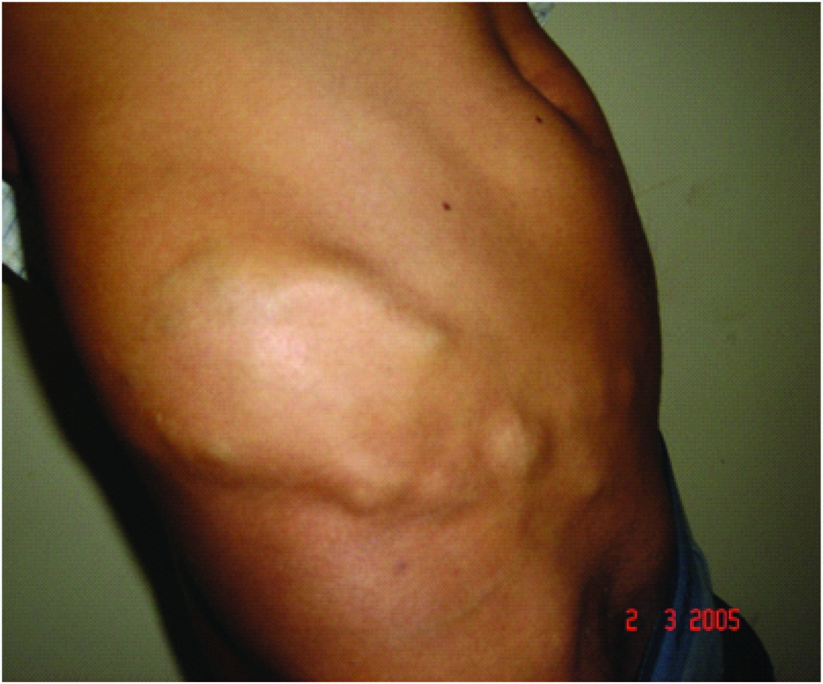
A 27-years-old male presented with a swelling around 10x8 cm on right side of lower abdomen mainly above iliac crest which was increasing in size involving right iliac and right lumbar region, swelling was soft, compressible, non pulsatile with indistinct borders, cough impulse was positive at inguinal region [Table/Fig-1].
Sonography was suggestive of a mass lesion with multiple hypoechoic areas lateral to right iliac crest in subcutaneous and intra muscular plane and another well defined hypoechoic mass measuring 1.8x1.2 cm at inner margins of iliac bone anteriorly, deep to the muscle and a presence of right inguinal hernia. MRI showed diffuse lesion of 9.7x9.1x13.3 cm in right lumbar and right iliac region in subcutaneous fat plane along right lateral abdominal wall and involving underlying anterior abdominal wall muscles with intra abdominal extension, feeding vessels could not be identified. MRI also kept possibility of rt inguinal hernia. Wide local excision of haemangioma with part of external oblique muscle, internal oblique and conjoint tendon was excised through two separate incisions [Table/Fig-2]. There was no hernia sac found intraoperatively. Meshplasty was done to cover the defect created by wide local excision.

Histopathology proved the diagnosis of intra muscular haemangioma as fibro fatty tissue with vessels of varying size and calibre, interconnecting with each other found without any malignant degeneration.
Postoperative period was uneventful. Sutures were removed on 10th day and patient went home without complication. Follow up after six months revealed no recurrence.
Discussion
Haemangiomas are common benign vascular soft tissue tumour especially present during the early years of life. Intramuscular haemangiomas are rare tumors comprising less than 1% of all [1,2]. The most frequent sites are extremities, head and neck whereas abdominal wall is a quiet rare location [3]. They are usually slow growing vague mass, with sign of compressibility and pain in some areas. Exact cause of haemangioma is not known but theories of congenital developmental anomaly with history of trauma has been listed as major causative factor [2,4]. Abdominal wall muscle is rare site for vascular swelling as less chance of trauma and less chance of any high flow arterial branch supplying the muscle. Based on predominant vascular pattern they are classified into Capillary, Cavernous and Mixed. Most reports of Intramuscular Haemangioma stress on its atypical clinical presentation [5]. Histopathologically the tumour contains various amount of fibro fatty tissue, smooth muscle, thrombus and bone beside blood vessels.
Ultrasonography confirms space occupying lesion and Colour Doppler sonography proves the vascularity of lesion. MRI is more specific in evaluating the extension of lesion. MR Angio can be used to identify feeding and draining vessels which can be an aid to operative planning and complete resection of tumour [1,3].
Till date we found only five reported cases of anterior abdominal wall haemangioma. Only two of them were found in internal oblique muscle, all the other three reported cases were in Rectus muscle. Our case was interesting as it involved External oblique, Internal oblique and transverse abdominus muscle. Mc Neill and Ray in their review of intramuscular Haemangioma revealed better outcome- if lesion is localized, good results with complete excision. Partial excision results in high recurrence rate and limited success if treated nonoperatively like sclerotherapy, radiotherapy and angioembolism. Spontaneous resolution has not been known to occur [2,4–5]. The defect in abdominal wall created by wide local excision can be covered by polypropylene mesh as in our case.
Conclusion
Intramuscular Haemangiomas are rare. Intramuscular Haemangioma of abdominal wall are the rarest. Diagnostic dilemma can occur if it extends along the fascia. Sub facial extension along muscle especially in inguinal region can present as cough impulse. MRI is investigation of choice. Wide local excision is the treatment of choice.
[1]. Goldberg SR, Halvorsen RA, Neifeld JP, Vascular tumours of the abdominal wallThe Am J of Surgery 2004 187:553-56. [Google Scholar]
[2]. Sharma D, Prasad RS, Puneet Shukla R, Kumar M, Shukla VK, Rectus muscle haemangioma: A case note with analysis of previously reported casesThe Internet journal of Surgery 2007 9:1 [Google Scholar]
[3]. Brown RA, Crichton K, Intramuscular haemangioma of thigh in a basket ball playerBr J Sports Med 2004 38:346-48. [Google Scholar]
[4]. Sunil TM, Intramuscular haemangioma complicated by Volkmans like contracture of the forearm musclesIndian Pediatrics 2004 41(3):270-73. [Google Scholar]
[5]. Choudhary N, Jain A, Gudwani S, Kapoor R, Motwani G, Intramuscular Haemangioma of Head and Neck regionJ. Laryngol. Otol 1998 112:1199-120. [Google Scholar]