Fine needle aspiration (FNA) biopsy is commonly used in the diagnosis of thyroid diseases. Serious complications are rare and this procedure is generally safe. Acute supurative thyroiditis (AST) after FNA has been seldomly reported. We report a case of a 57-year-old women with diabetes mellitus who developed AST with thyrotoxicosis after FNA. She was successfully treated by sonographically guided percutaneous drainage and antithyroid agent.
Abscess, FNA, Thyroiditis, Thyrotoxicosis
Case Report
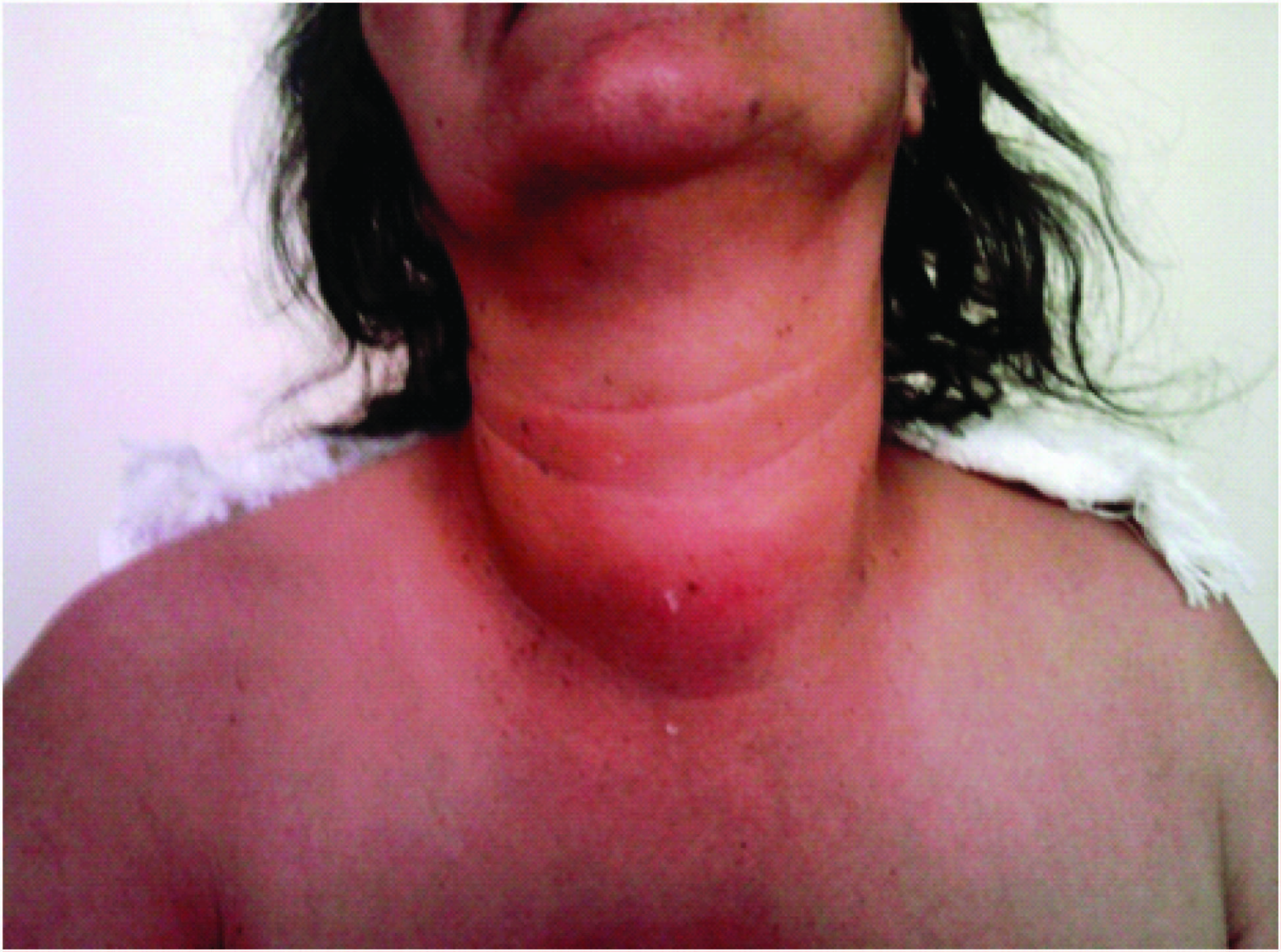
A 57-year-old female patient with symptoms of painful swelling in the neck, high fever, dyspnea and dysfagia that had appeared and worsened two days ago arrived at our clinic [Table/Fig-1]. Anamnesis revealed that thyroid nodule biopsy had been performed 15 days ago. Vital signs are temperature 39°C, blood pressure 100/70 mmhg, pulse 106/min. Examination revealed hoarseness with mild stridor and mild midline swelling and acute sensitivity in the neck above the thyroid gland. Ultrasound examination revealed an ill-defined hypoechoic area with central macroscopic cystic spaces, 2.0 x 1.5 x 1.6 cm in size at the junction of the thyroid isthmus and left lobe [Table/Fig-2a]. Cervical computarized tomography (CT) revealed a hypodense area in the thyroid isthmus [Table/Fig-2b]. The patient had a leukocytosis and elevated CRP levels. Abscess drainage with a 20 gauge needle accompanied by ultrasound was performed in three consecutive times, once a day. The aspirated material was brown and particulate. Thyroid abscess was diagnosed, and intravenous 1.5 g ampicillin sulbactam (1,000 mg ampicillin and 500 mg sulbactam, Duocid 1 g IV, Pfizer Inc., Istanbul, Turkey) four times daily was started. Methicillin-sensitive Staphylococcus aureus grew in abscess culture. No malignity was determined at pathological examination of the abscess material. HbA1c at 6.88% (Range: 4.80-5.90) and elevated blood sugar were determined, and insulin therapy was started. Thyrotoxicosis began developing on the 7th day of treatment. Tc 99m pertechnetate thyroid scintigraphy revealed a hypoactive nodule and increased activity in the tissue in which exception of the nodule. Propylthiouracil 150 mg/day (3 times a day) and propranolol 80 mg/day (twice daily) p.o. were started. Following 14-day parenteral antibiotherapy, clinical and ultrasound abscess findings completely resolved. Thyrotoxicosis began to improve, and the patient was discharged with ampicillin-sulbactam (750 mg) given orally twice daily, and antithyroid therapy on the 14th day of treatment.
a) A heterogeneous hypoechogenic area in the isthmus-left lobe junction at thyroid USG; b) an abscess within the isthmus and a part of the left lobe on neck CT c) a heterogeneous area with a hypo-anechogenic internal structure involving the right and left lobes at thyroid USG; d) the abscess drainage catheter (arrow) can be seen inside the thyroid gland at USG

At check-up on the 12th day after treatment, ultrasound examination revealed a hypo-anechogenic internal structure containing fine linear echogenic striations approximately 52x25 mm in size and heterogeneous ecogenity surrounding both lobes around the nodule on which FNA biopsy had been performed inside the thyroid gland [Table/Fig-2c]. Drainage was performed with a 6F percutaneous pigtail abscess drainage catheter [Table/Fig-2d]. The material removed was yellow, thick and non-odorous. Temperature, leukocytosis and CRP were normal. The patient’s laboratory findings are shown in [Table/Fig-3]. No bacteria were observed at Gram-staining of the abscess material. There was no growth in aerobic and anaerobic cultures or Lowenstein Jensen. No antibiotic treatment was started. Following drainage of the abscess cavity for 16 d with a 6 french catheter, with the application of negative pressure, ultrasound was performed when serous discharge appeared from the catheter and collections in both lobes were seen to have decreased. The drainage catheter was removed, and the patient was seen to be euthyroid. Antithyroid treatment was stopped, and the patient was discharged. At ultrasound performed on the 17th month after treatment, only a well-circumscribed, hypoechogenic nodular area 9 x 8 x 7 mm in diameter was determined in the left lobe [Table/Fig-4].
The patient’s laboratory findings., * Antithyroid treatment was started
| Biochemical parameters | First Hospitalization | Second Hospitalization | After discharge |
|---|
| 1st day | 2nd day | 7th day* | 10th day | 13th day | 1st day | 7th day | 14th day | 1 month | 3 months | 6 months | 12 months |
|---|
| Glucose (mg/dl) (74-109) | 129 | 153 | 284 | | | 137 | | | | | | |
| HbA1c (%) (4.8-5.9) | | | 6.88 | | | | | | | | | |
| WBC (103/μL) (4.5-11.0) | 13.3 | 7.6 | 5.1 | 7.5 | | 10.3 | 7.3 | 5.8 | 10.2 | 6.6 | 6.5 | 8.8 |
| Neutrophil (103/μL) (1.9-8.0) | 9.5 | 4.5 | 2.4 | 3.5 | | 5.8 | 3.7 | 2.5 | 5.1 | 2.9 | 3.7 | 4.6 |
| CRP (mg/L) (0-0.5) | 32.12 | 20.58 | 5.88 | | 3.02 | 3.16 | 0.63 | 0.12 | 0.13 | 0.06 | | |
| Sedimentation rate (1 hour) | 57 | | 73 | | | 75 | | 14 | 10 | 12 | 12 | 9 |
| AntiTGA (IU/ml) (0-115) | 52.97 | | | | | 11.07 | | | | | | |
| Free T3 (pg/ml) (2.0-4.4) | 3.95 | 4.90 | 9.69 | 7.99 | 4.56 | 3.07 | 2.45 | 2.21 | | 2.84 | | 2.84 |
| Free T4 (ng/dl) (0.93-1.70) | 2.18 | 3.47 | 6.99 | 7.69 | 6.80 | 2.20 | 1.37 | 1.11 | 0.88 | 1.05 | 1.00 | 1.15 |
| TSH (μIU/ml) (0.27-4.20) | 0.190 | 0.040 | 0.007 | 0.006 | 0.005 | 0.008 | 0.062 | 1.65 | 1.52 | 2.04 | 1.29 | 1.30 |
Well-circumscribed, hypoechogenic area in the left lobe on thyroid USG.

Discussion
Factors such as its possession of a high iodine content and widespread lymphatic drainage, the richness of its arterial blood flow and having a capsule that completely surrounds it, mean that the thyroid gland is resistant to bacterial infections [1,2]. Therefore, acute suppurative thyroiditis (AST) is a rare condition. The infection agent leading to AST generally reaches the thyroid gland through hematogenous or lymphatic spread from respiratory tract infection in adults and may be sinus fistula-mediated in children [3]. Underlying diseases including diabetes mellitus, tuberculosis, and human immunodeficiency virus infection, appear to increase the risk for developing of thyroid abscess [4]. The suppurative process generally develops in the presence of underlying thyroid pathology [5]. The most frequent pre-existing thyroid pathology is multinodular goiter [2,6]. Other causes of AST are neoplastic thyroid nodule, Hashimoto thyroiditis, subacute thyroiditis, and penetration of the thyroid gland by foreign bodies such as fish bones and trauma [2,7]. Fine needle aspiration (FNA) biopsy is a minimally invasive, economical and a reliable technique used in the diagnosis of thyroid pathologies. Local hematoma and pain are the most common complications of FNA [8]. AST can also develop (though rarely) after thyroid fine needle biopsy performed for diagnostic purposes [1,4,9,10]. Here, we report a case of thyrotoxicosis developing with AST after FNA . The patient has been newly diagnosed diabetes mellitus
AST onset is generally sudden, and the clinical picture progresses very rapidly. As in our patient, it frequently leads to symptoms such as painful swelling in the neck, hyperemia in the skin, hoarseness, dyspnea, dysfagia and high fever. It can also lead to life-threatening complications such as laryngeal oedema or tracheal compression-related airway obstruction [1].
As in our patient, during the course of the disease, leukocytosis, high erythrocyte sedimentation rate and elevated CRP level can be seen. Thyroid hormones may slightly rise in association with destruction of follicular cells, but thyrotoxicosis is rarely seen [4,5]. A few cases thyrotoxicosis developing with AST have been reported to date [3,4,11,12]. Moreover, to our knowledge, acute suppurative thyroiditis associated with thyrotoxicosis after FNA was reported only one case [4]. In this reported case of Nishihara E et al., antibiotic treatment for approximately one month did not improved to the inflammatory findings and left thyroid lobectomy performed. In our case, the patient was succesfully treated with catheter drainage without lobectomy. AST associated with thyrotoxicosisis generally temporary, and most patients will be improved without any specific treatment [12]. In the reported case of Nishihara E et al., thyroid hormone profile gradually returned to normal after surgery without antithyroid therapy [4]. Panamonta O et al., was reported the first child case of AST with thyrotoxicosis [11]. In this case, thyrotoxicozis was not treated with antithyroid therapy and thyroid function test returned to the normal levels five days after abscess drainage. In our case, antithyroid therapy was started a seven days after admission and thyroid hormones decreased immediately following the start of antithyroid therapy. In generally, at thyroid scintigraphy, thyroid gland activity is normal apart from a cold suppurative area [13]. In the reported cases of both Panamonta O et al., and Nishihara E et al., were not performed scintigraphy [4,11]. We fortunately performed scintigraphy. Because, in our case, the activity was increased outside a cold area in which abscess developed and antithyroid therapy was started. If we didn’t start to antithyroid therapy thyrotoxicosis may lead to thyroid storm, the worst complication of hyperthyroidism [14]. This can further exacerbate the existing picture.
As in our case, abscess in the thyroid gland can be confirmed with ultrasound [1] and tissue/fluid sampling can be performed with ultrasound guidance [2]. Computerized tomography or magnetic resonance imaging shows the abscess’ topographical relations with neighbouring structures, such as the trachea. They may also reveal causes such as pyriform sinus fistula and foreign bodies that may occur in young patients and recurrent cases in particular [6].
FNA is a gold standard in diagnosing thyroid abscess, and it renders the isolation of the pathogenic agent. FNA biopsy also makes it possible to determine underlying causes such as thyroid neoplasm [1]. The most frequently isolated agents in AST are Staphylococcus and Streptococcus species [2,5]. Fungal, parasitic and microbacterial species can sometimes also be identified [5,15]. In our case, Staphylococcus aureus is yielded in abscess culture as in the reported case of Nishihara E et al., [4].
Treatment is based on wide-spectrum antibiotherapy and abscess drainage. The widely accepted approach involves surgical drainage of the abscess. Since thyroid abscess can develop due to malignity, the abscess material must be examined for pathology, and any potential malignity must be excluded. If malignity is determined, an appropriate surgical approach must be performed if possible. Some authors have obtained successful results with using neddle aspiration or catheter drainage and have recommended it as an alternative to surgical drainage [2]. Ilyın A et al., treated thyroid abscess in two cases with neddle aspiration under USG guidance. [2]. In these cases, antibiotic was injected into the abscess cavity after the drainage of pus. In our patient the infection was not cured with simple needle drainage and we had to used drainage catheter.
Conclusion
In summary, in these patients, antibiotheraphy and drainage procedures should be performed urgently. Antithyroid therapy needs should be determined, if also thyrotoxicosis developes. In addition, it should be kept in mind that percutaneous drainage can be performed as an alternative to surgical drainage in patients developing AST.
[1]. Halenka M, Skodova I, Horak D, Kucerova L, Karasek D, Frysak Z, Thyroid Abscess as a Complication of Fine-Needle Aspiration BiopsyThe Endocrinologist 2008 18:263-65. [Google Scholar]
[2]. Ilyın A, Zhelonkina N, Severskaya N, Romanko S, Nonsurgical Management of Thyroid Abscess with Sonographically Guided Fine Needle AspirationJournal of Clinical Ultrasound 2007 35:333-37. [Google Scholar]
[3]. Kantorovich V, Patil N, Rassouli N, Acute suppurative thyroiditis with thyrotoxicosis caused by methicillin resistant staphylococcus aureusThe International Journal of Medicine 2010 13:350-54. [Google Scholar]
[4]. Nishihara E, Miyauchi A, Matsuzuka F, Acute suppurative thyroiditis after fine-needle aspiration causing thyrotoxicosisThyroid 2005 15(10):1183-87. [Google Scholar]
[5]. Herndon MD, Christie DB, Ayoub MM, Duggan AD, Thyroid abscess: case report and review of the literatureAm Surg 2007 73(7):725-28. [Google Scholar]
[6]. Dugar M, da Graca Bandeira A, Bruns J Jr, Som PM, Unilateral hypopharyngitis, cellulitis, and a multinodular goiter: a triad of findings suggestive of acute suppurative thyroiditisAm J Neuroradiol 2009 30(10):1944-46. [Google Scholar]
[7]. Chen CY, Peng JP, Esophageal fish bone migration induced thyroid abscess: case report and review of the literatureAm J Otolaryngol 2011 32(3):253-55. [Google Scholar]
[8]. Polyzos SA, Anastasilakis AD, Clinical complications following thyroid fine-needle biopsy: a systematic reviewClinical endocrinology 2009 71(2):157-65. [Google Scholar]
[9]. Sun JH, Chang HY, Chen KW, Lin KD, Lin JD, Hsueh C, Anaerobic thyroid abscess from a thyroid cyst after fine-needle aspirationHead Neck 2002 24:84-86. [Google Scholar]
[10]. Unlütürk U, Ceyhan K, Corapçıolu D, Acute suppurative thyroiditis following fine-needle aspiration biopsy in an immunocompetent patientJ Clin Ultrasound 2013 doi:10.1002/jcu.22077 [Google Scholar]
[11]. Panamonta O, Panombualert S, Panamonta M, Apinives C, Acute suppurative thyroiditis with thyrotoxicosisJ Med Assoc Thai 2009 92(10):1370-73. [Google Scholar]
[12]. Kobayashi A, Kuma K, Matsuzuka F, Thyrotoxicosis after needle aspiration of thyroid cystJournal of Clinical Endocrinology and Metabolism 1992 75:21-24. [Google Scholar]
[13]. Szabo SM, Allen DB, Thyroiditis. Differentiation of acute suppurative and subacute. Case report and review of the literatureClin Pediatr (Phila) 1989 28(4):171-74. [Google Scholar]
[14]. Chiha M, Samarasinghe S, Kabaker AS, Thyroid Storm: An Updated ReviewJ Intensive Care Med 2013 doi: 10.1177/0885066613498053 [Google Scholar]
[15]. Jacobs A, Gros DA, Gradon JD, Thyroid abscess due to Acinetobacter calcoaceticus: case report and review of the causes of and current management strategies for thyroid abscessesSouth Med J 2003 96(3):300-07. [Google Scholar]