An in Vitro Evaluation of Remineralization Potential of Novamin® on Artificial Enamel Sub-Surface Lesions Around Orthodontic Brackets Using Energy Dispersive X-Ray Analysis (EDX)
Pritam Mohanty1, Sridevi Padmanabhan2, Arun B Chitharanjan3
1 Senior Lecturer, Department of Orthodontics & Dentofacial Orthopaedics, Kalinga Institute of Dental Sciences, KIIT UniversityBhubaneswar, Odisha, India.
2 Professor, Department of Orthodontics & Dentofacial Orthopaedics, Faculty of Dental Sciences, Sri Ramachandra University, Porur, Chennai, Tamil Nadu, India.
3 Professor & Head, Department of Orthodontics & Dentofacial Orthopaedics, Faculty of Dental Sciences, Sri Ramachandra University, Porur, Chennai, Tamil Nadu, India.
NAME, ADDRESS, E-MAIL ID OF THE CORRESPONDING AUTHOR: Dr. Pritam Mohanty, N4/262, IRC Village, Nayapalli, Bhubaneswar, Odisha- 751015, India. Phone : 9777453388, E-mail : drpritammohanty@gmail.com
Objective: To evaluate and compare the Ca/P ratio of enamel samples around the orthodontic brackets for time periods of 0, 2 and 10 days in two groups (control group and study group).
Materials and Methods: Forty extracted teeth were randomly divided into control group and study group. All samples were demineralized and incubated in artificial saliva at 37°C for a period of 10 days after demineralization. During this phase the enamel samples in the study group were treated with remineralizing paste (NuproNusolution containing Novamin®-Dentsply) for 10 days. At the end of the incubation period, Ca/P ratios were analyzed for both the groupsby EDX analysis. Data obtained was subjected to statistical analysis using student t-test for paired samples and Student t- test for individual samples (p ≤ 0.05).
Results: It was found that the mean Ca/P ratio was significantly lower for the control group as compared to the study group (p-value < 0.05) after 10 d of incubation.
Conclusion: Novamin® containing remineralization toothpaste showed significant remineralizing potential in inhibition of artificial enamel sub-surface lesion around bracket after 10 days of remineralization phase. EDX element analysis was found to be an efficient method to quantify the changes in mineral content of a sample during in vitro caries studies.
Demineralization, Orthodontics, White spot lesions
Introduction
Though a wide variety of new materials and different treatment modalities have resulted in a remarkable improvement in the delivery of orthodontic treatment, orthodontists are still challenged by a significant clinical problem i.e. decalcification of the enamel surface adjacent to fixed appliances [1].
The term white spot lesion(WSL) was defined by Fejerskov et al., [2] as “the first sign of a carious lesion on enamel that can be detected with the naked eye”.Prevalence of WSLs in patients after orthodontic treatment ranges from 15% - 85% [1], with most studies reporting 50%-70% [3–5].
These WSLs during fixed appliance therapy are partly due to the surface irregularities caused by the appliances, which favour plaque accumulation. After active orthodontic treatment, the demineralization process normally decelerates due to alterations in local environmental factors. However, WSLs can still persist, resulting in an esthetically and structurally unacceptable condition [6].
Various methods have been recommended to minimize the incidence and severity of WSLs. Good oral hygiene regimen along with incorporation of fluoride in dentrifices [7,8], adhesives [9], elastomerics [10], varnishes [11], are some of the methods to prevent WSLs. In addition to preventive methods there has been a surge in the use of remineralization products such as those containing CPP-ACP (casein phosphopeptide-amorphous calcium phosphate) [12], nanocomplexes such as carbonate-hydroxyapatite nano-crystals [13] and calcium sodium phosphosilicate (Novamin®) [14].
At present, a wide range of both macroscopic and microscopic methods used in-vitro and in-vivo are available. It is desirable that the assessment method be accurate, reproducible,easy to use and should validate mineral loss from the enamel.For microscopic in-vitro evaluation, various methods like caries models, polarized light microscopy and Scanning Electron Microscope have been used.An improvement on the Electron microscope has been the EDX analysis with Scanning Electron Microscope (SEM) which is a micro-analytical technique employed to estimate quantitatively the amounts of mineral in a given tooth sample [15].
Thus, this study was designed to evaluate the effects of Novamin® on enamel demineralization around the orthodontic brackets with EDX analysis.
Materials and Methods
The sample consisted of 40 extracted human premolars obtained from patients requiring therapeutic extraction for orthodontic treatment. The inclusion criteria for teeth selected were intact buccal enamel without developmental defects, not subjected to any pre-treatment chemical agents, no physical damage due to extraction and any caries or WSLs.
The apical two third of the roots were removed by a diamond disc, for operational convenience. All the teeth were rinsed with normal saline and stored individually in closed pre-labeled plastic containers (Scintillation vial, Polylab Products, India) containing distilled water (Aquapure, Golechha Diagnostics, India) at room temperature (37oC).
Buccal surfaces of the premolars were polished and then etched with 37% phosphoric acid gel (ETCH, d- tech, SakhiChem Tech Pvt. Ltd., India) for 15seconds. The etchant gel was removed from the tooth surface with a water spray and dried followed by bonding of premolar brackets (Gemini Series, .022 MBT brackets, 3M Unitek, Monrovia, USA) using a light cure adhesive (Transbond® XT, 3M Unitek, Monrovia, USA). Subsequently the bonded teeth were coated with acid-resistant nail varnish (Colorbar cosmetics Pvt Lmt, USA) [16,17], leaving a 1 mm wide window around the bracket on the enamel surface. Samples were stored in individual pre-labeled plastic containers containing distilled water at room temperature till subjected to SEM scanning [Table/Fig-1] followed by EDX analysis [Table/Fig-2] for the evaluation of mineral content (calcium and phosphorus content in wt %).
Sample placed inside the SEM
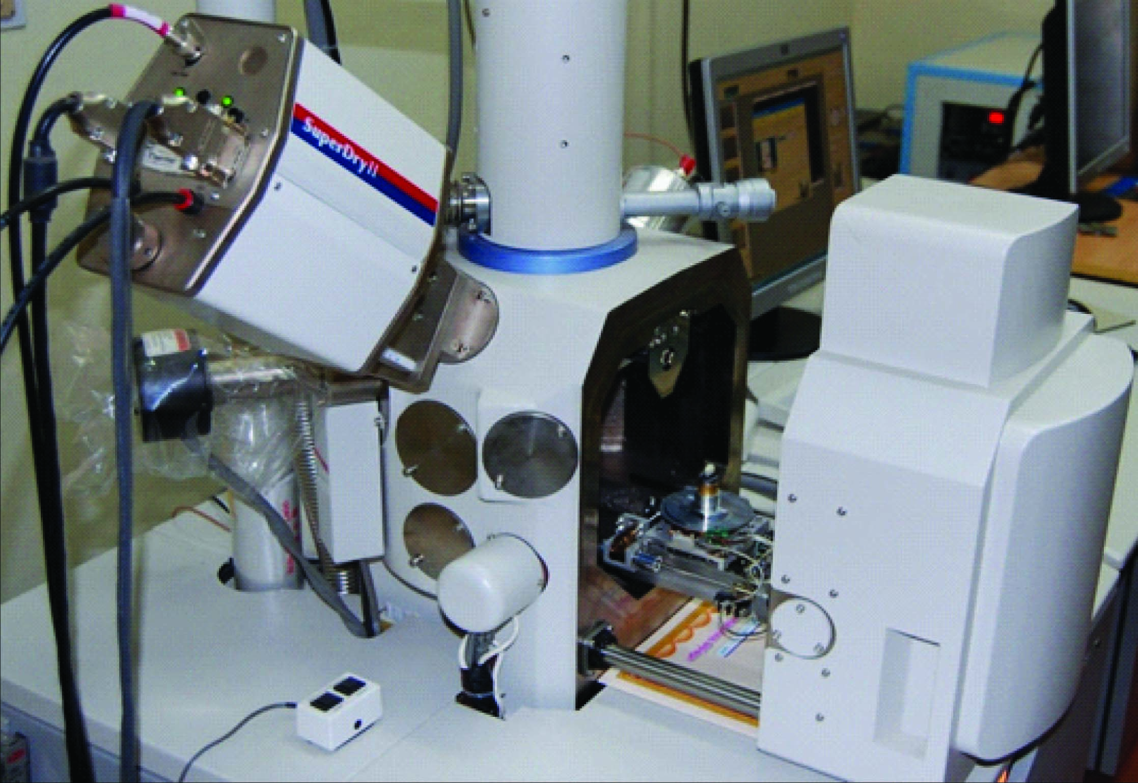
EDX software (Hitachi, Japan)

Each tooth was immersed in 10 ml of demineralizing solution for 48 h to produce artificial caries lesions. The demineralizing solution was made with 2.2mM (CaCl2), 2.2mM (NaH2PO4), 0.05 M acetic acid with the pH adjusted to 4.4 with 1 M KOH[18].
After demineralization, all tooth samples were washed with distilled water, randomly assigned to two groups of 20 each and all the samples were subjected to SEM scanning again and evaluated by EDX analysis.
The control group was incubated at 37°C for a period of 10 d in artificial saliva which contained 2.200 g/L gastric mucin, 0.381g/L NaCl, 0.213 g/L (CaCl2)2H2O, 0.738 g/L K2 (HPO4)3H2O and 1.114 g/L KCl.
The final pH was adjusted to 7.00 at 37°C with 85% lactic acid [19].
The teeth in the study group were also incubated in artificial saliva at 37°C for a period of 10 days.During this period they were treated with remineralizing paste (NuproNusolution containing Novamin®-DENTSPLY) for 3 min (manual brushing) every 12 h for a period of 10 days.
At the end of the incubation period all the samples were again evaluated by EDX analysis. Thus Ca/P ratios were analysed for both the groups at three time intervals: To representing 0 day just after the bonding was done, T1 after 48 h of demineralization and T2 representing 10 d post-treatment with remineralizing paste. Data obtained was subjected to statistical analysis.Student t – test for paired samples was used to compare the changes in mineral content between the groups. Student t–test for individual samples was used for comparison of mineral content at three time intervals, within the groups.The value of significance was set at p< 0.05.
Results
EDX analysis was used to determine calcium and phosphorus content (in weight %) of the samples at T0, T1 and T2 in each group. The mean Ca/P ratio of enamel at T0 was 1.79 ± 0.005 in control group and 1.79 ± 0.007 in study group. At T1mean Ca/P ratio fell to 1.49 ± 0.006 in control group and 1.50 ± 0.006 in study group. At T2 mean Ca/P ratio was 1.49 ± 0.005 in control group and 1.78 ± 0.006 in study group.
There was no significant difference in the Ca/P ratios between the groups at T0 and T1. However there was significant difference between the groups at T2. [Table/Fig-3].
Comparison of mean Ca/P ratios of samples between the groups using student t- tests for independent samples
| Groups | Samples | N | Mean | Sth. Error Mean | t – value | P - value |
|---|
| Control | To | 20 | 1.79 | 0.005 | 0.73 | 0.47* |
| Study | To | 20 | 1.79 | 0.005 | 0.73 | 0.47* |
| Control | T1 | 20 | 1.49 | 0.006 | 1.09 | 0.28* |
| Study | T1 | 20 | 1.49 | 0.006 | 1.09 | 0.28* |
| Control | T2 | 20 | 1.49 | 0.005 | -35.12 | 0.001** |
| Study | T2 | 20 | 1.78 | 0.006 | -35.12 | 0.002** |
* Statistically non-significant, ** Statistically significant
Comparison within the groups showed that there was a significant difference between T0 and T1 in both the groups.There was no significant difference in the control group between T1 and T2 [Table/Fig-4] whereas there was significant difference in the study group between T1 and T2 [Table/Fig-5].
Comparison of mean Ca/P ratios of samples within the group using student t- test for Paired sample incontrol group
| Sample | N | Mean | Std. Error mean | t - value | p – value |
|---|
| T | 20 | 1.79 | 0.003 | 103.38 | 0.0024** |
| T1 | 20 | 1.49 |
| T0 | 20 | 1.79 | 0.003 | 103.36 | 0.0024** |
| T2 | 20 | 1.49 |
| T1 | 20 | 1.49 | 0.001 | -0.809 | 0.43* |
| T2 | 20 | 1.49 |
* Statistically non-significant, ** Statistically significant
Comparison of mean Ca/P ratios of samples within the group using student t- test for Paired sample in Study group
| Sample | N | Mean | Std. Error mean | t - value | p – value |
|---|
| T0 | 20 | 1.79 | 0.007 | 45.54 | 0.0024 ** |
| T1 | 20 | 1.49 |
| T0 | 20 | 1.79 | 0.000 | 1.83 | 0.08* |
| T2 | 20 | 1.78 |
| T1 | 20 | 1.49 | 0.007 | -45.27 | 0.0022** |
| T2 | 20 | 1.78 |
* Statistically non-significant, ** Statistically significant
Discussion
In the oral environment, tooth structure undergoes continuous demineralization and remineralization [20]. During orthodontic treatment, tooth enamel is at a higher risk for caries which is due to food stagnation caused by the appliances and inadequate oral hygiene [21,22].
There are many possibilities to arrest or reverse the progression of the WSLs. The clinical use of calcium and phosphate ions for remineralization has not been successful in the past, due to low solubility of calcium phosphates particularly in the presence of fluoride ions. For every 2 fluoride ions, 10 calcium ions and 6 phosphate ions are required to form one unit cell of fluorapatite (Ca10(PO4)6 F2) [23]. Thus research has focused on the efficacy of delivery systems which can provide higher concentration of calcium and phosphate ions along with fluoride ions at the tooth surface.
A recent entrant into this scene is Novamin® (Calcium sodium phosphosilicate) which is a bioactive glass in the group of highly biocompatible materials, originally developed as bone regenerative materials [14]. These materials are reactive when exposed to body fluids, and deposit hydroxycarbonate apatite, a mineral that is chemically similar to the mineral in enamel and dentin [24] and thus enhances enamel remineralization [25,26]. This study sought to evaluate the effect of this molecule on artificial enamel subsurface lesion around brackets. Artificial caries-like lesions of enamel are more homogeneously reproducible than natural lesions and thus provide a reliable experimental model.
The results of this study showed that in both groups there was significant difference between T0 and T1 (p<0.05) which showed that significant demineralization occurred [Table/Fig-4&5]. However, when comparing between T1and T2 (p<0.05) there was significant difference only in the study group which showed that remineralization in response to Novamin® was significant as compared to the control group.
Comparison between the study and control group showed that there was no significant difference in the mean Ca/P ratio at both T0 and T1. Both groups had been subjected to the same treatment till then which is another testimony for the efficacy of the EDX analysis. However, at T2 (p< 0.05) there was significant difference in the study group as compared to the control. In the study group, the mean Ca/P ratio after remineralization phase was found to be comparable to the mean Ca/P ratio before demineralization. This showed that the remineralization potential of Novamin® was significant [Table/Fig-3]. Similar study by Mohan AG et al., also found that there was an increase in calcium/phosphate ratio for samples treated with fluoride enhanced hydroxyapatite gel (Remin Pro, VOCO GmbH Cuxhaven, Germany) irrespective of the radiation protocol [27].
Shetty S et al., [28] and Jayarajan j et al., [29] concluded that the addition of Fluoride to CPP-ACP shows improved remineralization of initial enamel caries when compared with CPP-ACP and NaF. In contrast to these study, mehta et al., [30] concluded that there was no significant difference when the remineralizing effect of CPP-ACP was compared with the remineralizing effect of CPP-ACFP.
Although the remineralizing capacity of various remineralizing agents on enamel is accepted, the evidence is insufficient to support the effectiveness of these agents for remineralization of postorthodontic WSLs around the orthodontic brackets. Consequently, further in vivo studies to verify the efficacy of Novamin® on WSLs during orthodontic treatment, would be benificial.
Conclusion
Based on the data from the present study it can be concluded that
Novamin® containing toothpaste showed significant remineralizing potential in inhibition of artificial enamel sub-surface lesion around bracket after 10 days of treatment.
EDX element analysis was found to be an efficient method to quantify the changes in mineral content of a sample during in vitro caries studies. It gives a validation of the effectiveness of in remineralization in the presence of mineral loss and justifies its use as an efficient method of preventing whitespot lesions during orthodontic treatment.
Further in vivo studies would help to determine the most effective way to use Novamin® to prevent or reverse demineralization during orthodontic treatment.
* Statistically non-significant, ** Statistically significant
* Statistically non-significant, ** Statistically significant
* Statistically non-significant, ** Statistically significant
[1]. Mitchell L, Decalcification during orthodontic treatment with fixed appliances- an overviewBritish Journal of Orthodontics 1992 19:199-205. [Google Scholar]
[2]. Fejerskov O, Nyvael B, Kidel EAM, Clinical and histological manifestations of dental cariesDental caries: the disease and its clinical management 2003 1Copenhagen, DenmarkBlackwell Munksgaard:71-99. [Google Scholar]
[3]. Mizrahi E, Surface distribution of enamel opacities following orthodontic treatmentAmerican Journal of Orthodontics and Dentofacial Orthopaedics 1983 84:323-31. [Google Scholar]
[4]. Ogaard B, Prevalence of white spot lesions in 19-year-olds; a study on untreated and orthodontically treated persons 5yrs after treatmentAmerican Journal of Orthodontics and Dentofacial Orthopaedics 1989 96:423-27. [Google Scholar]
[5]. Basdra EK, Huber H, Komposch G, Fluoride released from orthodontic bonding agents alters the enamel surface and inhibits enamel demineralization in vitroAmerican Journal of Orthodontics and Dentofacial Orthopaedics 1996 109:466-72. [Google Scholar]
[6]. Banks PA, Chdwick SM, AShurMcDade C, Wright JL, Fluoride-releasing elastomerics - a prospective controlled clinical trialEuropean Journal of Orthodotics 2000 22:401-07. [Google Scholar]
[7]. Ogaard B, Alm AA, Larsson E, Adolfsson U, A prospective, randomized clinical study on the effects of an amine fluoride/stannous fluoride toothpaste/mouthrinse on plaque, gingivitis and initial caries lesion development in orthodontic patientsEuropean Journal of Orthodotics 2006 28:8-12. [Google Scholar]
[8]. Alexander SA, Ripa LW, Effects of Self-Applied Topical Fluoride Preparations in Orthodontic PatientsThe Angle Orthodontist 2000 70:34-38. [Google Scholar]
[9]. Pascotto RC, Navarro MFL, Filho LC, Cury JA, In vivo effect of resin-modified glass ionomer cement on enamel demineralization around orthodontic bracketsAmerican Journal of Orthodontics and DentofacialOrthopaedics 2004 125:36-41. [Google Scholar]
[10]. Storie DJ, Regonnltter F, Fraunhofer JAV, Characteristics of a fluoride releasing elastomeric chainThe Angle Orthodontist 1994 64(3):199-210. [Google Scholar]
[11]. Gontijo L, Cruz RDA, Brandao PRG, Dental Enamel around Fixed Orthodontic Appliances after fluoride Varnish ApplicationBrazilian Dental Journal 2007 18(1):49-53. [Google Scholar]
[12]. Kumar VLN, Itthagarun A, King NM, The effect of casein phosphopeptide-amorphous calcium phosphate on remineralization of artificial caries-like lesions; an In-vitro studyAustralian Dental Journal 2008 53:34-40. [Google Scholar]
[13]. Roveri N, Battistella E, Bianchi CL, Foltran I, Foresti E, Iafisco M, Surface Enamel Remineralization: Biomimetic Apatite Nanocrystals and Fluoride Ions Different EffectsJournal of Nanomaterials 2009 2009:1-9. [Google Scholar]
[14]. Hench LL, Anderson O, Bioactive Glasses. In: Introduction to bioceramics. Singapore; World Scientific pp: 45-47.Hench LL, Andersson O. Bioactive glasses. In: Hench LL, Wilson J, edsAn Introduction to Bioceramics. Singapore: World Scientific 1993 1:45-47. [Google Scholar]
[15]. Kaczmarkek E, Surdacka A, Matthews T, Miskowiak B, Digital image analysis and visualization of early caries changes in human teethMaterial Science, Poland 2005 23(2):551-58. [Google Scholar]
[16]. Livas C, Jagtman AMK, Bronkhorst E, Derks Katsaros C, Quantification of white spot lesions around brackets with image analysisAO 2008 78(4):585-90. [Google Scholar]
[17]. Chu JS, Fox JL, Higuchi WI, Nash WP, Electron probe micro-analysis for subsurface demineralization and remineralization of dental enamelJ Dent Res 1989 68(1):26-31. [Google Scholar]
[18]. White DJ, Featherstone JBD, A longitudinal micro-hardness analysis of fluoride dentrifice effects on lesion progression in vitroCaries Research 1987 21:502-12. [Google Scholar]
[19]. Burwell AK, Litkowski LJ, Greenspan DC, Calcium Sodium Phosphosilicate(Novamin®): Remineralization PotentialAdvances in Dental Research 2009 21:35-39. [Google Scholar]
[20]. Matvienko A, Joen J, Mandelis A, Arvizu G, Gomez AE, Abrams SH, Antonio S, Dental biotherkmophotonics: A quantitative photothermal analysis of early dental demineralizationEuropean physics journal special topics 2008 153:463-65. [Google Scholar]
[21]. Burkland G, Hygiene and the orthodontic patientsJournal of Clinical Orthodontics 1999 33:443-46. [Google Scholar]
[22]. Burkland G, Hygiene and the orthodontic patientJournal of Clinical Orthodontics 1999 33:443-445. [Google Scholar]
[23]. Reynolds EC, Casein Phosphopeptide-Amorphous Calcium Phosphate: The scientific evidenceAdvanced Dental Research 2009 21:25-29. [Google Scholar]
[24]. Anderson OH, Kangasniemi I, Calcium phosphate formation at the surface of bioactive glass in vitroJournal of Biomedical Material Research 1991 25:1019-1030. [Google Scholar]
[25]. Stone AH, Schemehorn BR, Burwell AK. Enhanced enamel fluoride uptake from Novamin containing fluoride dentrificesJournal of Dental Research 2008 87(special Issue B):625-28. [Google Scholar]
[26]. Burwell AK, Greenspan DC, Potential for Dentifrice protection against Enamel Erosion in an In-vitroModelCaries Research 2007 41(4):268-334. [Google Scholar]
[27]. Mohan AG, Ebenezar AVR, Ghani MF, Martina L, Narayanan A, Mony B, Surface and mineral changes of enamel with different remineralizing agents in conjunction withcarbon-dioxide laserEur J Dent 2014 8:118-23. [Google Scholar]
[28]. Shetty S, Hegde MN, Bopanna TP, Enamel remineralization assessment after treatment with three different remineralizing agents using surface microhardness: An in vitro studyJ Conserv Dent 2014 17(1):49-52. [Google Scholar]
[29]. Jayarajan J, Janardhanam P, Jayakumar P, Deepika Efficacy of CPP-ACP and CPP-ACPF on enamel remineralization – An in vitro study using scanning electron microscope and DIAGNO dentIndian Journal of Dental Research 2011 22(1):77-82. [Google Scholar]
[30]. Mehta R, Nandlal B, Prashanth S, Comparative evaluation of remineralization potential of casein phosphopeptide amorphous calcium phosphate and casein phosphopeptide amorphous calcium phosphate fluoride on artificial enamel white spot lesion: An in vitro light fluorescence studyIndian Journal of Dental Research 2013 24(6):681-689. [Google Scholar]