Oral Submucous fibrosis (OSMF) is a chronic, progressive, scarring, disease that predominantly affects the people of South –East Asian origin [1]. OSMF is found to be of multifactorial aetiology including excessive chilli consumption, genetic susceptibility, autoimmunity, iron and vitamin deficiency [2], but it is strongly associated with areca nut chewing and pan masala [3]. Experiments have shown that ethanolic extracts of areca nut stimulate collagen synthesis in human dermal fibroblasts and also stabilize collagen fibrils and render them resistant to degradation by collagenase leading to fibrosis [4]. It has been found that some amount of copper is also present in areca nut which up regulates collagen production by increasing lysyl oxidase, involved in collagen synthesis and cross linking [5].
Areca nut is found to be the main cause of OSMF. But it has been found in the routine clinical practice that some individuals with the habit of areca nut chewing may not show any clinical evidence of OSMF [6]. While few individuals are found to have OSMF without the habit of areca nut chewing [7], So there must be some other factors associated with OSMF.
Recently, plasma fibrinogen degradation products (FDPs) have been identified as an early indicator of disease in OSMF patients [2]. FDPs also termed as Fibrin split products are components of blood, produced by clot degeneration. Whenever injury occurs to any blood vessel, thrombin is produced in the first stage of coagulation cascade. In the second stage, fibrinogen, a glycoprotein synthesized by liver is converted to fibrin which forms a fibrin meshwork. As the site heals, this clot is broken by the enzyme plasminogen which gets converted into plasmin, that further splits fibrin resulting in release of fibrin split products termed as FDPs. Principal FDPs X,Y,A,B,C,D and E, particularly fragment Y and to a lesser extent fragment X, are known to produce anticoagulant effects. These FDPs are detected only when their plasma levels rise above normal levels. In normal healthy individuals, plasma FDPs levels are in very low concentrations, and thus can’t be detected [2]. Excessive FDPs are released in plasma in three main conditions: Disseminated intravascular coagulation, Thrombolytic therapy, Primary fibrinogenolysis.
It has been found that fibrin precipitating factor (FPF) is present in the saliva of OSMF patients, which behaves like Thrombin. When this FPF encounters fibrinous exudates in the oral cavity, it clots the exudates, and the body in response to this clotting produces more fibrinogen and its degradation products. Hence, as the severity of disease increases, more amount of fibrin precipitating factor is produced and in turn more amount of FDPs is produced [2]. As no haemorrhagic manifestations are encountered in OSMF, FDPs are labeled as molecules immunologically similar to fibrinogen [2].
As the FDPs are found to be early indicator of OSMF, this study was undertaken to know the plasma FDPs levels in the individuals with the habit of areca nut chewing without any clinical evidence of OSMF and comparing these to the plasma FDPs levels of the individuals with the habit of areca nut chewing with OSMF.
Materials and Methods
The patients, with the habit of areca nut chewing with and without any clinical evidence of OSMF served as the sample for our study. Total of 95 subjects were included in the study and divided into three groups. Group I: comprised of 35 subjects with the habit of areca nut chewing with OSMF, Group II: comprised of 30 subjects with the habit of areca nut chewing without OSMF and Group III: comprised of 30 individuals without any habit of areca nut chewing without OSMF, (control group).
Exclusion Criteria: Individuals with any systemic disease or on any medication and with presence of other oral mucosal lesions were excluded from the present study.
Before conducting the study, informed consent was obtained from all the subjects. After review of detailed case history, all the cases of OSMF were graded clinically according to a working classification on the basis of clinical symptoms and mouth opening which is as follows: The Clinical signs and symptoms were graded from S1 to S4 and Mouth opening from M1 to M4 and eventually a cumulative score was formulated.
A. Grading of Clinical signs and symptoms:[
8]
S1 – Stomatitis and / or blanching of oral mucosa.
S2 – Presence of palpable bands in buccal mucosa and / or oropharynx, with or without stomatitis.
S3 – Presence of palpable bands in buccal mucosa and /or oropharynx and in any other parts of oral cavity with or without stomatitis.
S4 – a. Any one of the above stage along with other potentially malignant disorders e.g. oral leukoplakia, oral erythroplakia, etc
b. Any one of the above stage along with oral carcinoma.
B. Grading of Mouth opening
M1 - Inter- incisal mouth opening upto or > 35 mm
M2 - Inter- incisal mouth opening between 26 mm – 35 mm
M3 - Inter- incisal mouth opening between 15 mm – 25 mm
M4 - Inter- incisal mouth opening < 15 mm
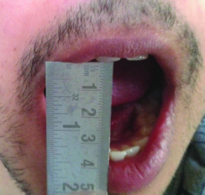
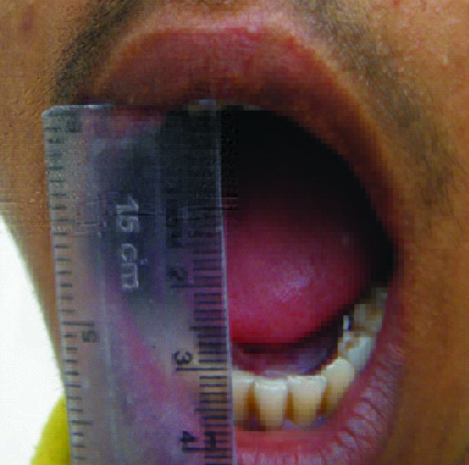
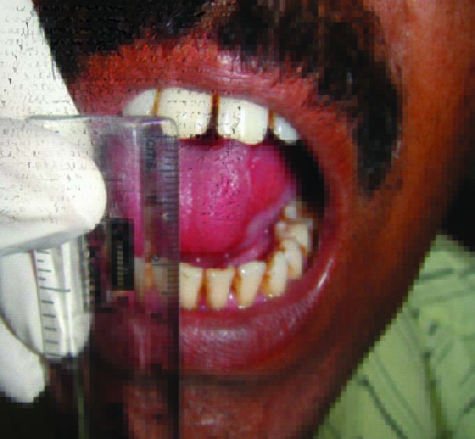
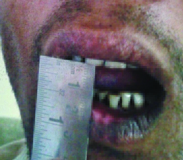
According to above parameters, the patients were divided into four Grades (with the Grade I being the least severe and Grade IV being the most severe) [Table/Fig-1,2,3,4] as follows:
Clinical picture of OSMF patient with grade I
Clinical picture of OSMF patient with grade II
Clinical picture of OSMF patient with Grade III
Clinical picture OFOSMF patient with grade IV
Cumulative Grade
Grade 1: S1M1, S1M2
Grade II: S1M3, S2M2
Grade III: S2M3
Grade IV: S3M3, S1M4, S2M4, S3M4, S4M4
Collection of Sample
Under all aseptic precautions, 5 ml of fresh venous blood was withdrawn from all the subjects by venipuncture and collected in EDTA coated test tubes. The test tubes were allowed to stand for 2 hrs at room temperature and were centrifuged at 4000 rpm to separate the plasma which was quantitated for FDPs levels.
Estimation of plasma FDPs
Plasma FDPs levels were estimated by using a diagnostic kit, Tulip XL-FDP. The quantitation was based on the principle of agglutination. The test specimen (plasma) was mixed with XL FDP latex reagent. Reagent with a sensitivity of 200 ng/ml was used, below which samples were negative and above which samples gave a positive agglutination reaction. Using Phosphate buffer saline (PBS) solution, serial dilutions of the plasma sample were prepared that is; 1:2, 1:4,1:8,1:16,1:32,1:64. Agglutination in the highest plasma dilution corresponded to the approximate amount of plasma FDPs level in ng/ml.
To calculate plasma FDPs level in ng/ml in the sample, the following formula was used.
Plasma FDPs (ng/ml) = 200 x d; Where, d was the highest dilution of plasma showing agglutination during the semi quantitative test.
Statistical Analysis
The data obtained in this study was systematically tabulated and subjected to statistical analysis. [Table/Fig-5]. To compare the mean values of plasma FDPs of all the three groups, Kruskal-Wallis Test and Mann-Whitney Test were applied and p-value <.001 was considered to be significant. For the statistical analysis median was used by the statistician.
Vmean values of plasma FDPS (ng/ml) in all the three major groups.
| Group | | Min | Max | Mean ± SD | Median |
|---|
| Group I (areca nut chewers with OSMF) | Grade I | 400 | 800 | 533.33 ± 200 | 400 |
| Grade II | 800 | 800 | 800 ±0 | 800 |
| Grade III | 800 | 1600 | 1230.77 ± 415 | 1600 |
| Grade IV | 800 | 3200 | 2755.56 ± 1 | 3200 |
| Group II (areca nut / pan chewers without OSMF) | | .0 | .0 | 0 | .0 |
| Group III (Control group) | | .0 | .0 | 0 | .0 |
Results
It was observed that all the groups were showing highly significant differences (<.001) between their mean values except when Group II (Areca nut chewers without OSMF and Group III (control group) were compared, where the difference was statistically insignificant (1.000).
Out of 35 cases in Group I, 9 were categorized with Grade I OSMF, 4 were with Grade II OSMF, 13 were with Grade III OSMF and 9 were with Grade IV OSMF. It was also observed that as the clinical grades increased in OSMF patients, levels of FDPs also increased [Table/Fig-5].
When comparison was done between the mean values of plasma FDPs levels of all the four grades of Group I, using Kruskal Wallis test and Mann Whitney test, a statistically significant difference was found among all comparisons except when Grade II OSMF was compared with Grade III OSMF, where the difference was statistically insignificant.
Discussion
OSMF is a chronic insidious disease progressed by early and advanced clinical stages. The early prodromal symptoms include burning sensation in the mouth while consuming spicy food, appearance of blisters especially on palate, ulcerations, excessive salivation, defective gustatory sensations and dryness of mouth, Petechiae formation on tongue followed by labial and buccal mucosa with no signs of blood dyscrasias. As the disease progresses, the oral mucosa becomes blanched and opaque with white fibrous bands. Buccal mucosa and lips are affected early. Faucial pillars, uvula, hard and soft palate are also affected [1]. Difficulty in mouth opening occurs with progression of the disease. The main aetiological agent of this disease is areca nut and its products. In the present study, detailed history of habits was taken and it was found that majority of the patients had a habit of chewing mixture of areca nut and other products like pan and tobacco, whereas only areca nut chewers were few in number. The duration of the habit in all the patients was almost more than 5 y and time of placement of the areca nut and products in the oral cavity was between 15 -20 min.
In the present study, it was observed that plasma FDPs were detected in all the subjects of OSMF. Similarly Phatak AG [9], Koshti SS et al., [2], Kiran G et al., [10], Gharat et al., [11] also detected the presence of these plasma FDPs in all the subjects of OSMF. The reason for could be the presence of thrombin like substance termed as FPF, present in the saliva of OSMF patients. Phatak AG [9] suggested that individuals with presence of FPF in saliva would be susceptible or the actual candidates of OSMF. Phatak AG [2] further postulated that this FPF has a thrombin like behaviour. According to him when this FPF enters the submucosal zone of oral cavity, it acts on the available fibrinogen, and clots the fibrinous exudates resulting in the local fibrosis and in the production of soluble circulating fibrin monomers. The body in response to this clotting produces more fibrinogen and its degradation products. As the severity of this disease increases, more amount of FPF is produced and in turn more FDPs are also increased. This results in excessive deposition of fibrin which further leads to progressive restriction in mouth opening. However, in contrary to our study, Kadani M et al., [12] found that out of total 30 cases, only 14 cases of OSMF with areca nut chewing habit showed the presence of plasma FDPs.
In our study it was also observed that the plasma FDPs could not be detected in individuals without OSMF [Table/Fig-5]. Similar results were showed in the study conducted by Kiran G et al., [10]. As per our knowledge, ours is second study done to detect FDP in areca nut chewers without OSMF. The reason for the absence of FDPs in this group could be the absence of thrombin like factor termed as FPF in the normal saliva. Other aspect may be that these products could be present in the plasma of these patients but we could not find them as the sensitivity of the reagent used in our study was 200 ng / ml below which it could not be possible to detect these FDPs values.
In Control Group also, the plasma FDPs could not be detected [Table/Fig-5]. As the sensitivity of the reagent used in present study was ~200 ng / ml, therefore they can be detected in plasma only when the level rises above 200 ng / ml. Similarly Koshti SS et al., [2], Kiran G et al., [10], Kadani M et al., [12] also could not detect any plasma FDPs in normal healthy individuals, as the sensitivity of the reagent used in their study was similar to our study.
When mean values of plasma FDPs levels were compared between all the three groups, statistically highly significant difference was observed. Although in both Group I and Group II, all individuals were areca nut chewers, but FDPs were detected in Group I individuals with the clinical evidence of OSMF. This shows that the presence of these plasma FDPs serve as an early indicator of OSMF.
Further, when the mean values of plasma FDPs between specific groups were compared, a statistically highly significant difference was observed between Group I (areca nut chewers with OSMF) and Group II (areca nut chewers without OSMF and Group I (areca nut chewers with OSMF) and Group III (Control group). This shows that the FDPs are an early indicator for the OSMF. But when the mean values of plasma FDPs were compared between Group II (areca nut chewers without OSMF) and Group III (control group), the results were not significant statistically (p<1.000).
When comparison was done between the mean values of plasma FDPs levels of all the four grades of OSMF, a statistically significant difference (p<.000) was found. Similarly Koshti SS et al., [2] in their study also observed a statistically significant (p <.000) increase of plasma FDPs levels with an increase in clinical grades of the OSMF, but there was no significant difference between the various histological grades of OSMF. However, in contrary to our study, Kiran G et al., [10] and Kadani M et al., [12] in their study observed no statistically significant increase of plasma FDPs with an increase in clinical as well as histological grades of OSMF.
As the plasma FDPs are an early indicator of fibrin deposition, the increase in their level with the increase in clinical grade suggests that there is increased fibrin deposition in OSMF, thus leads to severity of the disease. Also these FDPs have been detected in various malignant conditions and their level increases with the progression of the disease, which indicates that it can be an early valuable sign in the diagnostic and prognostic evaluation of carcinoma. As the clinical Grade IV of OSMF is also associated with other potentially malignant disorders or oral carcinoma, therefore the levels of plasma FDPs can be elevated with the increased severity of disease. Similarly Ghosh M et al., [13] also observed the significant increase in mean values of plasma FDPs levels with the advancement of stages of oral cancer patients as compared to normal individuals. Okholm M et al., [14] in their study also observed the increased levels of FDPs in plasma of colorectal adenocarcinoma patients as compared to individuals with non neoplastic lesions in the colon.
These plasma FDPs produce some systemic effects also in the patients with OSMF. High levels of these products can cause Glomerulosclerosis, Cerebral contusion, Pulmonary embolism, Deep vein thrombosis, Liver cirrhosis, Disseminated intravascular coagulation, Arterial thromboembolism, Tachycardia/palpitation, Tachypnea/dyspnea, Hypotension, Acute myocardial infarction, Ventricular fibrillation, Arteriosclerosis and related effects.
When the comparison was made between the Grade II OSMF and Grade III OSMF, an increase in mean values of plasma FDPs levels was observed, but it was statistically in significant in our study. The reason for this could be the lesser number of subjects in Grade II OSMF for comparison or there was less difference in the amount of fibrin deposition between Grade II OSMF and Grade III OSMF. In contrast to our results, Koshti SS et al., [2] observed a statistically significant increase in the mean values of plasma FDPs levels between Grade II OSMF and Grade III OSMF.
The main concern in OSMF is the management of trismus and burning sensation of the oral mucosa [15]. A large number of treatment modalities have been tried by both non surgical and surgical approach. The preventive measures should be in the form of discontinuation of habit, which can be encouraged through education & advocacy. Vitamins, iron and mineral rich diet should be advised. Hyaluronidase, Chymotrypsin, Interferon-Gamma, Immune Milk, turmeric powder, Injection of Gold, Vitamin A & Collagenase is helpful as medical aids. Physiotherapeutic measures such as forceful mouth opening and heat therapy have been tried to improve mouth opening. Surgical treatment is indicated in patients with severe trismus and/or biopsy results revealing dysplastic or neoplastic changes. Surgical modalities that have been used include simple excision of the fibrous bands, with major limitation being contracture of the tissue and exacerbation of the condition [15]. Split-thickness skin grafting following bilateral temporalis myotomy or coronoidectomy are also done.
The present study reveals a non invasive technique to diagnose the clinical stage of OSMF, so that in the early stage only patient can be treated without the need of invasive surgical approach.
Conclusion
Plasma FDPs were detected in all the patients of OSMF with the habit of areca nut chewing. But the Plasma FDPs were not detected in the individuals with areca nut chewing without OSMF and in control group. The plasma levels of FDPs increased, as the disease progressed from Grade I to Grade IV in the OSMF, indicating that there is increase in deposition of fibrin, thus leading to increased severity of the disease characterized by restricted mouth opening. Two cases were associated with Leukoplakia and Erythroplakia patches and levels of Plasma FDPs were higher in these cases as compared to other cases. (3200 ng/ml).
From our study, it can be concluded that plasma FDPs could not be detected in the individuals without clinical evidence of OSMF, so these products are an early valuable indicator of OSMF. Early blood investigation done to evaluate the levels of these products can give the idea of stage of the OSMF, thus helping in preventing the progression of further symptoms by motivating the patients and treating the disease in early stage only without any surgical approach. However, more studies are still required on a large scale to reveal their role in OSMF.