Chylolymphatic Cyst of Mesentery of Terminal Ileum: A Case Report in 8 Year-old Boy
Rajendra K Ghritlaharey1, Santosh More2
1Associate Professor, Department of Paediatric Surgery, Gandhi Medical College and Associated Kamla Nehru and Hamidia Hospitals Bhopal, Madhya Pradesh, India.
2MCh Student, Department of Paediatric Surgery, Gandhi Medical College and Associated Kamla Nehru and Hamidia Hospitals Bhopal, Madhya Pradesh, India.
NAME, ADDRESS, E-MAIL ID OF THE CORRESPONDING AUTHOR: Dr. Rajendra K Ghritlaharey, Associate Professor, Department of Paediatric Surgery, Gandhi Medical College and Associated Kamla Nehru and, Hamidia Hospitals Bhopal, Madhya Pradesh-462 001, India. Phone : 0-9425009272 , + 91-755-4050571(R), 4050261(O) ,
E-mail: drrajendrak1@rediffmail.com
Mesenteric cysts are rare benign intra peritoneal tumor and more than half of the mesenteric cysts involve the mesentery of the terminal ileum. We present 8 year-old boy, who presented with features of acute intestinal obstruction. Ultrasonography (USG) of the abdomen revealed a cystic mass in the peritoneal cavity with dilated loops of bowel. Exploration of the abdomen revealed a solitary cyst of the mesentery of the terminal ileum measured 10 x 8 cm. There was twisting of the part of the ileum (volvulus) due to the cyst. It also involved the wall and lumen of the adjacent ileum and there were dilated bowel loops proximal to the cyst. Complete cyst excision and resection of the part of the ileum involved with the cyst was done en bloc. An ileostomy was created due to gross disparity in the lumen of the ileum, which was closed two and half month later. Histopathology of the excised cyst was consistent with the chylolymphatic cyst (mesenteric cyst).
Abdominal cysts, Children, Chylolymphatic cysts, Intestinal obstruction, Mesenteric cysts
Case Report
Eight year-old boy was admitted with complaints of abdominal pain, vomiting, abdominal distension and constipation (features of acute intestinal obstruction) of five days duration. He had a history of recurrent abdominal pain for last six months. He had no history of passing blood or worms in stool. His abdominal examination revealed that it was distended, visible bowel loops were seen, tense, and tender with absent bowel sounds. Other systemic examination was within normal limits. His haematological investigations (Hb%, TLC, Electrolytes, urea, creatinine) all were within normal limits. Standing skiagram of the abdomen showed multiple air fluid levels without free gas under diaphragm, suggestive of intestinal obstruction. USG of the abdomen revealed a solitary, cystic mass of 10 x 8 cm in the peritoneal cavity with dilated loops of bowel. Exploratory laparotomy revealed a large, solitary cyst involving the mesentery of the terminal ileum and was measuring 10 x 8 cm. There was twisting of the part of the ileum (volvulus of the ileum) due to the cyst and dilated bowel loops proximal to the twist [Table/Fig-1a]. It also involved the adjacent ileum and occluding the lumen [Table/Fig-1b]. This mesenteric cyst was 10 cm proximal to the ileo caecal junction. Complete cyst excision and resection of the part of the ileum involved with the cyst was done en bloc [Table/Fig-2]. Ileal continuity was not possible for restoration because of gross disparity in the lumen of ileum, and an ileostomy was created [Table/Fig-3a,Table/Fig-3b]. The rest of the viscera were normal, and there was no mesenteric lymphadenopathy. Intra peritoneal drain was inserted and abdomen was closed. Transillumination of the excised mesenteric cyst was positive [Table/Fig-4]. His post - operative recovery was uneventful. The excised mesenteric cyst, part of the ileum and mesenteric lymph nodes were submitted for histology. The cyst contained clear serous fluid. Histopathology of the excised cyst wall was composed of compressed connective tissues with single layer of flattened cell and it was consistent with the chylolymphatic cyst (mesenteric cyst) [Table/Fig-5]. Histology of the intestine revealed intestinal structure with non-specific chronic inflammatory infiltrate, and histology of the lymph node revealed reactive hyperplasia only [Table/Fig-5]. An ileostomy was electively closed two and half month later. His post-operative recovery following an ileostomy closure was excellent and uneventful.
Discussion
The first report of the mesenteric cyst date back to 1507 and it was revealed during an autopsy of an 8-year- old-boy by Benevieni, an anatomist [1]. Mesenteric cysts are rare intra-abdominal tumor with prevalence is about 1:100,000 in adults and 1:20,000 in pediatric hospitals admission [1]. Mesenteric cysts may occur anywhere in the mesentery of the gastrointestinal tract from the duodenum to the rectum, but most commonly localized in the mesentery of the small intestine, the mesentery of the large intestine and retroperitoneum [1-5]. In a review of 162 cases of mesenteric cysts by Kurtz et al., they found that 60% were located in the mesentery of small bowel, 24% located in the mesentery of large bowel, and 14.5% were retroperitoneal [1]. Mesenteric cysts are most commonly solitary and multiloculated, but may occur at multiple positions within the peritoneal cavity. One third of the mesenteric cysts occur in the children younger than 15 years of age and it is reported slightly more often in males [1,2]. The exact etiology for the development of the mesenteric cysts is not known. The most commonly accepted theory was proposed by Gross, and it is the result of benign proliferation of ectopic lymphatics in the mesentery that lack communication with remainder of the lymphatic system [1].
Clinically mesenteric cysts may present as an asymptomatic abdominal mass, incidental finding during laparotomy for other abdominal conditions, or it may present as an acute abdomen. Mesenteric cysts may cause acute abdomen from the cyst rupture, infection, hemorrhage, intestinal obstruction, and volvulus [1-9]. USG and computed tomography scan of the abdomen are the investigations of choice and may helpful in the pre-operative diagnosis of mesenteric cysts, but not possible to confirm it in all the case [2,4-6,9]. Mesenteric cysts may vary in size from 4 cm to 30 cm [3,8].
Other cystic lesions in the abdomen that resemble mesenteric cysts are cystic lymphangioma (hygroma), cystic teratoma, ovarian cyst, intestinal duplication cyst, hydatid cyst, etc [1,9]. The pathological features of these cysts can vary considerably. They can be single or multiple, unilocular or multilocular; can have serous, chylous, hemorrhagic or mixed fluid. There lining can also vary from a flattened endothelium to patchy fibrosis [1]. Mesenteric cysts are most commonly single and multilocular and the fluid are generally serous when the cyst involves the distal small bowel or colonic mesentery and chylous when it is located in the proximal small bowel mesentery [1]. Histopathological examination is confirmatory and differentiates chylolymphatic cysts from all these lesions [1,9]
Complete surgical excision remains the mainstay for the treatment of the mesenteric cysts with an excellent result [1-9]. In approximately 20% to 60% of the cases bowel resection and anastomosis is needed along with the excision of the mesenteric cysts [2,4-6,8]. Excision of the mesenteric cysts may also be accomplished successfully with laparoscopically [3,7,8]. Tran NS, et al., reviewed laparoscopic management of abdominal lymphatic cyst in 47 children. Amongst 47 cases; laparoscopic cyst excision was possible in 36 cases, laparoscopy-assisted bowel resection en bloc with the cyst in 8 cases, and 3 cases required conversion to open surgery. They concluded that laparoscopic management is safe, feasible, and effective for the management of abdominal mesenteric cysts in children [8]. Prakash A, et al., reported their experience with 17 cases of mesenteric cysts in children below 10 year of the age. The most common mode of presentation among above cases was acute small intestinal obstruction. The cysts were localized in the mesentery of the small intestine in 14 cases, and 3 were in the sigmoid mesentery. During exploration, 5 of these cases had volvulus of the intestine. Four of the above cases required bowel resection along with excision of the cysts during the management. Histologically, all the cases were confirmed as lymphangiomatous mesenteric cysts [4] . Rattan KN, et al., reported their retrospective review on chylolymphatic mesenteric cysts in pediatric age group and included 8 cases, aged below 10 year. All the cases were treated by exploratory laparotomy and complete excision of the cyst along with resection of the involved bowel. All the 8 cases required bowel resection. All of these cases were multiloculated and localized at the mesentery of the small intestine with a maximum size of 9 cm. All the cases were confirmed on histological examination as chylolymphatic cysts, and there were no recurrence [9]. The recurrence following complete excision of the mesenteric cyst is very rare, but may occur [8].
This present case the cyst was localized in the mesentery of the terminal ileum, and it also involved the adjacent ileum. He was presented as an acute intestinal obstruction due to the volvulus of the terminal ileum due to the mesenteric cyst. The diagnosis of the cyst was suspected on the USG of the abdomen and was confirmed during laparotomy. En bloc complete surgical excision of the cyst involving the mesentery of the terminal ileum along with the involved adjacent ileum was done. The ileal continuity was not possible for restoration due to the disparity in the lumen and needed an ileostomy, which was closed later.
Operative photograph showing cyst involving the mesentery of the ileum,

Operative photograph showing mesenteric cyst, volvulus of the ileum, and dilated ileum,

Excised specimen showing mesenteric cyst and part of resected ileum

Operative photograph showing disparity in the ends of ileal lumen,

Operative photograph showing an ileostomy and the right para median incision (sutured)

Positive transillumination of the excised mesenteric cyst

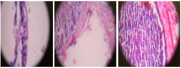
Histopathology of the mesenteric cyst (a), resected ileum (b), & mesenteric lymph nodes (c)
Conclusion
Children with chylolymphatic cyst (mesenteric cyst) involving the mesentery of terminal ileum may present with features of acute intestinal obstruction. Surgical excision of the mesenteric cyst remains a mainstay of treatment with an excellent result. Bowel resection may be needed if involved by the cyst and may rarely need an ileostomy.
[1]. RR Ricketts, Mesenteric and omental cysts. In: Grosfeld Jay L, O’Neill JA, Fonkalsrud EW, Coran AG, Caldamone AA (eds), Pediatric Surgery. Chapter 89. Mosby Inc 2006 Vol II6th Edition:1399-1406. [Google Scholar]
[2]. MA Chung, ML Brandt, D St-Vil, S Yazbeck, Mesenteric cysts in childrenJ Pediatr Surg 1991 26:1306-08. [Google Scholar]
[3]. JJ Tan, KK Tan, SP Chew, Mesenteric cysts: an institution experience over 14 years and review of literatureWorld J Surg 2009 33:1961-65. [Google Scholar]
[4]. A Prakash, A Agrawal, RK Gupta, B Sanghvi, S Parelkar, Early management of mesenteric cyst prevents catastrophes: a single centre analysis of 17 casesAfr J Paediatr Surg 2010 7:140-43. [Google Scholar]
[5]. ME Senocak, H Gündodu, N Büyükpamukçu, A Hiçsönmez, Mesenteric and omental cysts in children. Analysis of nineteen cases Turk J Pediatr 1994 36:295-302. [Google Scholar]
[6]. M Fernández Ibieta, J Rojas Ticona, I Martinez Castaño, P Reyes Rios, V Villamil, O Giron Vallejo, Mesenteric cysts in childrenAn Pediatr (Barc) 2014 [Google Scholar]
[7]. A Pampal, A Yagmurlu, Successful laparoscopic removal of mesenteric and omental cysts in toddlers: 3 cases with a literature reviewJ Pediatr Surg 2012 47:e5-8. [Google Scholar]
[8]. NS Tran, TL Nguyen, Laparoscopic management of abdominal lymphatic cyst in childrenJ Laparoendosc Adv Surg Tech A 2012 22:505-07. [Google Scholar]
[9]. KN Rattan, VJ Nair, M Pathak, S Kumar, Pediatric chylolymphatic mesenteric cyst - a separate entity from cystic lymphangioma: a case seriesJ Med Case Rep 2009 3:111 [Google Scholar]