Femoral shaft fractures in children are usually associated with long periods of morbidity. Traditional methods do give satisfactory results in younger children but older children may have complications such as malunion, delayed union, rotational deformities and psychological problems [1]. Choice for fixation is based on many factors, including the age and size of the child, associated injuries, the location and pattern of the fracture, and the social situation of the child [2]. Treatment of the femoral shaft fracture in children is controversial especially in children of age 6-12 years [3]. The aim of fracture treatment in children is rapid healing without complications, easy nursing care, rapid rehabilitation and minimal negative psychological impact on children and their families and keeping the cost effectiveness [4]. Keeping this in mind, the recent treatment modality has evolved, from conservative to operative approach. Operative methods for femoral shaft fractures in children include external fixation [5,6] plating [7], rigid or flexible intramedullary nailing [8–10]. Flexible intramedullary nailing has revolutionized the treatment of femoral shaft fractures in children. The idea of using multiple flexible intramedullary nails (Ender nails) was first conceptualized by Ender and Simon Weidner [11]. More recently Ligier and Métaizeau advocated the use of elastic titanium nails (Nancy nails) for femoral shaft fractures in children [9,10]. Their technique is known as Elastic Stable Intramedullary Nailing (ESIN) or Métaizeau technique. Qidwai et al., and Al-Zaharani et al., advocated the use of more cost effective closed intramedullary K-wire fixation for femoral fractures in children with the same technique [4,12]. We compared the results of femoral shaft fractures in children aged 6-14years treated by intramedullary titanium elastic nails (TENS nails) with intramedullary K-wire fixation.
Materials and Methods
This was a prospective randomized study conducted in Orthopaedic Department of a tertiary care teaching hospital in northern India from March 2010 to March 2012. It included consecutive 52 children with displaced diaphyseal fractures of the femur irrespective of the degree of comminution or associated injuries (head injury and multiple fractures) in children between 6 years to 14 years of age. The fractures were treated with closed reduction and percutaneous internal fixation. The cases were randomly divided in two groups and each group had 26 cases. K-wire fixation was done in Group I and titanium elastic nail in Group II. The case was allocated to Group I or Group II following the “Computer Generated Random Number Technique”. Informed written consent was obtained from each case. The study was approved from ethical committee of the institute.
The diameter of the individual nail/K-wire was selected as per Flynn et al’s formula (Diameter of nail = Width of the medullary canal at Isthmus x 0.4 mm) [13].
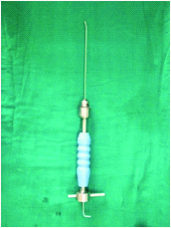
With the patient supine on a fracture table and under general anesthesia, the fracture was reduced using longitudinal traction applied through a traction boot under fluoroscopic guidance. The surgical technique for both K-Wire and titanium elastic nail was similar to what Métaizeau described [10]. The flexible rod is initially bent or curved (plastically deformed). The sharp entry tips of the K-wire were nibbled and a proximal bent of 30 degrees was made same as that in flexible nails [Table/Fig-1]. Two nails/K-Wire of identical diameter were used in a single fracture [13]. During intramedullary insertion, this is typically retrograde in the femur. Small longitudinal skin incisions are made at the medial and lateral distal femoral metaphysis. By using an awl, two holes are made on each side of distal metaphysis about 1.5 cm proximal to growth plate. Through the holes, two long standard K-wires/TENS nail of 2.5–3.5 mm in diameter (depending on femoral isthmus and patient age) are introduced with bent proximal end (300) by using a T-handle chuck one after the other. Once the K-wires/TENS nails have passed the fracture site, the tips were driven up to the level of proximal metaphysis with divergence one towards the neck and one towards greater trochanter to provide three-point fixation. At this stage, traction is released and the wires pushed further, fixing their tips at the proximal metaphysis without perforating the physis. Care is taken during the wire insertion to avoid rotational deformity. The outer wire ends are bent, cut to the desired length and buried under the skin.
Showing k -wire loaded on insertion device with bend tip (Arrow)
Postoperatively, knee movements were allowed on the next day or as soon as pain was tolerable. Non-weight bearing walking with bilateral axillary crutches was started as soon as the pain was tolerable, usually by the end of the third day. Partial weight bearing was started after 3-4 weeks of surgery and full weight bearing after 6-10 weeks. The timing of full weight bearing was guided by the clinico-radiological stage of union. All the patients were followed up till consolidation of fractures. Clinico-radiological union is defined as non-tender, non-mobile fracture with presence of bridging callus in three cortices at fracture site in plain radiographs. The average duration of hospital stay was 4-6 days.
Results
In this study the mean age of the patients was 9.15 years (range: 6-14). There were 36 male children and 16 female children. The left femur was fractured in 24 cases and right femur in 28 cases. Road traffic accident was the mode of injury in 30 cases and fall from a height in 22 cases. The fracture was located in the proximal third in 10 cases (6 in Group I and 4 in Group II) and middle third in 42 cases (20 in group I and 22 in Group II). There were 30 transverse (14 in Group I and 16 in Group II), 16 oblique (10 in Group I and 6 in Group II), 4 spiral (2 in each Group), 2 comminuted (Group II) and no segmental fracture.
Associated injuries were present in 8 cases, including head injury in 2 cases, epiphyseal injury of the distal radius in 1 case, ipsilateral humerus shaft fracture in 1 case and both bone forearm fracture in 4 cases. The procedures were performed within 2 days (range, 0-17 days) of hospital admission. Average duration of operative time in Group I was 59.31 min and that in Group II was 54 min. The average period of follow up was 23 weeks and no case was lost in follow-up. All the fractures healed satisfactorily. The average time of clinico-radiologic union in Group I was 6.08 weeks and for Group II was 6.00 weeks. Clinical evaluation revealed full range of motion of the hip, knee, and ankle in all patients at final follow-up. In addition, no case showed a gait abnormality other than a mild limp till full weight bearing walking was achieved. Cases in Group I achieved full weight bearing by 6.38 weeks (mean) while in Group II full weight bearing was achieved at 6.54 weeks (mean).
In Group I two cases developed superficial infection at entry point, and two cases showed leg length discrepancy of 1-2 cm. In Group I four patients had varus mal alignment, three of them had varus of less than 10°, and fourth case had varus of more than 10°
In Group II two patients had pain at entry site. In Group II three patients developed leg length discrepancy, two of them had a shortening of 2 cm or less, third case had lengthening >2 cm. In Group II two cases had varus mal-alignment, one of them had varus <10° and another case had varus > 10° [Table/Fig-2].
| Group I | Group II |
|---|
| Nail-tip irritation | 0 | 2 |
| Varus Mal alignment | Less than 10 0 - 3 cases More than 100 - 1case | Less than 10 0 - 1case More than 100 - 1case |
| Entry site Infection | 2 | 0 |
| Limb Length Discrepancy | Lengthening > 2cm - 0 case Shortening < 2cm - 2 case | lengthening > 2cm - 1 case Shortening < 2cm - 2 case |
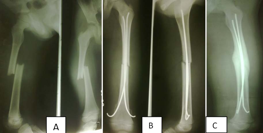
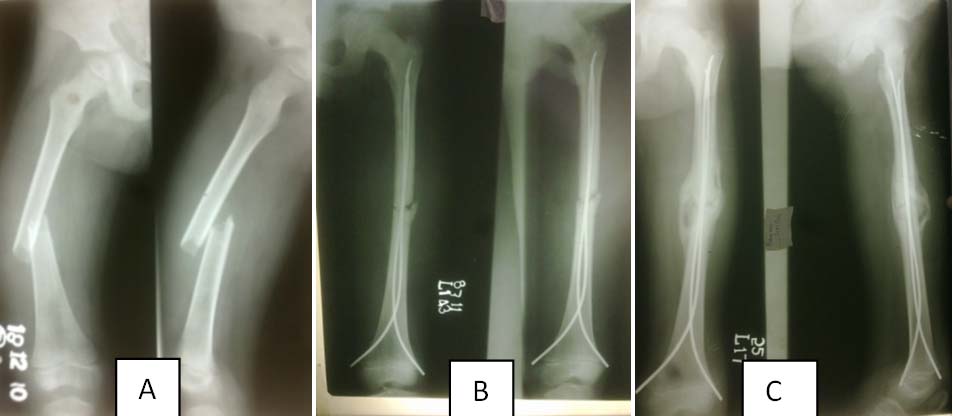
The outcome scores were graded with the help of Flynn et al., criteria [13]. In Group I, 20 cases were graded as excellent, 5 cases as good and 1 case had poor outcome. In Group II, 20 cases were graded as excellent, 3 as good and 5 patients had poor outcome [Table/Fig-3]. [Table/Fig-4A-C, 5A-C] showing pre and post operative radiographs of two patients treated with K wire and TENS, respectively.
Showing final functional results
| Groups | Excellent | Good | Poor |
|---|
| Group I (K- Wire fixation) | 20 | 5 | 1 |
| Group II (Titanium elastic nail fixation) | 19 | 4 | 3 |
Showing pre-operative (A), immediate post-operative (B) and follow –up x-ray (C) films showing union in fracture shaft femur treated with K wires
Showing pre-operative (A), immediate post-operative (B), and follow –up x-ray (C) films showing union in fracture shaft femur treated with TENS
Discussion
Conservative treatment has been the treatment of choice for pediatric femur shaft fracture, union was usually achieved which was associated with extended period of morbidity and hospital stay [14–16]. However to avoid prolonged immobilization, loss of school days and for better nursing care the operative approach has been gaining popularity for last two decades [17]. There are many options for operative fixation including external fixators, flexible and locked intramedullary nails, and compression and bridge plating [2].
Compression plating recommended by Hansen [7] has the disadvantages of larger soft tissue dissection, a large scar and a second major operation for removal of the plate. External fixation advocated by Krettek et al., [6] has been associated with problems of pintrack infection and refracture through the pin tracks [1]. Rigid intramedullary nailing is associated with problems of physeal damage and coxa valga or epiphysiodesis of the greater trochanter, avascular necrosis of the femoral head and growth disturbances [18].
After publication of good outcomes by the Nancy group in the early 1980s, elastic stable intramedullary nailing (ESIN) has become a well accepted method of surgical treatment of long bone fractures in children and adolescents [19, 20]. Recently many authors have come up with the good results by intramedullary K-wire in paediatric femur shaft fractures [4,12]. There is no study in the past comparing the efficacy of K-Wire and Titanium Elastic Nail. In our study we compared the results of intramedullary K-wire with intramedullary Titanium Elastic nail in treatment of femoral shaft fracture in children between 6 and 14 years of age.
We used the same principles of elastic nailing as described by Métaizeau [10] for fixation in both the groups. Full range of motion at knee was achieved in all the cases in both groups. Entry site infection was seen in two cases of Group I which resolved by early removal of K-wires. In Group II no case had entry site infection but two cases had nail irritation at entry site, this type of complication arrived in our initial cases which was later corrected by bending the Wires/Nail at 90° and turning it away from the skin. Limb length discrepancy was seen in five cases, three of them had shortening of less than 2 cm (two in Group I and one in Group II) and two cases had lengthening of more than 2cm (both in Group II). A total of 6 patients developed varus mal-alignment at fracture site, four of them had varus of acceptable limit [21] i.e. less than10°, 2 patients had unacceptable mal-alignment (one of each group). All the cases of malalignment had a strong association with the use of smaller diameter nail being used.
Average duration of surgery in Group I was slightly longer than that in Group II, this was mainly because the K-wire technique was initially new to us and was associated with a slight learning curve.
According to Flynn’s scoring system in Group I, 20 out of 26 patients had excellent results, five patients had good results and one patient had poor result. In Group II, 19 out of 26 patients had excellent results, four patients had good results and three patients had poor result. Results of both the groups were good and are in correlation with the studies done on the both the treatment modalities in the past.
The cost of the treatment in both the groups was a major differentiating feature; with direct cost of TENS nail being approximately 1500 Rs for each nail and a single K-wire to cost around 100 Rs each.
Conclusion
After looking the results of the present study and the economical aspect of both the treatment options, we conclude that the K-wire technique provides a cost-effective treatment option with equally good results. In the developing countries like ours, closed intramedullary K-wiring may provide a good option for those patients who opt for conservative treatment due to lack of adequate financial resources.