Occipital Condyle Syndrome in a Young Male: A Rare Presentation of Cranio-Vertebral Tuberculosis
Chaudhry Neera1, Patidar Yogesh2, Puri Vinod3, Khwaja Geeta A4
1Professor, Department of Neurology, G.B.Pant Hospital, New Delhi, India.
2Consultant, Department of Neurology, Bhaktivedanta Hospital Mira Road (E), Thane, Maharashtra, India.
3Dircetor Professor & Head, Department of Neurology, G.B.Pant Hospital, New Delhi, India.
4Dircetor Professor, Department of Neurology, G.B.Pant Hospital, New Delhi, India.
NAME, ADDRESS, E-MAIL ID OF THE CORRESPONDING AUTHOR: Dr. Neera Chaudhry, B-142, SaritaVihar, New Delhi 110076, India.
E-mail: neerachaudhry@gmail.com
Occipital condyle syndrome (OCS) is a rare syndrome characterized by severe, unilateral, occipital headache and ipsilateral 12th nerve palsy. Tumors are a common cause of OCS. Inflammatory lesions causing OCS is however rare. We describe a young male with OCS as the only manifestation of cranio-vertebral tuberculosis.
Craniovertebral junction, Headache, 12th Nerve palsy
Case Report
A 25-year-old male labourer presented in outpatient department of a tertiary care hospital with a six-month history of severe, constant, left-sided pain over back of the head and neck. The pain originated in the left occipital region, was associated with neck stiffness, and worsened on turning the neck to the right and on bending forward. There was no history of worsening of pain on coughing or straining. Four weeks after the onset of pain,he experienced tongue weakness in the form of difficulty in clearing food from the left cheek and in another two weeks noticed thinning of the left half of tongue.There was no history of any other cranial nerves involvement, dysarthria, any motor or sensory deficit, or any in-coordination in the limbs. There was no nausea, vomiting, giddiness, visual symptoms, fever, and loss of appetite or weight. The patient was a chronic smoker and consumed alcohol occasionally but there was no past history of any chronic illness.
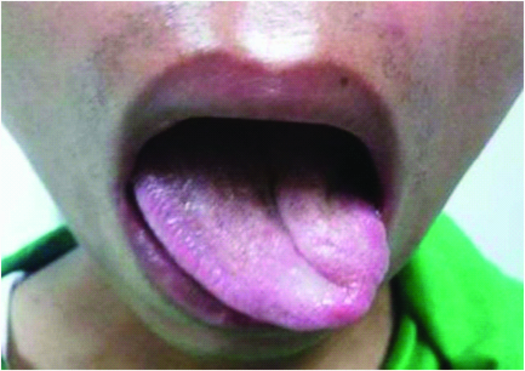
His vitals were stable and general physical examination revealed firm lymph nodes, three in number in the anterior chain in neck on the left side. These ranged from 1cm to 3cm in size. Rest of the general physical and systemic examination was normal. There was no neck stiffness, spinal deformity or tenderness, but the left occipital area was tender. The sole abnormality on neurological examination was left sided 12th nerve palsy with left-sided tongue atrophy and deviation of the tongue to the left side [Table/Fig-1].
ESR by Westegrens method was 86 mm in the 1st hour (normal 0-18 mm in the 1st hour). Rest of the hematological and biochemical profile was normal. HIV 1 and 2 (ELISA), HBsAg and Anti HCV were non-reactive. X-ray chest and ultrasound abdomen were normal. FNAC from the cervical lymph node revealed presence of epithelioid cell granuloma without necrosis. The AFB staining and culture were negative; however the aspirate was positive for PCR using hupB gene as target.
MRI brain with cervical spine [Table/Fig-2a-c] revealed a well-defined 3.5x2.5x1.5 cm collection at C2-C3 left prevertebral region involving ipsilateral prevertebral muscles (Longus Colli and Rectus Capitis). It was hyperintense on T2/FLAIR images, and showed mild rim enhancement on post contrast images. The C2-C3 vertebral bodies, odontoid process and clivus showed multiple lytic areas without any reactive sclerosis.Craniovertebral Junction (CV) junction area showed mild heterogenous enhancing granulation tissue at around odontoid process with mild indentation of anterior thecal sac. Brain parenchyma was normal. Contrast MRI sections showing non-enhancing soft tissue infiltration at region of left hypoglossal canal [Table/Fig-3a-b].CT skull base and head [Table/Fig-4a-b] showed multilevel erosions and lytic lesions involving bodies and posterior A4surfaces of C2-C4 vertebres with evidence of enhancing soft tissue posterior to the vertebral bodies and anterior to thecal sac. Facets of atlas and axis and all the intervertebral discs appeared normal. There was evidence of retropharyngeal and left parapharyngeal abscess extending from C1-C4 levels.
A possibility of cranio-vertebral tuberculosis was entertained. Considering Tuberculosis as the underlying etiology was enforced by the results of FNAC from the lymph nodes. As there was no motor sensory deficit and no radiologically appreciable instability of spine, a trial of antitubercular treatment (ATT) prior to surgical treatment was contemplated, along with anexternal orthosis in form of a cervical collar. Four drug regime (Isoniazid, Rifampicin, Pyrazinamide and Ethambutol) with oral steroids was started, to which the patient showed a good response. On ATT the patient became pain free over 2-3 wk. Repeat MRI (six months later) showed resolution of prevertebral abscess [Table/Fig-5a-c]. An 18 mnth therapy was given. At a follow up six months after stopping ATT, patient continues to remain asymptomatic except for persisting tongue atrophy.
Discussion
Occipital condyle syndrome (OCS) consists of unilateral pain in the occipital region along with an ipsilateral paresis of the 12th cranial nerve [1]. The 12th cranial nerve arises from the motor nucleus in medulla, exits the base of the skull through the hypoglossal canal in the occipital bone; it then traverses the neck and innervates the tongue muscles.
OCS was first recognized by Greenberg in 1981 [1]. The occipital pain is associated with neck stiffness and occipital tenderness; and is typically worsened by neck flexion and rotation to opposite side, whereas neck movement to same side typically ameliorates the pain, a pattern that was observed in our patient also. In some patients the pain may radiate to the mastoid, vertex and/or frontal region as well [2]. In the largest series of 11 cases of OCS,by Capobianco et al., 2/3rd of the patients had occipital region pain preceding 12th nerve paresis by several days to ten weeks [2]. In our case pain preceded the 12th nerve palsy by four weeks.
The commonest association of OCS is with metastases to skull-base and primary head and neck tumors[1-5]. In the largest published series of 100 patients with 12th nerve paresis (Keane et al.,), malignancy accounted for 49% cases; whereas trauma, infection, stroke and GBS were responsible for the remaining cases [6].
Spinal tuberculosis (TB) accounts for only 1% of all tuberculosis cases.Cranio-vertebral junction (CVJ) TB is still rarer, the reported incidence being 0.3-1% of all spinal TB [7]. Isolated occipital tuberculosis is rare because there is a paucity of the cancellous component in the occipital bone as compared to the frontal and parietal bones which have a greater area of diploic space and cancellous bone making them more vulnerable to tuberculosis [8,9]. In addition paucity of lymphatics in the occipital bone may also account for the rarity of tuberculosis as a cause of OCS.
CVJ TB usually occurs secondary to TB elsewhere in the body such as pulmonary TB, lymph nodes (cervical or mediastinal) or other sites. Involvement of the ligaments (transverse and alar) and osteolytic lesionsin the anterior and posterior arches of C1 vertebra, odontoid process, lateral mass and body of C2 vertebraand the occipital condyle scan lead to a markedinstability of the CVJ and compression of the neuraxis. In his review Rajkumar, reported that spastic quadriparesis was present in 60-100% cases of CVJ TB [7]. In a case series of 25 patients with CVJ TB documented over 12 y, by Bhojraj et al., none of the patients had occipital condyle involvement [10]. In a radiological series of 29 cases of CVJ TB by Krishnan etal., the skull was involved in 19, clivus in 11 patients, and occipital condyle in 14 patients [11].
Our patient however is a rare case of CVJ TB clinically manifesting as OCS. CVJ involvement due to tuberculosis usually presents with other clinical manifestations such as involvement of lower cranial nerves, corticospinal and sensory signs. Isolated hypoglossal palsy as a manifestation of CVJ TB is rare and described only in few case reports [12-14]. It is however important to be aware of this entity because early diagnosis can prevent significant motor disability. To the best of our knowledge Wegner’s granulomatosis is the only other non-malignant cause of OCS that has been reported in the literature [15].
The management of CVJ TB has to be individualized depending upon neurological status, compression of spinal cord, extent of bony destruction, abscess formation and atlanto-axial dislocation, but irrespective of whether surgery is needed or not, all cases of CVJ TB should receive ATT for a period of 18-24 month [7].
Our patient had typical clinical features of an OCS but had no clinical manifestation of cervical spine involvement or myelopathy though imaging showed lytic changes in C1-C4 vertebrae along with a paravertebral collection and involvement of the left occipital condyle. So, conservative management was considered and he responded well, both clinically and radiologically
Atrophy and weakness of left half of tongue
MRI cervical spineSagittal T2W image showing a well defined collection at C2-C3 prevertebral regi on involving ipsilateral prevertebral muscles (Longus Colli and Rectus Capitis). Axial T2W image showing left prevertebral collection with mild rim enhancement on post-contrast images

MRI brain and cervical spineAxial and Sagittal T1W contrast images showing involvement of hypoglossal canal (left side)

CT skull base and head showing multilevel erosions and lytic lesions involving bodies and posterior surfaces of C2-C4 vertebres. Post contrast CT showing enhancing soft tissue posterior to the vertebral bodies and anterior to thecal sac along with retropharyngeal abscess extending from C1-C4 levels

MRI cervical spineT2W (Sagittal and Axial) and post contrast images showing complete resolution of the collection, after 6 months of antitubercular therapy

Conclusion
In conclusion an isolated OCS without clinical involvement of the spine or features of myelopathy can be a rare presentation of CVJ TB.
[1]. HS Greenberg, MDF Deck, B Vickram, FCH Chu, JB Posner, Metastasis to the base of the skull: clinical findings in 43 patientsNeurology 1981 31:530-37. [Google Scholar]
[2]. DJ Capobianco, PW Brazis, FA Rubino, JN Dalton, Occipital condyle syndromeHeadache 2002 42:142-46. [Google Scholar]
[3]. G Moris, C Roig, M Misiego, A Alvarez, J Berciano, J Pascual, The distinctive headache of the occipital condyle syndrome: a report of four casesHeadache 1998 38:308-11. [Google Scholar]
[4]. JIM Salamanca, C Murrieta, J Jara, JL Munoz-Blanco, F Alvarez, JG de Villoria, Occipital condyle syndrome guiding diagnosis to metastatic prostate cancerInt J Urology 2006 13:1022-24. [Google Scholar]
[5]. JJ Moeller, S Shivakumar, M Davis, CE Maxner, Occipital Condyle Syndrome as the First Sign of Metastatic CancerCan. J. Neurol Sci 2007 34:456-59. [Google Scholar]
[6]. JR Keane, S Mehta, R Kalra, Twelfth- nerve palsy: analysis of 100 cases Arch Neurol 1996 53:561 [Google Scholar]
[7]. Rajkumar Cranio-vertebral Junction tuberculosisIndian Journal of Neurosurgery 2012 1(1):61-5. [Google Scholar]
[8]. MyoungSoo Kim (2012). Skull Osteomyelitis, Osteomyelitis, Prof. Mauricio S. Baptista (Ed.), ISBN: 978-953-51-0399-8, InTech, Available from:http://www.intechopen.com/books/osteomyelitis/skullosteomyelitis Accessed on 14 Aug 2013. [Google Scholar]
[9]. B Bhandari, SL Mandowara, H Joshi, Tubercular osteomyelitis of skullIndian J Pediatr 1981 48(390):113-15. [Google Scholar]
[10]. SY Bhojraj, N Shetty, PJ Shah, Tuberculosis of the craniocervical junctionJ Bone Joint Surg [Br] 2001 83:222-25. [Google Scholar]
[11]. A Krishnan, D Patkar, T Patankar, J Shah, S Prasad, T Bunting, Craniovertebral Junction Tuberculosis: A Review of 29 CasesJournal of Computer Assisted Tomography 2001 25(2):171-76. [Google Scholar]
[12]. IM Richards, AM White, MM O'Sullivan, JD Jessop, BD Williams, Unilateral palsy of the hypoglossal nerve in a patient with tuberculosis of the first cervical vertebraBr J Rheumatol 1989 28(6):540-42. [Google Scholar]
[13]. PP Chakraborty, Deviated tongue: the presenting manifestation of spinal tuberculosisIndian J Pediatr 2009 76(9):967-69. [Google Scholar]
[14]. H Ebadi, D Fathin, Unilateral Hypoglossal Nerve Palsy: As the Only Presentation of TuberculosisActa Med Iran 2012 50(10):717-20. [Google Scholar]
[15]. A Hornik, F Rodriguez-Porcel, CH Ersahin, R Kadanoff, J Biller, Wegener's disease presenting with occipital condyle syndromeFront Neurol 2012 3:53 [Google Scholar]