Lamellar Icthyosis – A case Report
Pranitha V1, Thimma Reddy B.V22, Daneswari V3, Sudhanwan N Deshmukh4
1Professor, Department of Pediatric and Preventive Dentistry, Mamata Dental College, Khammam, Telangana, India.
2Professor & Head of Department, Department of Pediatric and Preventive Dentistry, Mamata Dental College, Khammam, Telangana, India.
3Reader, Department of Pediatric and Preventive Dentistry, Mamata Dental College, Khammam, Telangana, India.
4Senior Lecturer, Department of Pediatric Dentistry, Sharad Pawar Dental College, Wardha, Maharashtra, India.
NAME, ADDRESS, E-MAIL ID OF THE CORRESPONDING AUTHOR: Dr. Pranitha V, Professor, Department of Pediatric and Preventive Dentistry, Mamatha Dental College, Khammam, Telangana-507002, India. Phone : 9440587228,
E-mail: drjsr27@gmail.com
Autosomal recessive congenital ichthyosis is a heterogenous group of disorders that are present at birth with generalized involvement of skin and lack of other organ systems. Clinical presentation, pattern of inheritance, and laboratory evaluation may establish a precise diagnosis, which can assist in prognosis and genetic counseling. There is a little knowledge about the oral manifestations of these disorders.This case report presents management and complete oral rehabilitation of a rare case of lamellar ichthyosis.
Caries, Dental management, Ectropion, Lamellar ichthyosis (LI), Photophobic
Case Report
A 6-year-old male child reported to the Department of Pediatric and Preventive Dentistry, Mamata Dental College, Khammam, with the compliant of decay in lower left back tooth region since one year which is not associated with any history of pain. Detailed medical history revealed an operated patent ductus arteriosus and lamellar ichthyosis persisting since birth. There was consanguinity and his younger brother also suffers with similar condition. Opthalmologist elicited multiple plate like large brown, firmly adherent scales, variable in size all over the body including flexural surfaces and cornea [Table/Fig-1,2]. There was Grade III ectropion in lower eyelids and conjunctiva was congested, dry, photophobic. The child followed light and responded to sound stimuli briskly. There was short, dry and sparse hair and showed no nail abnormalities.
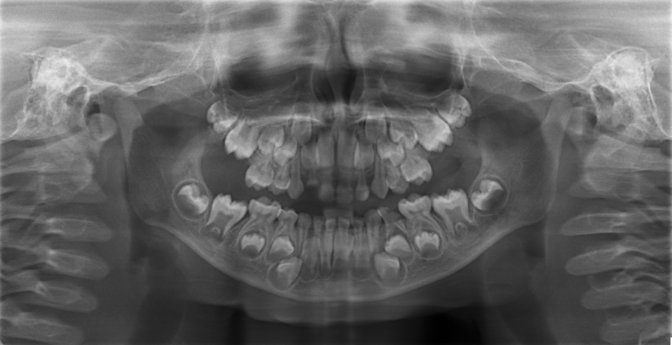
Intraoral examination revealed presence of grossly decayed #74,#52 and dental caries extending into the pulp in relation of #64.Panoramic radiograph confirmed chronic irreversible pulpitis in relation to #64 [Table/Fig-3] and treatment plan was formulated and explained to the parents. Pediatrician referral was made keeping in view the patient’s medical status.
In first appointment, under sterile aseptic environment, band pinching was done for #75 to fabricate a band and loop space maintainer and single visit pulpectomy was performed in relation to #52 followed by biological post placement [Table/Fig-4].
In next visit, Local anaesthesia 2% lignocaine containing 1:100,000 epinephrine (Lignox 2%;Indoco Remedies Ltd, Mumbai, India) was administered in relation to #64. Under rubber dam isolation, access cavity was prepared and formocresol pulpotomy was done.Grossly mutilated #74 was extracted after profound anaesthesia was obtained followed by cementation of the band and loop spacemaintainer in relation to #75 [Table/Fig-5].
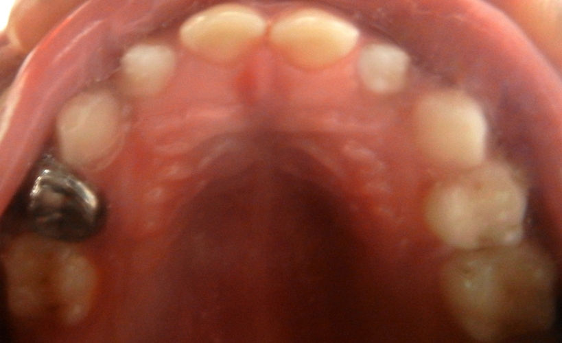
After 15 days followup, #64 was found assymptomatic, preformed stainless steel crown was selected and cemented with GC Fuji 1(GC Corporation,Tokyo,Japan) [Table/Fig-6].
We recommended the necessity of pit and fissure sealants, topical fluorides as preventive measure. Our patient is now in mixed dentition phase and reports for a recall visit of every six months.
Discussion
The term ichthyosis is derived from the Greek word ‘ichthys’ meaning “fish” and refers to the similarity in appearance of the skin to fish scales. Early reports of ichthyosis in the Indian and Chinese literature date back to several hundred years [1]. The ichthyoses form part of a large ,clinically and etiologically heterogenous group deshmukh4of mendelian disorders of cornification and typically involve all or most of the integument [2]. Lamellar ichthyosis is the rarest form with an incidence of less than one in 3 lacs. It has autosomal recessive inheritance and there is a defect on chromosome 14q11 causing transglutaminase-1 (TG) defect . Autosomal recessive ichthyosis with hypotrichosis (ARIH), mutation results in a Glycine → Arginine substitution in residue 827 of the matriptase protein resulting in thickened scaling, and grayish skin and curly, sparse, fragile, brittle, dry, lusterless, and slow growing hair. Patients with LI often have profound hypohydrosis and are at risk for hyperpyrexia in hot climates. Other manifestations linked to homozygosity for the c.2672G → A ST14 allele included corneal opacity, photophobia, and abnormal deciduous and permanent teeth [3]. Miteva noted both hair and dental abnormailities in his patient [4]. Cremers et al., observed early childhood deafness, congenital nonbullous ichthyosiform erythroderma, corneal involvement, photophobia, hypotrichosis, anhidrosis, hyperkeratosis of the nails and dental dysplasia [5]. Ramer et al., reported scarring alopecia, dark brown scaly lesions prominent in palmoplantar region in their patient [6].
The clinical features of patients with lamellar ichthyosis are generally similar to our present case, which is typical. An expertise of dermatologist, paediatrician, genetist, opthamologist and physiotherapist play an important role in managing such cases. The management is aimed at decreasing symptoms and include emollients (petrolatum, coconut oil, alpha hydroxyl acetic acid),Keratolytics containing salicylates with propylene glycol and local and systemic retinoids [2].
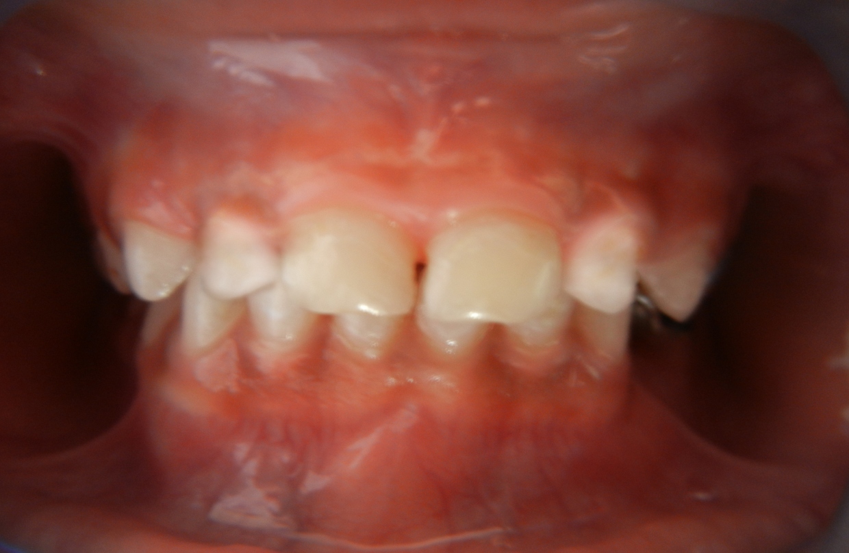
As the patient was photophobic, patient was asked to wear eye protection glasses for smooth rendering of dental treatment [Table/Fig-7]. In some patients, teeth are normally developed as seen in our case but in others they may be defective and likely to develop caries as the oral cavity tends to be inoculated with high numbers of bacteria leading to excessive tooth plaque formation [7].
Dentist must take care to avoid any trauma and limit the mouth opening period as the perioral skin is scaly, tender and friable. As these patients are on retinoids,choice of local anaesthetic agents is important as there is possibility of hepatic toxicity.
Multiple plate like large brown, firmly adherent scales all over the body

Panoramic radiograph showing grossly decayed #52, #74 and chronic irreversible pulpitis in relation to #64
Intraoral postoperative photograph #52 with biological post,
Band and loop space maintainer

Stainless steel crown #64

Conclusion
Dental management and rehabilitation of such rare cases poses a challenge to the paediatric dentist. Hence, patient should be offered genetic counselling , psychological support and preventive dental care. Patient should be offered genetic counselling and provided with psychological support.
[1]. A Hemandez-Martin, N Cuadrado-Corrales, S Cirla-Abad, D Arias-Palomo, JM Mascaro-Galy, MJ Escamez, X-linked ichthyosis along with recessive dystrophic epidermolysis bullosa in the same patientDermatology 2010 221:113-16. [Google Scholar]
[2]. V Oji, G Tadini, M Akiyama, C Blanchet Bardon, C Bodemer, E Bourrat, Revised nomenclature and classification of inherited ichthyoses: Results of the First Ichthyosis Consensus Conference in Sorze 2009J Am Acad Dermatol 2010 63(4):607-41. [Google Scholar]
[3]. B Sandler, K Hashimoto, Collodion baby and lamellar ichthyosisJ Cutan Pathol 1998 25(2):116-21. [Google Scholar]
[4]. B Bolgul, N Hamamci, S Akdeniz, S Colenk, Oral manifestations of lamellar ichthyosis –A case reportIran J Pediatr 2009 19(3):298-302. [Google Scholar]
[5]. NV Rathi, SM Rawlani, KR Hotwani, Oral manifestations of lamellar ichthyosis: A case report and reviewJ Pak Assoc Derma 2013 23(1):99-102. [Google Scholar]
[6]. K Ramar, S Annamalai, VP Hariharavel, R Aravindhan, C Ganesh, K Leshwaryah, Oral manifestations of autosomal recessive congenital ichthyosis in a 2-year-old patient Case Reports in Dentistry 2014 7(1):1-3. [Google Scholar]
[7]. S Pallagatti, S Sheikh, A Kaur, A Aggarwal, R Singh, X-linked icthyosis along with epidermolysis bullosaContemp Clin Dent 2012 3:S96-98. [Google Scholar]