Herpes Gestationis after Ovodonation: Is Placenta the only Target of the Immune Reaction?
Isabella Piva1, Giuseppe Lo Monte2, Angela Graziano3, Roberto Marci4
1 Faculty, Department of Morphology, Surgery and Experimental Medicine, University of Ferrara, Ferrara, Italy.
2 Faculty, Department of Morphology, Surgery and Experimental Medicine, University of Ferrara, Ferrara, Italy.
3 Faculty, Department of Morphology, Surgery and Experimental Medicine, University of Ferrara, Ferrara, Italy.
4 Professor, Department of Morphology, Surgery and Experimental Medicine, University of Ferrara, Ferrara, Italy.
NAME, ADDRESS, E-MAIL ID OF THE CORRESPONDING AUTHOR: Dr. Roberto Marci, Professor, Department of Morphology, Surgery and Experimental Medicine, Ferrara University, Corso Giovecca 183, 44100, Ferrara, Italy. Phone : +393386960872, Fax +390532203844, E-mail : roberto.marci@unife.it
Herpes Gestationis (HG) is an autoimmune disorder that rarely complicates pregnancy. Its pathogenesis has not been clarified yet, as it derives from a complex interplay between immunologic, genetic, and hormonal factors. We present a case of HG occurring in a surrogate mother whose pregnancy was achieved via egg donation and in vitro fertilisation (IVF). The disease appeared at 26 weeks and 6 days of gestation and subsequently worsened despite the administration of oral and topic corticosteroids. A healthy baby was delivered at 35 weeks and 2 days of gestation by elective cesarean section and the clinical manifestations resolved in the postpartum period.
Autoimmunity, Dermatoses of pregnancy, Herpes gestationis, Prurigo gestationis, Ovodonation
Case Report
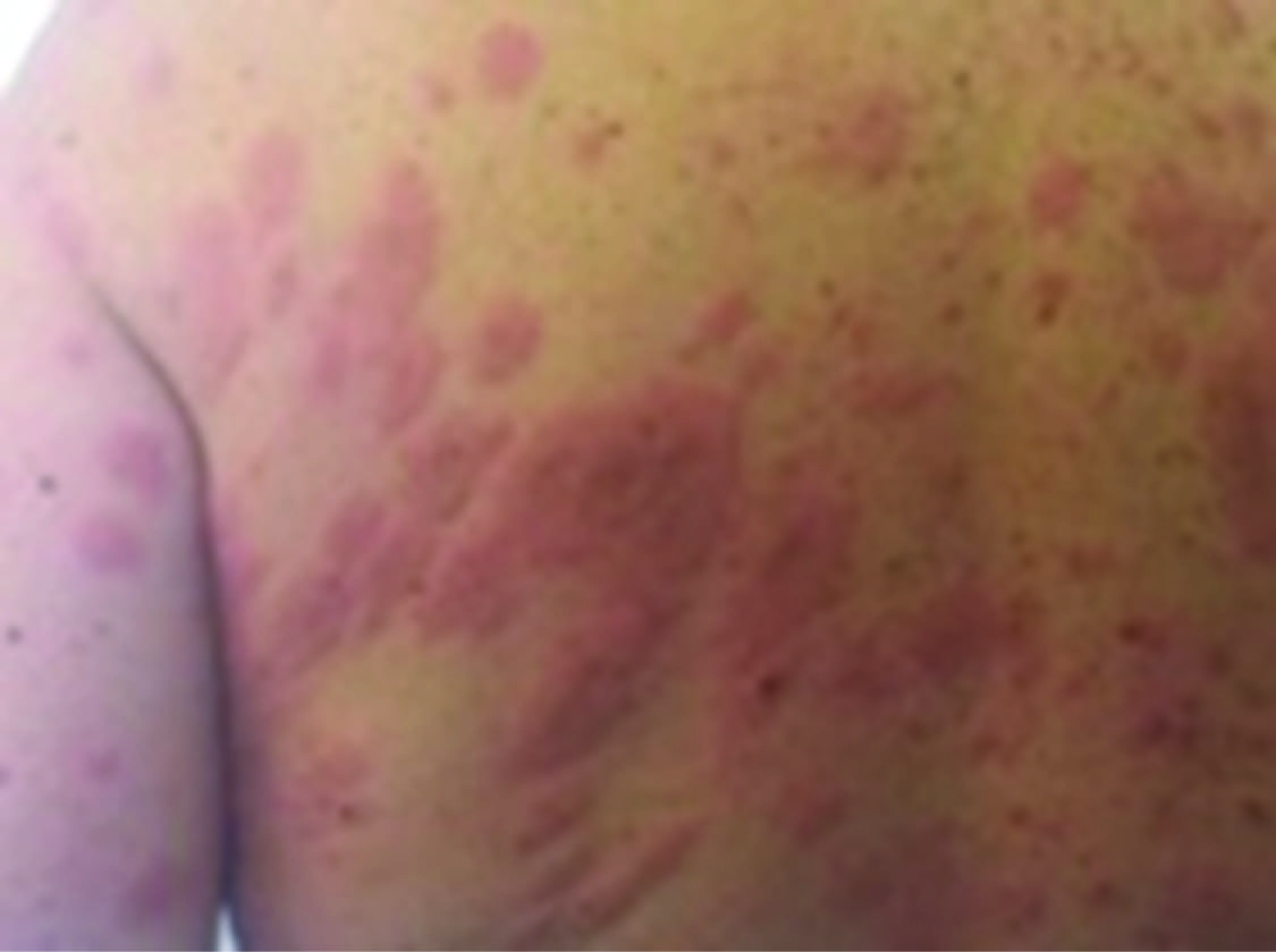
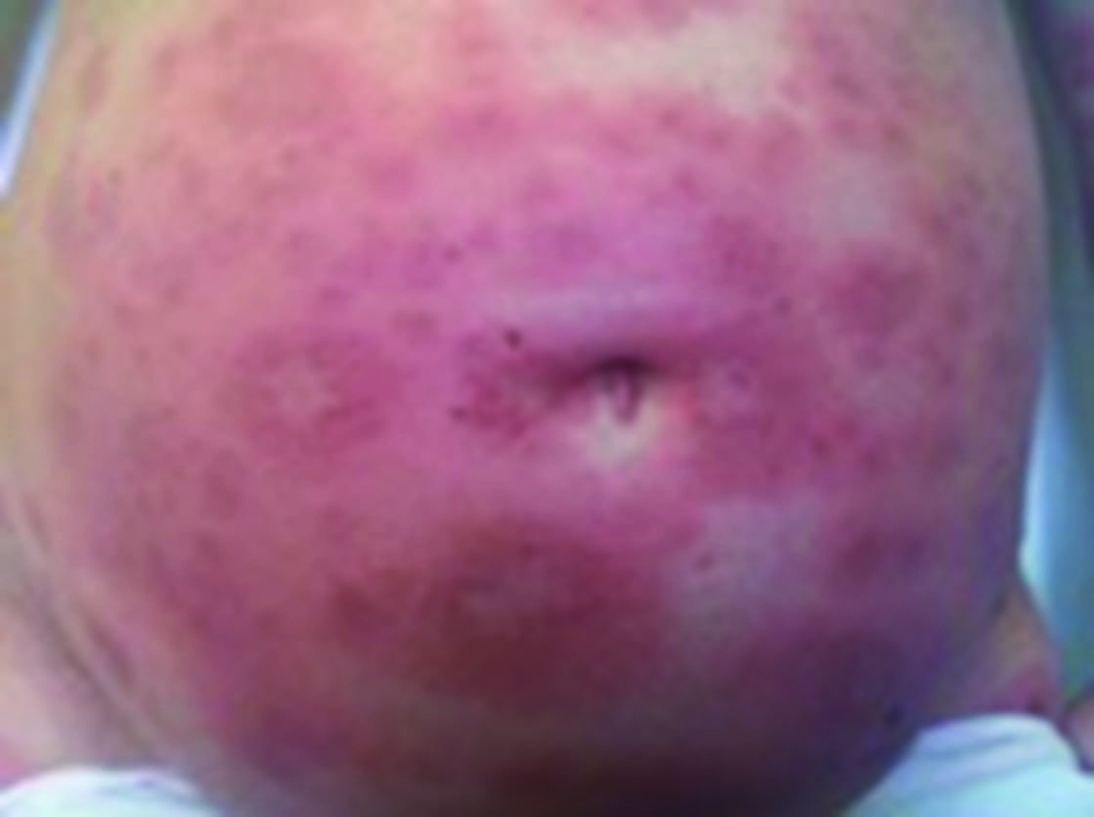
The surrogate mother, a 43-year-old asymptomatic white woman, underwent ovodonation because of infertility (gravida 0). The oocyte was fertilized by the surrogate mother’s husband (the biologic father). At 26 wk and 6 d of gestation the woman was admitted to the emergency room with a complaint of acute-onset papulourticarial eruption associated with intense pruritus. The patient did not report any other dermatosis or allergy in her medical history. The initial clinical examination showed erythematous urticarial patches and plaques on the periumbilical region, the trunk and the extremities yet vital signs were normal [Table/Fig-1, 2]. Ultrasound and pelvic examination performed in the emergency room revealed normal findings and were both compatible with the woman’s gestational age. After dermatological consultation, the patient was diagnosed with “prurigo gestationis” and referred to the Dermatology clinic, where she subsequently underwent punch biopsy of the skin lesions. The exam documented the presence of achantosic and orthokeratotic epidermis with mild spongiosis, subcorneal serous collections and occasional necrotic keratinocytes, without dermal-epidermal detachment. Moreover, the papillary dermis showed oedema and lymphocytic infiltration predominantly at perivascular level, with rare eosinophils. Immunofluorescence performed with anti-C3 serum revealed linear microgranular reactivity at the dermal-epidermal junction, while anti-IgG serum stained the subcorneal blisters. These findings were indicative of autoimmune bollous dermathitis, probably due to Herpes Gestationis (HG). Thus, the patient was administered oral prednisone (40 mg/day) in addition to a topical corticosteroid cream. However, as the clinical manifestations worsened and the woman complained about reduced active fetal movements, she was finally admitted to the hospital at 35 weeks of gestation. Complete blood count, serum biochemical tests and urine analysis were all within normal limits. After rheumatologic consultation, the patient was tested for ANA, anti-dsDNA, ENA, C3, C4, LAC and antiphospholipids, which were all negative. Since systemic corticosteroid therapy provided moderate relief from pruritus and did not prevent the spreading of the lesions, two doses of betamethasone 12 mg were given intramuscularly 24 h apart in order to prevent respiratory distress syndrome and elective cesarean section was performed at 35 wk and two days of gestation. A 2320 g healthy male infant was delivered. The amniotic fluid was meconium stained. On histologic exam of the placenta a grade 1-stage 1 chorioamnionitis was noted. Following delivery, the lesions started to resolve and the patient was prescribed metilprednisolone (8 mg daily) and antihistaminic, which she was advised to continue even after hospital discharge. On the tenth postoperative day the lesions had disappeared.
Erythematous urticarial patches and plaques on the back
Erythematous urticarial patches and plaques on the abdomen
Discussion
HG is a rare autoimmune disorder that usually begins in the second or third trimester and spontaneously regresses in the postpartum period [1]. It is characterized by pruritic, urticarial and vesiculobullous lesions that spare face, mucous membranes, palms, and soles [2]. This generalized bullous reaction mirrors subepidermal vesicle formation, due to deposition of complement three along the basement membrane zone. In fact, the disease is believed to depend on the presence of an antibody belonging to the immunoglobulin G1 subclass, which reacts against a hemidesmosomal protein. The activation of the complement through the classic pathway leads to chemo attraction and degranulation of the eosinophils, which are responsible for the skin lesions [3]. Even though the etiology is presently unknown, both hormonal factors and immune compatibility seem to be involved in the pathogenesis [1, 2]. As to the former, recurrences with menses, use of oral contraceptives and the worsening of the disease in subsequent pregnancies have been reported [3]. There is only one report in literature of pemphigoid gestationis developing after in vitro fertilization and intracytoplasmic sperm injection [4]. In that case, the disease followed a severe clinical course and flared up in the postpartum period. The Authors speculated that an exaggerated hormonal response following an in vitro fertilization cycle may have a role in predisposing the patient to the disease in a first pregnancy [4]. As to immune compatibility, HG has been linked to the presence of HLA-DR3 (61%-80%), and HLA-DR4 (52%) or both, which would support the role played by autoimmune factors in HG1. Accordingly, the patients frequently develop other autoimmune diseases such as Graves’ disease [1]. Furthermore, a rare association with molar pregnancies [5] and choriocarcinoma [6] has been reported, stressing the possible causal role of paternal antigens as the target of anti-HLA antibodies that subsequently to cross-react with the skin.
Even though fetal mortality and morbidity is not significantly higher, fetal risks include premature deliveries and small for gestational age newborns [5]. These complications have been attributed to mild placental failure, as the placenta can be targeted by the immune response. Nevertheless, in our report low birth weight was compatible with gestational age at delivery and did not necessarily depend on placental insufficiency. Besides, the histologic exam of the placenta did not point out any sign of placental failure, but it revealed mild chorioamnionitis. Up to now, an autoimmune origin of chorionamnionitis has not been demonstrated, however, immunologic factors have been suggested to play a role in the pathogenesis of recurrent placental villitis and maternal chronic autoimmune diseases have been proved to predispose to the infection of the placenta [7]. The presence of urticarial, vesicular or bullous lesions reported in 5-10% offspring [5] is worthy of note, as this phenomenon may suggest a direct insult to the fetus. Even though the newborn baby was healthy in our report, we can hypothesize that pregnancies achieved by ovodonation can further trigger the immune response of the patient because they can lead to the development of additional antibodies against the fetus, whose genetic material is completely foreign. Accordingly, it has been recently proved that severe diseases, such as congenital heart block, can occur in genetically unrelated children obtained by ovodonation and exposed to maternal anti-SSA/Ro antibodies [8]. Further studies are needed to characterize the immune response against the placenta in HG and to verify whether maternal antibodies can damage the fetus directly.
Conclusion
HG is still an open challenge and pregnancies achieved by ovodonation represent a unique condition to study the causal role of hormonal, genetic and immunologic factors.
[1]. Kroumpouzos G, Cohen LM, Specific dermatoses of pregnancy: an evidence-based systematic reviewAm J Obstet Gynecol 2003 88:1083-92. [Google Scholar]
[2]. Giugliano E, Cagnazzo E, Servello T, Pruritic urticarial papules and plaques of pregnancyJ Obstet Gynaecol 2012 32(3):301-02. [Google Scholar]
[3]. Carruthers JA, Ewins AR, Herpes gestationis: studies on the binding characteristics, activity and pathogenetic significance of the complement-fixing factorClin Exp Dermatol 1978 31:38-44. [Google Scholar]
[4]. Guven S, Erkin G, Tuncer ZS, Pemphigoid gestationis complicating a pregnancy following in vitro fertilization and intracytoplasmic sperm injectionInt J Dermatol 2006 45(9):1120-21. [Google Scholar]
[5]. Kolodny RG, Herpes gestationis: a new assessment of incidence and foetal prognosisAm J Obstet Gynecol 1969 104:39-45. [Google Scholar]
[6]. Do Valle Chiossi MP, Silva Costa R, Ferreira Roselino AM, Titration of herpes gestationis factor fixing to C3 in pemphigoid herpes gestationis associated with choriocarcinomaArch Dermatol 2000 136:129-30. [Google Scholar]
[7]. Redline RW, Abramowsky CR, Clinical and pathologic aspects of recurrent placental villitisHum Pathol 1985 16(7):727-31. [Google Scholar]
[8]. Brucato A, Ramoni V, Penco S, Passively acquired anti-SSA/Ro antibodies are required for congenital heart block following ovodonation but maternal genes are notArthritis Rheum 2010 62(10):3119-21. [Google Scholar]