Prevalence and Mophometry of Os Peroneum amongst Central Indians
Pragati S. Mittal1, Sharda S. Joshi2, Rupa Chhaparwal3, Subhash D. Joshi4
1 Assistant Professor, Department of Anatomy, Sri Aurobindo Medical College and Postgraduate Institute, Indore, Madhya Pradesh, India.
2 Professor & Head, Department of Anatomy, Sri Aurobindo Medical College and Postgraduate Institute, Indore, Madhya Pradesh, India.
3 Assistant Professor, Department of Anatomy, Sri Aurobindo Medical College and Postgraduate Institute, Indore, Madhya Pradesh, India.
4 Professor & Dean, Department of Anatomy, Sri Aurobindo Medical College and Postgraduate Institute, Indore, Madhya Pradesh, India.
NAME, ADDRESS, E-MAIL ID OF THE CORRESPONDING AUTHOR: Dr. P. S. Mittal, Assistant Professor, Department of Anatomy, Sri Aurobindo Medical College and Postgraduate Institute, Indore-453555, Madhya Pradesh, India. Phone : +91 9827395622, E-mail : pragatisheelmittal@gmail.com
Introduction: Os Peroneum is round or oval shaped sesamoid within the substance of the Peroneus longus tendon as it plays on the Cuboid bone.
Materials and Methods: Thirty six embalmed cadavers were dissected bilaterally. Lateral part of foot and sole was dissected to expose Peroneus longus tendon and Os Peroneum. Measurements of Os Peroneum and articular surfaces of cuboid (calcaneum) were taken by the Digital Vernier Caliper. Histological and Radiological Examinations of Os Peroneum were also done.
Observation: A flattened oval enlargement i.e. Os Peroneum was found in all the tendons examined. The deep surface of Os Peroneum (i.e. articular surface) was concave, smooth and shiny; sometimes divided into two parts. There was a well defined convex articular facet on the Cuboid at the proximal end of the peroneal sulcus, which sometimes extended proximally on to the Calcaneum forming a synovial joint.
Results: Average length of Os Peroneum was found to be 13.35 mm (Right - 13.35 mm, Left - 13.35 mm). Average breadth of Os Peroneum was 8.96 mm (Right - 8.87 mm, Left - 9.05 mm). Average thickness of Os Peroneum was 4.11 mm (Right - 4.13 mm, Left - 4.10 mm). Incidence of double articular facets on cuboid and Calcaneum was more on the right side (25.80%) than the left (16.20%).
Conclusion: Findings suggest that Os Peroneum is present at the site of angulations of Peroneus Longus tendon with attendant change in direction and the exposure to various stresses and strains leading to its thickening and formation of Os Peroneum.
Os peroneum, Sesamoid bone, Peroneus longus
Introduction
The sesamoid bones are one of Mother Nature's finest anatomical creations [1]. Embedded within certain tendons the principal function of the sesamoids is to allow tendons to glide over joints without getting caught or impinged.
Sesamoid bones tend to develop within tendons in areas that experience both tensile strain and hydrostatic compressive mechanical stresses [2]. Some sesamoids ossify, whereas others remain fibrous or cartilaginous. Galen (A.D. 129-199) is usually credited for giving the sesamoids their name, or at least, popularizing the name. Patil V et al., have stressed that, this development is mediated epigenetically by local mechanical forces associated with skeletal geometry, posture, and muscular activity [2]. In the Peroneus longus (PL) tendon a sesamoid is found, as it courses underneath the peroneal groove of the Cuboid. This particular sesamoid is called Os Peroneum (OP). The origin of the OP has given rise to much controversy. It may be due to a response to the intense mechanical stresses involved at the angulated part of the tendon of PL, other workers regard OP as a vestigial structure of phylogenetic significance [3]. Waddington proposed the theory of genetic assimilation stating that a phenotypic character initially produced in response to an environmental stimulus can be stabilized, or "canalized”, through natural selection to the point where the adapted phenotype is assimilated into the genome and is found to occur even in the absence of the initial external environmental stimulus. This may explain the occurrence and prevalence of OP in the tendon of PL [4].
When ossified, it is visible in 20% of foot radiographs [5]. Investigators studying cadaveric specimens find more sesamoids than when the data is obtained from just x-ray evaluation. This is an important fact to be kept in mind by the podiatric surgeon, particularly in cases where there may be symptoms of involvement of sesamoid and no radiographic evidence is found [1].
Due to the clinical significance of OP in the form of Painful Os Peroneum syndrome (POPS), which includes OP fracture or a diastasis of multipartite OP, the present study has been conducted to ascertain the prevalence and the various parameters like Length, Breadth and Thickness of OP amongst central Indians. Details of the articular surface of OP and its reciprocal articular surface on Cuboid or its extension on Calcaneum were also studied.
Materials and Methods
The present study was conducted on 36 embalmed cadavers bilaterally in the Department of Anatomy, Sri Aurobindo Medical College & P.G. Institute, Indore, India. No consideration was given to the age and sex of the specimens examined. The study was conducted over the period of two years (2012-2014). Lateral part of foot and sole was dissected to expose PL tendon and OP. Various dimensions of OP (max. length, max. breadth and max. thickness in accordance of its axis) and PL tendon (max. breadth and max. thickness, 1 cm proximal and distal to OP) were measured with the help of Scale and Digital Vernier caliper. The PL tendon approximately 10-12 cm long was removed which included OP. The specimens were submitted for radiological and Histological study.
Observations
A flattened oval enlargement (OP) was observed in all the PL tendons examined. The deep surface of OP was articular, concave, smooth and shiny [Table/Fig-1a]; sometimes divided into two parts [Table/Fig-1b]. Reciprocal convex articular facet was present on the Cuboid [Table/Fig-1a] at the proximal end of the peroneal sulcus (cuboid tunnel). In a few cases the articular surfaces extended on to the Calcaneum [Table/Fig-1b].
(a) Single large facet on OP & cuboid, (b) Showing double facets on OP (arrow heads) and reciprocal facets on Cuboid {Yellow arrows} & Calcaneum {white arrows}

On the right the breadth of proximal tendon (4.92 mm) was greater than the distal part (4.55 mm); whereas, the thickness in the distal part was more (2.44 mm) than the proximal part (2.23 mm). While on the left side the breadth & thickness of PL tendon was the same, both in the proximal and distal parts [Table/Fig-2].
Breadth and thickness of peroneus longus tendon
| Breadth (mm) | Thickness (mm) |
|---|
| 1 cm proximal to OP | 1 cm distal to OP | 1 cm proximal to OP | 1 cm distal to OP |
|---|
| Right | 4.92 | 4.55 | 2.23 | 2.44 |
| Left | 4.70 | 4.64 | 2.28 | 2.28 |
| Mean | 4.81 | 4.59 | 2.25 | 2.36 |
The average length and breadth of OP was lesser on right side as compared to the left but the average thickness on the right was found to be slightly greater than the left side [Table/Fig-3].
Average length, breadth and thickness of os peroneum
| Average Length of OP (mm) | Average Breadth of OP (mm) | Average Thickness of OP (mm) |
|---|
| Right | 12.93 | 8.87 | 4.13 |
| Left | 13.35 | 9.05 | 4.10 |
| Mean | 13.14 | 8.96 | 4.11 |
The anterio-posterior diameter of articular surface on Cuboid was greater when there was single facet (average 11.43 mm) as compared to when double facets were present (average 9.59 mm) [Table/Fig-1a,b].
On the right double facets on Cuboid & Calcaneum were more (25.8%) as compared to left (16.2%). When double facets were present; the facet on Cuboid (average anterio-posterior diameter - 9.59 mm & transverse diameter – 8.82 mm) was larger than the facet on Calcaneum (average anterio-posterior diameter – 7.30 mm & transverse diameter – 7.57 mm) [Table/Fig-1b].
In this study, OP was present in all the PL tendons examined. When OP was examined radiologically it was found as non-calcified thickening in 65% & Calcified thickening in 35% [Table/Fig-4,5].
Radiograph showing ossific nodule in OP

Incidence of Non-calcified & Calcified thickening
| Non-Calcified (%) | Calcified (%} |
|---|
| Right | 72.8 | 27.2 |
| Left | 57.2 | 41.8 |
| Mean | 65.0 | 35.0 |
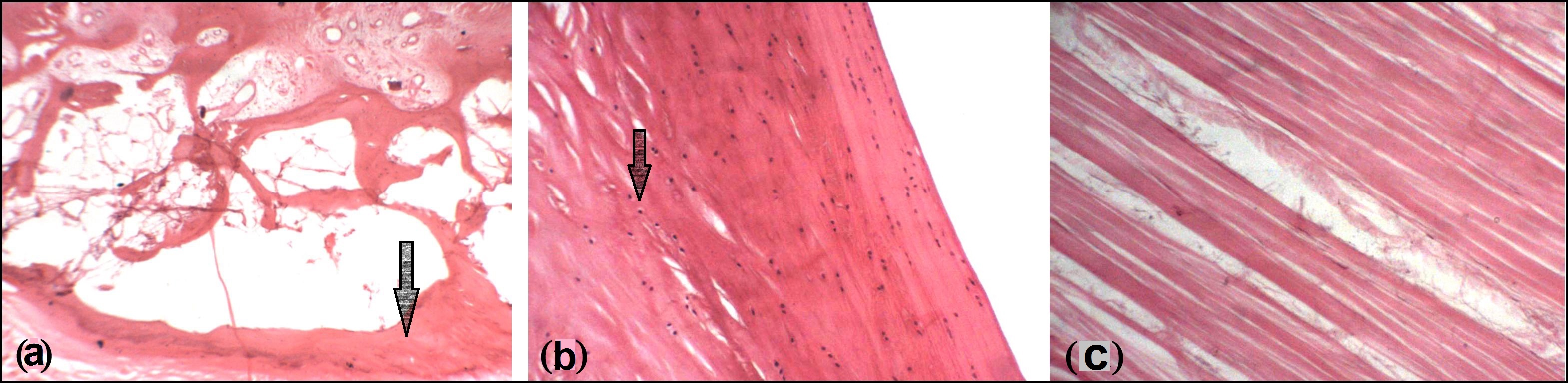
Histologically, OP were either cancellous bone [Table/Fig-6a], fibrocartilage [Table/Fig-6b] or only thickened collagen fibres [Table/Fig-6c].
6]: Photomicrographs of OP showing (a) Cancellous bone (b) Fibrocartilage (c) Collagen fibres [H&E (10X)]
Discussion
Le minor while investigating the comparative anatomy and significance of the OP has stated that during evolution of higher apes and man OP is associated with a substantial decline in the frequency. In man, the OP may be interpreted as a regressive form of that bone constantly found in the Cercopithecidae and in the Hylobatidae [3].
The function and origin, as well as incidence of OP is controversial, and Girnbaum et al., have stated that it ranges from 2.3% to 9% in radiological studies while in anatomical research it varies from 8.5 to 26% as reported in the literature [4]. Many support the idea that the OP is always present, but is completely ossified in only about 20% of the population [6]. Seon Jeong et al., while stating about Painful Os Peroneum Syndrome (POPS) presenting as lateral plantar foot pain have reported the presence of an OP between 4.7% and 30% of normal feet, and in some cases they were unilateral [7]. In the present study OP was found as a thickening on PL tendon where it was angulated and passed through peroneal sulcus of cuboid; and was present bilaterally in all the limbs examined.
Xiao-Tian Wang et al., stated that the prevalence of the OP is unknown. When ossified, it is visible in 20% of foot radiographs [5]. When the tendons were subjected to radiological investigations, we could observe calcification of these nodules in 35% of the OP only.
Muehleman et al., stated that 30% tendons displayed an OP both radiologically and histologically [8]. We've found that the OP was either cancellous bone or fibrocartilage or mere thickening of collagen fibres. Where OP was present as a hypertrophy of collagen bundles, the deep surface was covered by fibrocartilage or hyaline cartilage. In the present study, the average length of OP was 13.14 mm, average breadth of OP was 8.96 mm & average thickness of OP was 4.11 mm; but as the literature reviewed does not show similar parameters being studied by other workers; hence, no comparison can be drawn.
The OP is almost always single but may be multipartite in some individuals [6]. They are sometimes be mistaken for a fractured sesamoid. Fracture lines in the sesamoids usually run transversely, resembling bipartite sesamoids. A fracture line, however, will be irregular, whereas the margin of a bipartite sesamoid is smooth and regular [1].
The Painful Os Peroneum Syndrome (POPS), a term coined by Sobel et al., includes OP fracture or a diastasis of a multipartite OP. Clinical diagnosis of the POPS can be facilitated by the Single stance heel rise and Varus inversion stress test [9]. The presence of an OP can predispose to rupture of the PL tendon at the cuboid level, with or without, concomitant fracture [10,11].
Investigators studying cadaveric specimens find more sesamoids than when the data is obtained from just x-ray evaluation. This is an important fact for the Podiatric surgeon to keep in mind, particularly in cases where there may be symptoms of POPS and no radiographic evidence of OP [1].
The play of PL on the Cuboid can be easily appreciated when one finds presence of a synovial joint with the articular surfaces which may be single or double. On the right double facets (on Cuboid & Calcaneum) were more (25.8%) as compared to the left (16.2%). When double facets were present; the facet on Cuboid was larger than on Calcaneum [Table/Fig-1b]. We did not come across a reference to double articular facet of OP in the literature reviewed.
Conclusion
Every time when we see the nature's ingenuity in the structural and consequently in the functional adaptation one is awestruck by it and OP is one such example. It is present at the site of angulations of PL tendon with attendant change in direction and the exposure to various stresses and strains leading to the thickening and formation of OP. Although present in all the limbs studied; it was fibrous, cartilaginous or bony. Thus, it seems that Os Peroneum is a misnomer and should be called as: Peroneal sesamoid.
[1]. Mercado OA, Mercado OK, Mercado-Ciessau CM. Those Magnificent Sesamoids. www.oamercado.com/aos/tmsaa.pdf [Google Scholar]
[2]. Patil V, Frisch NC, Ebraheim NA, Anatomical Variations in the Insertion of the Peroneus (Fibularis) Longus TendonFoot Ankle Int 2007 28(11):1179-82. [Google Scholar]
[3]. Le Minor JM, Comparative Anatomy and Significance of the Sesamoid Bone of the Peroneus Longus Muscle (Os Peroneum)J Anat 1987 151:85-99. [Google Scholar]
[4]. Grinbaum CEA, Abreu AVD, Aguiar ROCD, Radiomorphological Study of the Peroneus Longus Tendon Adjacent to the Cuboid BoneRadiol Bras 2009 42(3):151-54. [Google Scholar]
[5]. Wang XT, Rosenberg ZS, Mechlin MB, Normal Variants and Diseases of the Peroneal Tendons and Superior Peroneal Retinaculum: MR Imaging FeaturesRadiographics 2005 25(3):587-602. [Google Scholar]
[6]. Sesamoids and Lateral Ankle Pain. Musculoskeletal and Orthopedic MRI Saturday, July 18, 2009. http://musculoskeletalmri.blogspot.in/2009/07/sesamoids-and-lateral-ankle-pain.html [Google Scholar]
[7]. Seon Jeong Oh, Kim YH, Kim , Painful Os Peroneum Syndrome Presenting as Lateral Plantar Foot PainAnn Rehabil Med 2012 36(1):163-66. [Google Scholar]
[8]. Muehleman C, Williams J, Bareither ML, A Radiologic and Histologic Study of the Os Peroneum: Prevalence, Morphology, and Relationship to Degenerative Joint Disease of The Foot and Ankle in a Cadaveric SampleClin Anat 2009 22(6):747-54. [Google Scholar]
[9]. Sobel M, Pavlov H, Geppert MJ, Painful Os Peroneum Syndrome: A Spectrum of Conditions Responsible for Plantar Lateral Foot PainFoot Ankle Int 1994 15(3):112-24. [Google Scholar]
[10]. Maurer M, Lehrman J, Significance of Sesamoid Ossification in Peroneus Longus Tendon RupturesJ Foot Ankle Surg 2012 51(3):352-55. [Google Scholar]
[11]. Verma P, Arora AK, Comparative anatomical study and incidence of os peroneum in peroneus longus tendon and its clinical significanceRev Arg de Anat Clin 2014 6(1):15-19. [Google Scholar]