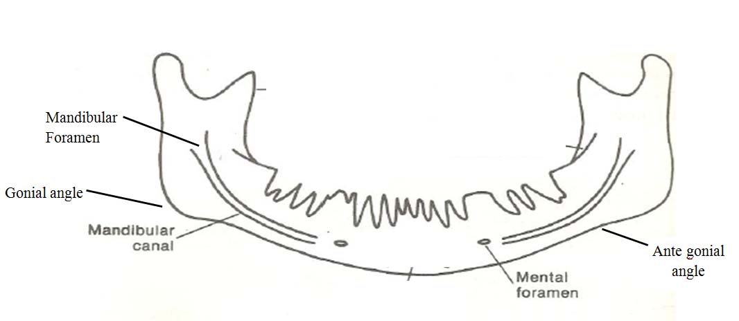
In human civilization the face and hands are the only remnants of the original individuality. As such, they are more accessible, more familiar socio biologically, and have been extensively studied. It is not surprising that these parts have been used commonly for identification purposes, the teeth and the bones of the craniofacial skeleton being also usually the best preserved parts of human remains. Furthermore, their inherent complexity is expressed by a large variability in size, shape, and proportions, which leads to individualization [1].
The identification and actual location of the various anatomical structures is of great importance in clinical dentistry for instance in the administration of local anaesthesia for surgery, operative or diagnostic purposes, in endodontic treatment as well as for predicting age. The majority of the mandibular changes are expected to occur in the alveolar process; however changes in the basal bone also occur throughout life [2].
Thus remodelling of the mandible with age, gender and dental status also occurs throughout the life in many parameters such as Gonial angle, antegonial angle, mental foramen, mandibular foramen mandibular canal. These changes can be easily evaluated in dried mandible as well as on radiographs [3].
Materials and Methods
The present study was prospective conducted in the Department of Oral Medicine and Radiology at Kanti Devi Dental College and Hospital, Mathura, India. The study population and there radiographs were taken depending on exclusion and inclusion criteria from OPD of the institution. Exclusion criteria are individuals giving history of any surgical procedure of mandible, Micrognathia, any skeletal or dental malocclusion and TMJ disorders, mandibular arch associated with any pathologies, mixed dentition, systemic diseases. Evident radiographic error, presence of bifid mandibular canal. Inclusion criteria include clear visibility of all parameters on digital panoramic radiographic image (gonial angle, antegonial angle, mandibular foramen, mandibular canal, mental foramen) bilaterally.
The study was approved by the Institutional ethical committee and written consent was taken from all patients. The study group comprised of 300 patients which were divided into Group A (25-34 years), Group B(35-44 years) and Group C(45-54 years). Each group consists of 50 males and 50 females. Digital panoramic radiographs were taken from ProMax II Digital panoramic X-ray unit (Planmeca, Helsinki, Finland) with HCL computer with Pentium D CPU 2.66 GHz 512 RAM and monitor, Planmeca Dimaxis Pro 4.1.4 software to evaluate the age changes in various parameters of mandible.
The interpretation of digital panoramic radiograph was done using Planmeca Dimaxis Classic 4.1.4 version software. Using the digital ruler, the selected five parameters were determined and measured bilaterally by two observers. The details of the five parameters are as follows:
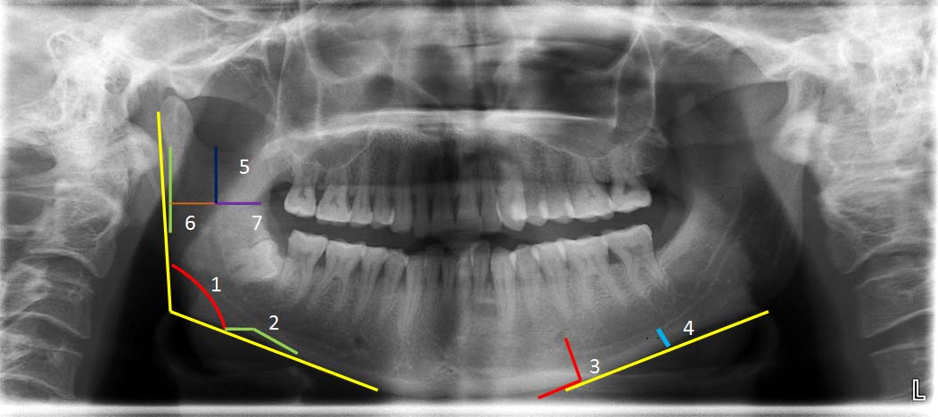
Gonial Angle: The gonial angle was assessed by tracing a line tangent to the lower border of the mandible and another line tangent to the distal border of the ramus on each side. The intersection of these lines formed the mandibular angle [Table/Fig-1,2].
Antegonial angle: The antegonial angle was measured by tracing two lines parallel to the antegonial region that will intersect at the deepest point of the antegonial notch [Table/Fig-1,2].
3. Mandibular foramen: This is been measured in three directions. a) Superio inferior position (X1),b) Posterioanterior position (X2),c)Anterioposterior direction (X3) Superio inferior position was measured by Vertical distance of the most inferior point of the image of the mandibular notch to the image of mandibular foramen. The distance of the foramen from the P-A position, was determined by drawing a perpendicular line from the MF to the tangent of the posterior border of the ramus it was measured from posterior border till the mandibular foramen. The A-P position of the Mandibular foramen on the ramus was measured from mandibular foramen till anterior aspects of ramus [Table/Fig-1 & 2] [3].
Mental foramen: The mental foramen was measured by drawing tangents along the lower border of body of mandible A perpendicular line was drawn from the lowest part of outline of the mental foramen to the tangent drawn along the lower border of the body of the mandible and the distance is measured from the mental foramen till the perpendicular line touching at the tangent at lower border of the mandible [Table/Fig-1,2] [4].
Mandibular canal: The mandibular canal was measured by drawing a Vertical distance of the most inferior point of the image of the inferior cortical of the mandibular canal to the inferior limit of the mandible [Table/Fig-1,2].
Diagrammatic representation of the parameters in mandible
1. Gonial angle, 2. Antegonial angle, 3. Mental foramen, 4. Inferior most border of mandibular canal, 5. Mandibular foramen(X1), 6. Mandibular foramen (X2), 7. Mandibular foramen (X3)
Statistical Analysis
A data was obtained and subjected to statistical analysis using Student T test, ANOVA, Post Hoc Turkey’s [5] which was prepared using SSPS version 14 for windows 7.
Results
In present study Gonial angle and Mandibular foramen (X2) were statistically highly significant when compared among all groups [Table/Fig-3].
Comparison of changes in various parameters between groups
| Sr. no | Parameters | Groups | f-value | p-value |
|---|
| 1 | Gonial angle (degrees) | A+ B+ C | 7.648 | 0.01 |
| 2 | Antegonial angle (degrees) | A+ B+ C | 0.851 | 0.42 |
| 3 | Mental foramen (mm) | A+ B+ C | 0.298 | 0.74 |
| 4 | Mandibular canal (mm) | A+ B+ C | 4.198 | 0.16 |
| 5(a) | Mandibular foramen(X1) (mm) | A+ B+ C | 0.461 | 0.63 |
| 5(b) | Mandibular foramen(X2) (mm) | A+ B+ C | 16.33 | 0.01 |
| 5(c) | Mandibular foramen(X3) (mm) | A+ B+ C | 0.86 | 0.91 |
p-value ≤0.01 is highly significant, p-value≤0.05 is significant, p-value ≥0.05 is non significant
When comparison was done among gender Gonial angle, Antegonial angle, Mandibular canal, Mandibular foramen (X1), (X2), (X3), was found to be statistically highly significant among genders [Table/Fig-4].
Although when inter group comparison was done among gender it was found that Gonial angle Mandibular foramen (X1), in group B & C were found to be statistically highly significant Comparison of male and female irt to Antegonial angle in group C was found to be statistically highly significant. Comparison of male & female irt to Mental Foramen was found to be statistically highly significant. Comparison of male and female irt to Mandibular canal in group A was found to be statistically highly significant Comparison of male and female irt to Mandibular foramen (X2) & mandibular foramen (X3), in group A, B & C was found to be statistically highly significant [Table/Fig-5]. When comparison was done side wise it was found that Gonial angle in group A,B&C were found to be statistically highly significant. Comparison of right and left irt to Antegonial angle in group A and C was found to be statistically highly significant whereas in group B it was statistically significant. Comparison of right & left irt to Mental foramen in group A was found to be statistically highly significant [Table/Fig-5].
Mean of variables and comparison of all parameters among gender
| Parameters | Gender | Mean± SD | p-value |
|---|
| Gonial angle (in degrees) | M | 117.66±6.54 | 0.01 |
| F | 122.10±6.04 |
| Antegonial angle (in degrees) | M | 157.63±7.55 | 0.01 |
| F | 160.66±7.34 |
| Mental foramen (in mm) | M | 13.31±2.27 | 0.01 |
| F | 12.51±1.88 |
| Mandibular canal (in mm) | M | 8.03±2.24 | 0.01 |
| F | 7.29±1.72 |
| Mandibular foramen (X1) (in mm) | M | 18.62±2-91 | 0.01 |
| F | 17.25±2.96 |
| Mandibular foramen (X2) (in mm) | M | 16.63±2.47 | 0.01 |
| F | 14.33±2.11 |
| Mandibular foramen (X3) (in mm) | M | 15.23±2.32 | 0.01 |
| F | 14.09±1.77 |
pvalue ≤0.01 is highly significant, p-value≤0.05 is significant, p-value ≥0.05 is non significant
Comparison of right and left among groups.
| Sr. no | Parameters | Age groups | Side | t-test | p-value |
|---|
| 1 | Gonial angle (degrees) | Group A | R | L | 12.516 | 0.01 |
| Group B | R | L | 7.492 | 0.01 |
| Group C | R | L | 6.678 | 0.01 |
| 2 | Ante gonial angle (degrees) | Group A | R | L | 4.623 | 0.01 |
| Group B | R | L | 2.130 | 0.03 |
| Group C | R | L | 2.840 | 0.01 |
| 3 | Mental foramen (mm) | Group A | R | L | 2.834 | 0.01 |
| Group B | R | L | 1.490 | 0.13 |
| Group C | R | L | 0.422 | 0.67 |
| 4 | Mandibular canal (mm) | Group A | R | L | 1.806 | 0.07 |
| Group B | R | L | 0.202 | 0.84 |
| Group C | R | L | 0.358 | 0.72 |
| 5a | Mandibular foramen(X1) (mm) | Group A | R | L | 3.013 | 0.01 |
| Group B | R | L | 0.159 | 0.87 |
| Group C | R | L | 0.062 | 0.95 |
| 5b | Mandibular foramen(X2) (mm) | Group A | R | L | 0.318 | 0.75 |
| Group B | R | L | 0.415 | 0.67 |
| Group C | R | L | 0.854 | 0.39 |
| 5c | Mandibular foramen(X3) (mm) | Group A | R | L | 1.198 | 0.23 |
| Group B | R | L | 1.603 | 0.11 |
| Group C | R | L | 0.403 | 0.68 |
p-value ≤ 0.01 is highly significant, p-value≤0.05 is significant, p-value ≥0.05 is non significant
On evaluation of the accuracy of parameters to predict age Mandibular canal was found to be highly significant and Mandibular foramen (X1) was found to be statistically significant to evaluate the accuracy for predicting age in group C [Table/Fig-6].
Evaluation of the accuracy of parameters to predict age
| Groups | Parameters | Gonial angle | Antegonial angle | Mental foramen | Mandibular canal | Mandibular Foramen (X1) | Mandibular Foramen(X2) | Mandibular Foramen(X3) |
|---|
| Group A | Pearson coefficient | 0.01 | -1.11 | -0.06 | 0.72 | 0.023 | -0.271 | .028 |
| p-value | 0.86 | 0.27 | 0.95 | 0.47 | 0.82 | 0.06 | 0.78 |
| Group B | Pearson coefficient | 0.04 | -0.10 | 0.02 | -0.05 | -0.18 | 0.02 | -0.101 |
| p-value | 0.66 | 0.31 | 0.81 | 0.58 | 0.06 | 0.84 | 0.31 |
| Group C | Pearson coefficients | -0.04 | 0.12 | 0.18 | 0.24 | 0.19 | 0.14 | 0.15 |
| p-value | 0.65 | 0.20 | 0.72 | 0.01 (HS) | 0.05 (S) | 0.16 | 0.11 |
Discussion
The results of our study suggests that, mean Gonial angles becomes more obtuse in all three groups and were comparable with Kasat V et al., [2] and Shendakar AT et al., [6] who opine that as age advances, there is change in angle of the mandible from more obtuse to less obtuse and again to more obtuse [2].
However, the mean values of the Gonial angle are more in females than in males. This can be correlated with the study by Xie QF et al., [7] and Huumonen S et al., [8] where women had greater values of gonial angle than men. According to Casey DM et al., [9], usually mean angle is 3-5 degrees larger in females than in males.
This could be because subjects with maximum masticatory force have a small Gonial angle; on average men have greater masticatory force than do women.
Mean value of left side was greater in the present study in all age groups which was comparable with that of Raustia MA [10], possibly because of more use of right side.
In present study when inter group comparison was done for Gonial angle it was found to be highly significant in A Vs B and B Vs C. the Antegonial angle when compared among three age groups, it was non significant, however, Antegonial angle for females were higher than males was comparable to Enlow DH et al., [11] and Dutra V et al., [4].
Bone deposition takes place throughout inferior border except in the Antegonial region, where resorption takes place which would decrease the Antegonial angle and increase antegonial depth. Males have significantly smaller antegonial values than females. This might be due to gender hormonal differences testosterone in males and estrogen in females affecting bone metabolism and thereby showing visible changes in radiographs [4].
Mean Antegonial angle at left side was more compared to right side in all age groups was consistent with Dutra V et al., [4]. This could be due to increased function on preferred chewing side.
The accuracy of parameters were assessed to predict age for Ante Gonial and was found to be non significant in all groups.
Mean value of the distance between Mental foramen and tangent drawn to base of mandible had no statistically differences between the analysed age groups, was consistent with Amorin M et al., [12], Kasat V et al., [2] and Shendarkar et al., [6]. The mean values were more for males which can be compared to Enlow DH et al., [11] and Amorin M et al., [12]. In adult phase, the rate and speed of growth are bigger in men with the result that craniofacial dimensions in this gender are 5-9% bigger when compared to women. Sex hormones, such as estrogen and progesterone can influence in the speed of bone growth in this phase, contributing to the development of craniofacial morphologic differences between genders [12].
The accuracy of parameters were assessed to predict age from mandibular canal and was found to be highly significant in group C. Mean values of mandibular canal compared among all age groups was found to be non significant. This observation is consistent with Amorim M et al., [12], who states that measurements are directly related to the curvature of mandibular canal that an individual presents, independent of variation of gender or age groups and so the absence of statistical differences between the studied groups [12].
Mean mandibular canal values in males were found to be greater than in females in all age groups, also the values were greater on right side than left. This is the only study of its kind where comparison among the gender and side wise in same age range done.
In the present study inter group comparison was done for mandibular canal between B Vs C and it was found to be significant.
The accuracy of parameters were assessed to predict age for Mandibular Canal and was found to be highly significant in group C. three dimensions were taken for mandibular foramen i.e., X1, X2 and X3. When all three parameters of mandibular foramen were compared among all age groups, no significance was seen in all age groups at X1, X3 but at X2 position it was found to be highly significant.
The difference in mean values i.e. movement of mandibular foramen to a more anterior position can be explained by the fact that mandibular growth of ramus in an anterior position is mainly by bone apposition in the posterior border of ramus and remodeling in anterior border [13]. Although in our study Mandibular foramen moves posteriorly this observation is in consistent with Mabjiorgu EF [14], Thangavelu K et al., [15] and Samantha PP et al., [16] Mean X1 in the present study were found to be more for males than females in group Band C which were comparable to that of Amarghan et al., [17] and Amorim M et al., [12].
Also mean X2 and X3 for males were more than females. Muscular tension is considered an inductive factor of bone formation and in mandible the combination of elevating muscles during masticatory movement exerts tension throughout the ramus. In general, men have stronger masticatory muscles than women [12]. Mean value of X1 on left side is more in group A and C and more on right side in group B. In the present study inter group comparison was done for Mandibular Foramen at X1, X2, X3 between A Vs B, C Vs A and were found to be highly significant.
The accuracy of parameters were accessed to predict age for Mandibular Foramen at X2, X3 position and were found to be non significant in Group A, B & C, Although X1 was found to be significant in group C.
Conclusion
There were significant changes in Mandibular canal and Mandibular foramen as age advances. Age was less clearly related for Gonial angle, Antegonial angle and Mental foramen.
p-value ≤0.01 is highly significant, p-value≤0.05 is significant, p-value ≥0.05 is non significant
pvalue ≤0.01 is highly significant, p-value≤0.05 is significant, p-value ≥0.05 is non significant
p-value ≤ 0.01 is highly significant, p-value≤0.05 is significant, p-value ≥0.05 is non significant