Microscopy is a basic essential technique which is used in both pathology and microbiology laboratories for the diagnostic and research works [1,2]. The advent of digital imaging has enhanced the role of microscopy in the academic and research fields, as well as for consultation and assessment of slides [1,2]. Obtaining images of slides viewed by a microscope can be invaluable for both diagnosis and teaching.They can be transferred among technologically-advanced hospitals for further consultation and evaluation [2,3].
Photography by a microscope normally requires a specially adapted microscope with a camera port, a specialized camera, an adaptor to attach the camera to the port and a computer system [3,4].In many places this required equipment is either unavailable or insufficiently portable. It is difficult to fit it in every microscope and also not affordable for individual instrument in all the sections of laboratory, especially in the developing world [1–3].
The advent of affordable compact box type digital cameras with displays that reflect precisely the image as seen through the camera lens has made possible a simplified method of taking photographs through a microscope [1,2]. Almost any combination of light microscope and box type digital camera with optical zoom (including some camera phones) can be used without the need for specialized equipment [1,2].
Materials and Methods
When we were trying to do photomicrography, we found that our system of photomicrography(MIPS) was not available due to some reason. At that moment a box type digital camera (Nikon Coolpix S6150) was available. We used it and took photomicrographs.
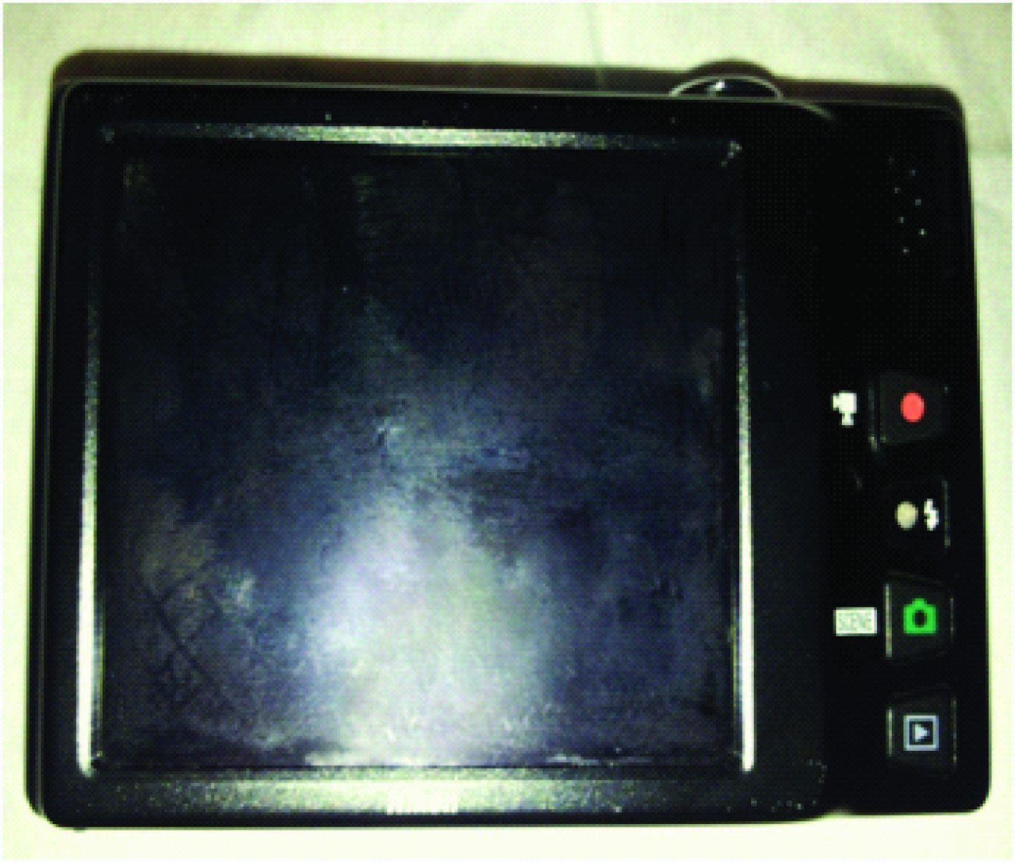
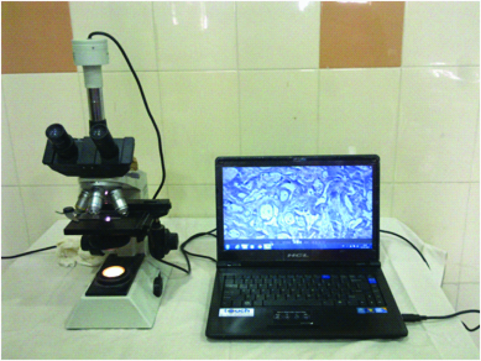
1. A box type digital camera [Table/Fig-1&2]
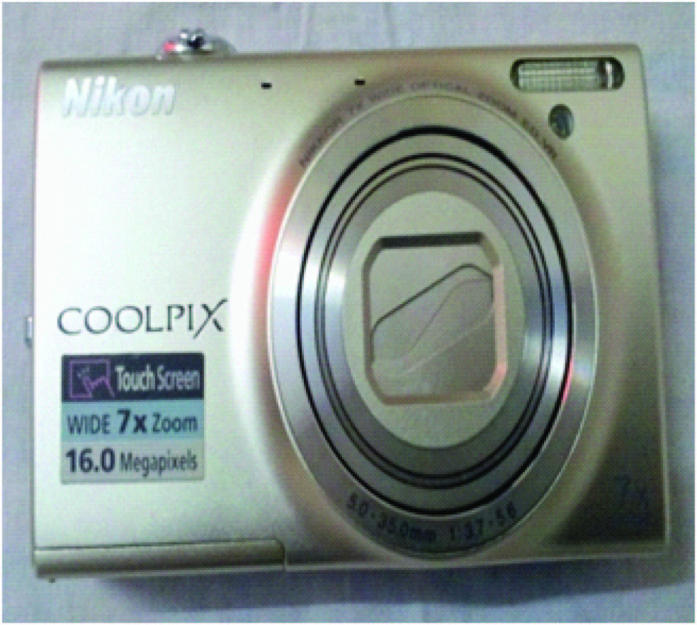
Company – NIKON
Model – COOLPIX S6150
Lens – NIKKOR Glass lens
With a focal length of 5.0 - 35.0 mm and f/3.7 - f/5.6 aperture
Zoom - 7X optical zoom, 4X digital zoom
Resolution - 16 megapixels
LCD screen - 3 inch touch screen
PRIZE – Rs. 6500 to 8500 0nly
2. Microscope
Company – Olympus
Model - CH20i
Type – Binocular or Trinocular Microscope
Achromatic objectives - Olympus iNE4x, iNE 10x, iNE 40x (spring), iNE 100x (spring, oil). All antifungal coated.
Eyepiece - Olympus iCWHK10x LB wide field, antifungal coated.
Light Source - Built-in 6V, 20W illuminator base with halogen lamp
The technique we used for taking digital photographs of specimens visualized through a light microscope is as follows:
Using the microscope, we examined the slide and selected the area of interest and the magnification required.
We adjusted the light source to get adequate intensity of light.
We then adjusted the focus to maximise clarity of image.
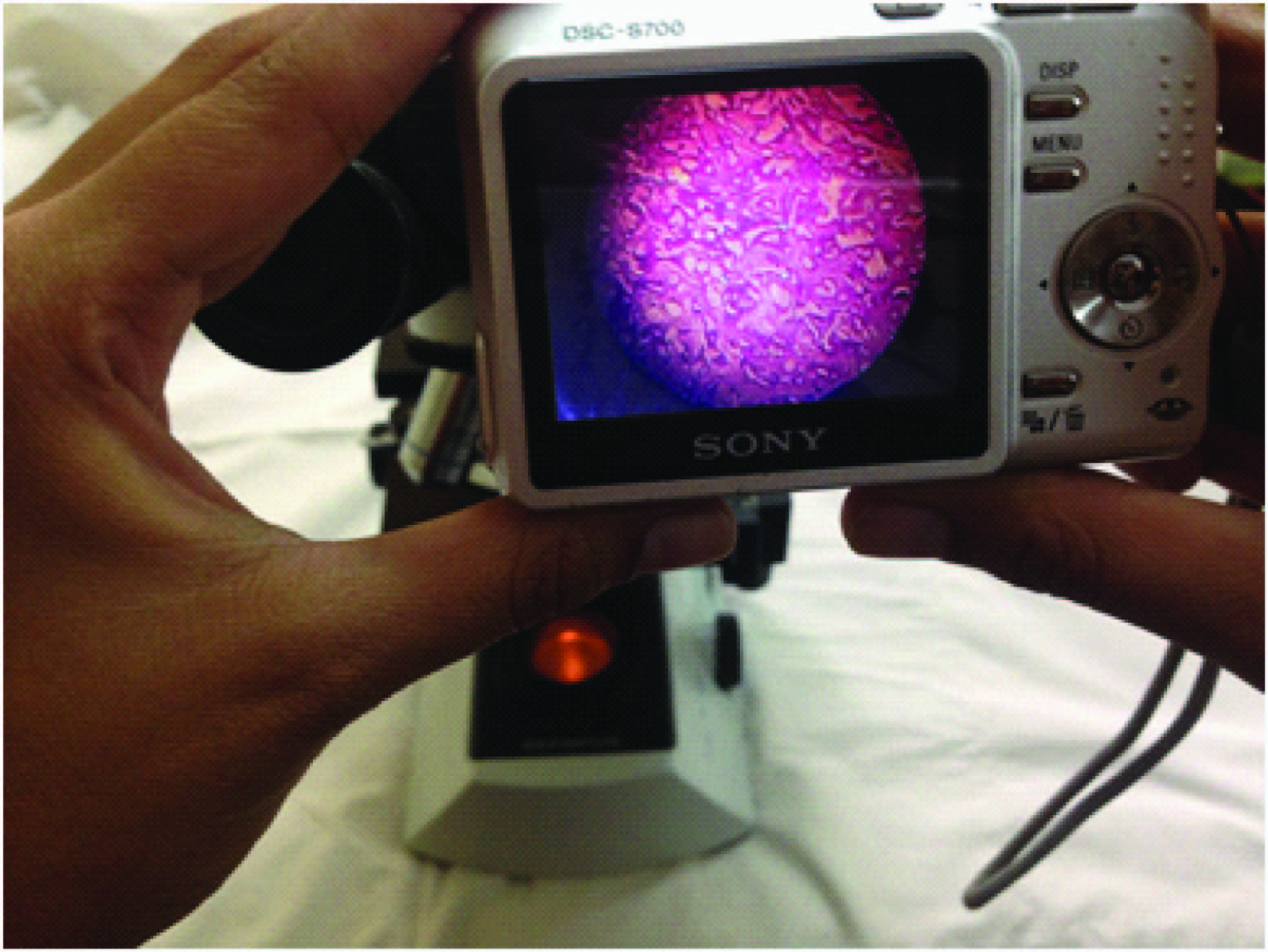
We held the camera lens against the microscope eyepiece. (On doing so a small circle of light would be seen on the camera’s LCD screen) [Table/Fig-3,4].
The zoom function of the camera was used to increase the size of the circle as required. The most difficult step was moving the camera lens small distances across the eyepiece to centre the circle. The autofocus of the camera would then self-adjust to give a clear image.
The camera was held very still, a photograph taken, and the image examined to see if it was satisfactory.
Hold the camera lens against the microscope eye-piece
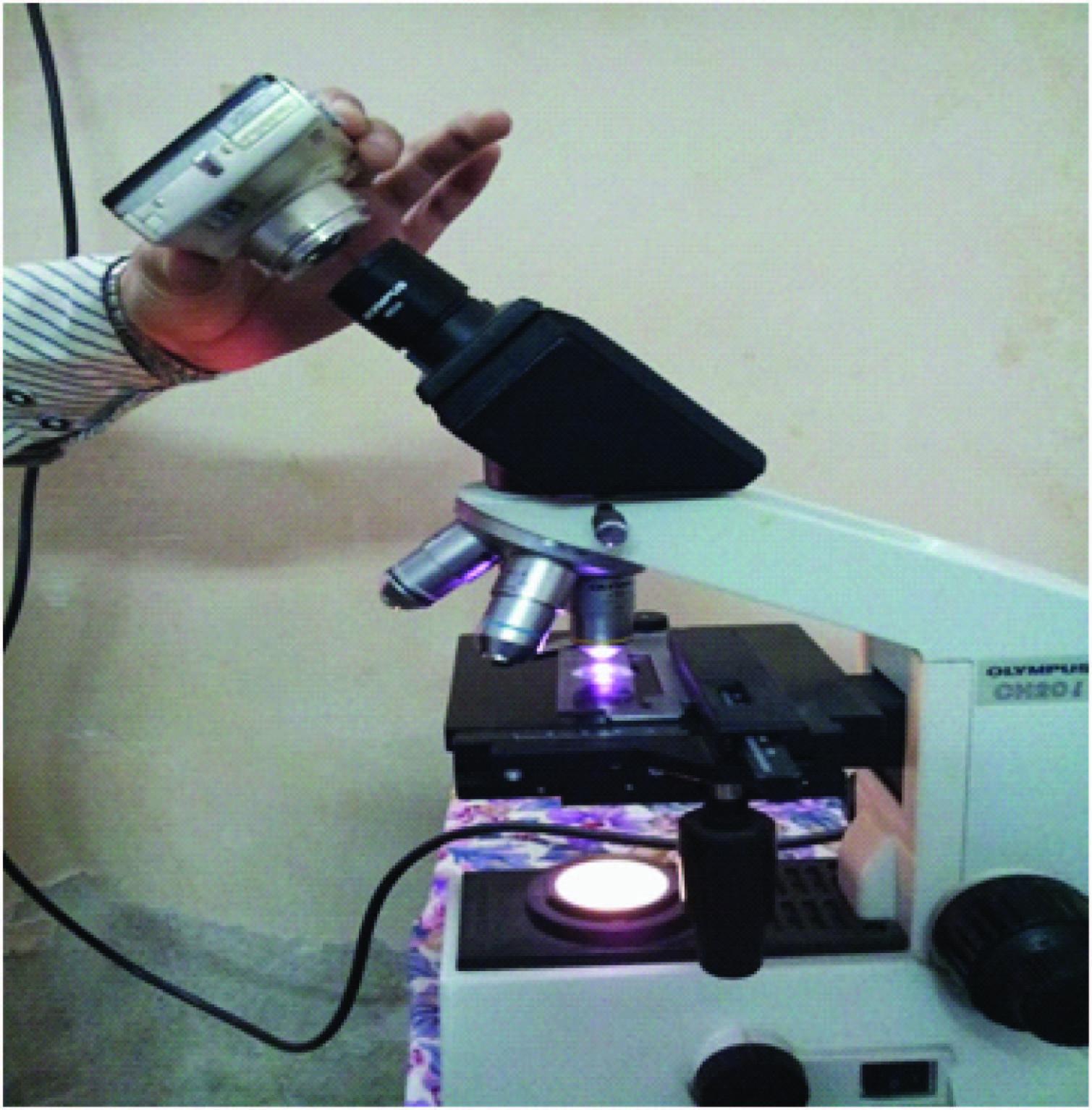
A small circle of light would be seen on the camera’s LCD screen
3. A standard microscopic photography camera unit [Table/Fig-5].
A standard microscopic photography camera unit,
After some time, our MIPS became available & we took photomicrographs by that system also, which includes:
Olympus CH20i Trinocular Microscope (Specifications are same as above)
Microscope Image Projection System (MIPS)
Company – Magnus
Model - MIPS-USB 2.0
CCD Sensor - 1/4” Interline Transfer CCD
Active pixels (Hx V) - 752 x 582 (PAL)
Recommended PC Specifications - Intel Pentium P IV 2.0 GHz or better with at least 256 MB RAM, 20 GB Hard Disk, USB 2.0 ports Windows XP Operating System
Power supply - 5VDC via USB bus
PRIZE– Rs. 30,000 – 40,000
Computer system compatible with MIPS
Results
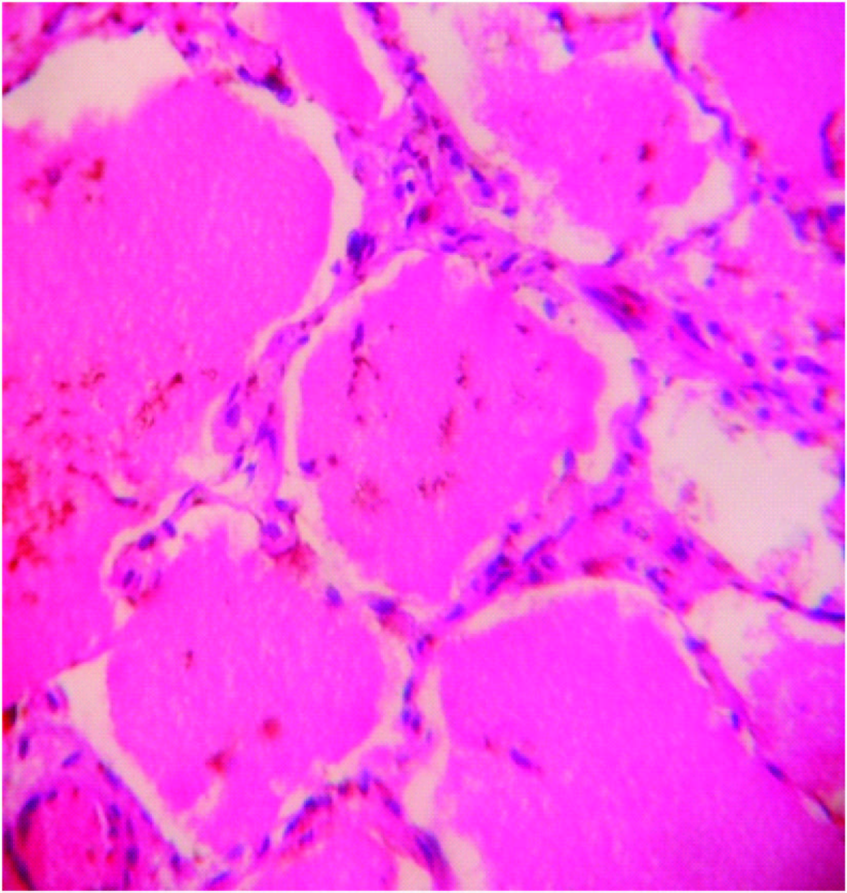
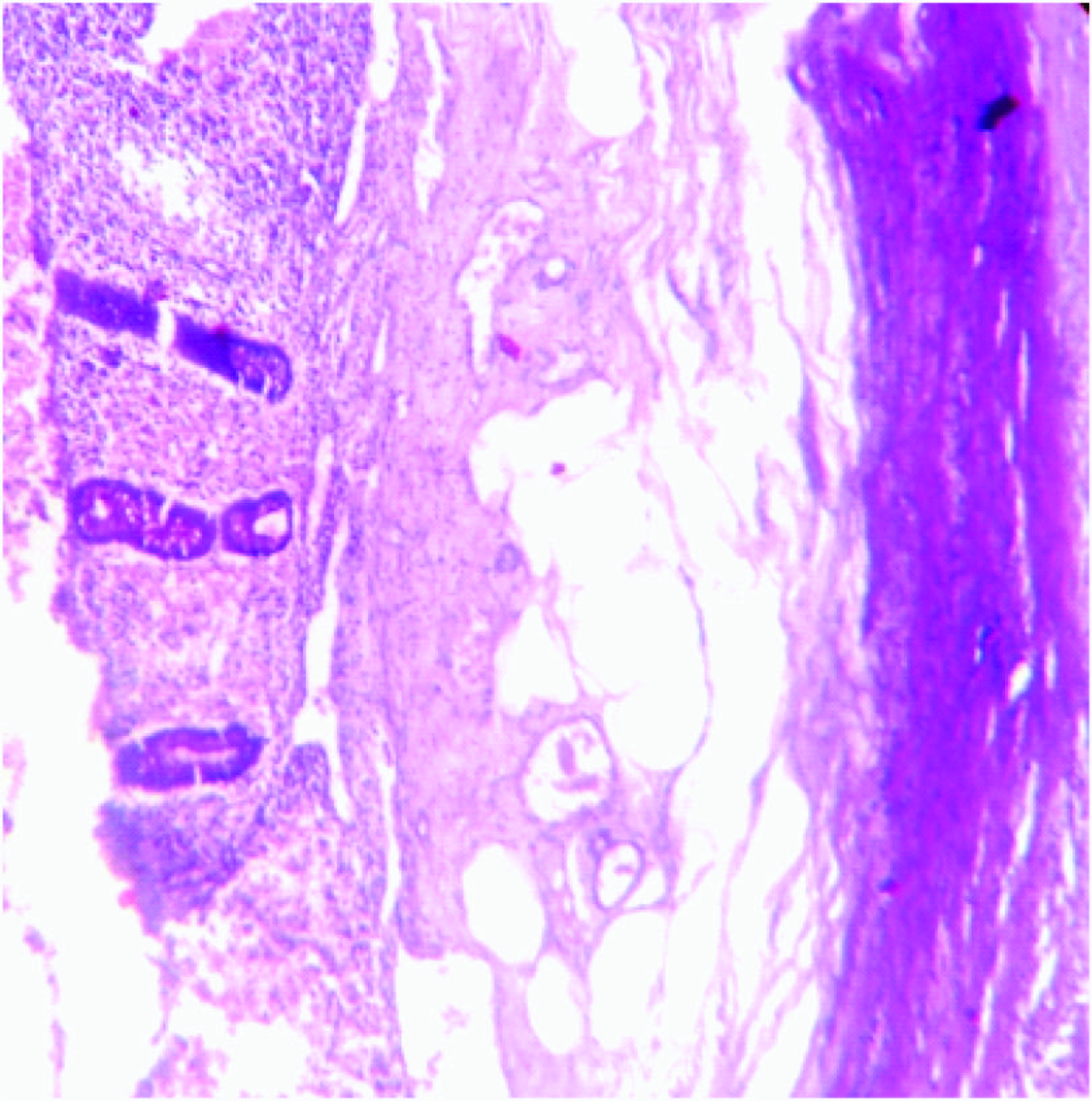
The photomicrographs were taken with different objective lenses (10x, 40x and 100x) of light microscope using both the systems in different slide preparations (Wet mount preparation of stool, Peripheral blood smear, Histochemical stains – H & E, PAS) and then transferred to laptop for editing using photograph editing software PICASA. Both sets of photographs were compared with each other.
Images captured even by a low-cost box type digital camera produced brilliant images, the quality being comparable with expensive camera especially designed for the microscope (MIPS – USB system) [Table/Fig-6,7,8,9,10,11,12,13,14,15,16,17,18and19].
HE stain, 10x field,box type digital camera,

HE stain, 10x field, MIPS,

HE stain, 40x field, box type digital camera
HE stain, 40x field, MIPS,

PAS stain, 10x field, box type digital camera,
PAS stain, 10x field, MIPS,

PAS stain, 40x field, box type digital camera

PAS stain, 40x field, MIPS

Field stain, 10x field, box type digital camera
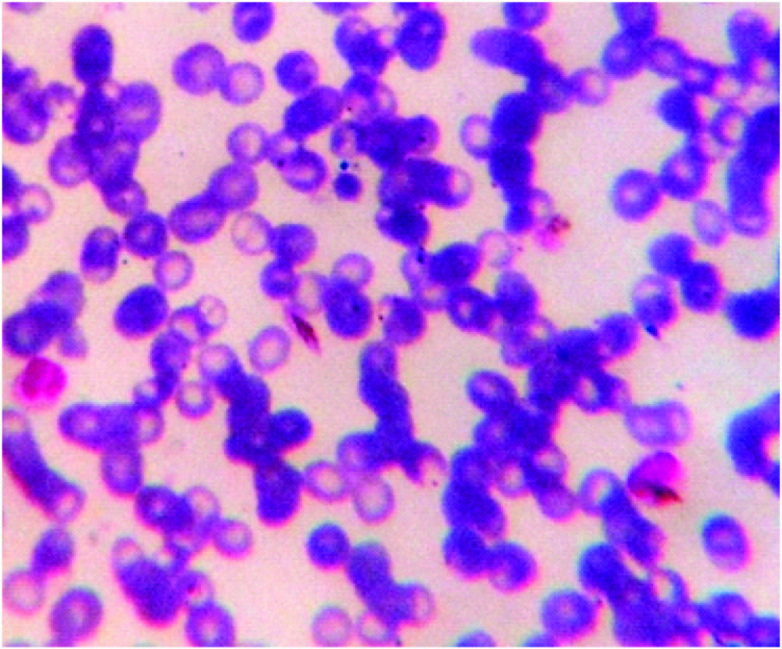
Field stain, 10x field, MIPS

Field stain, 40x field, box type digital camera
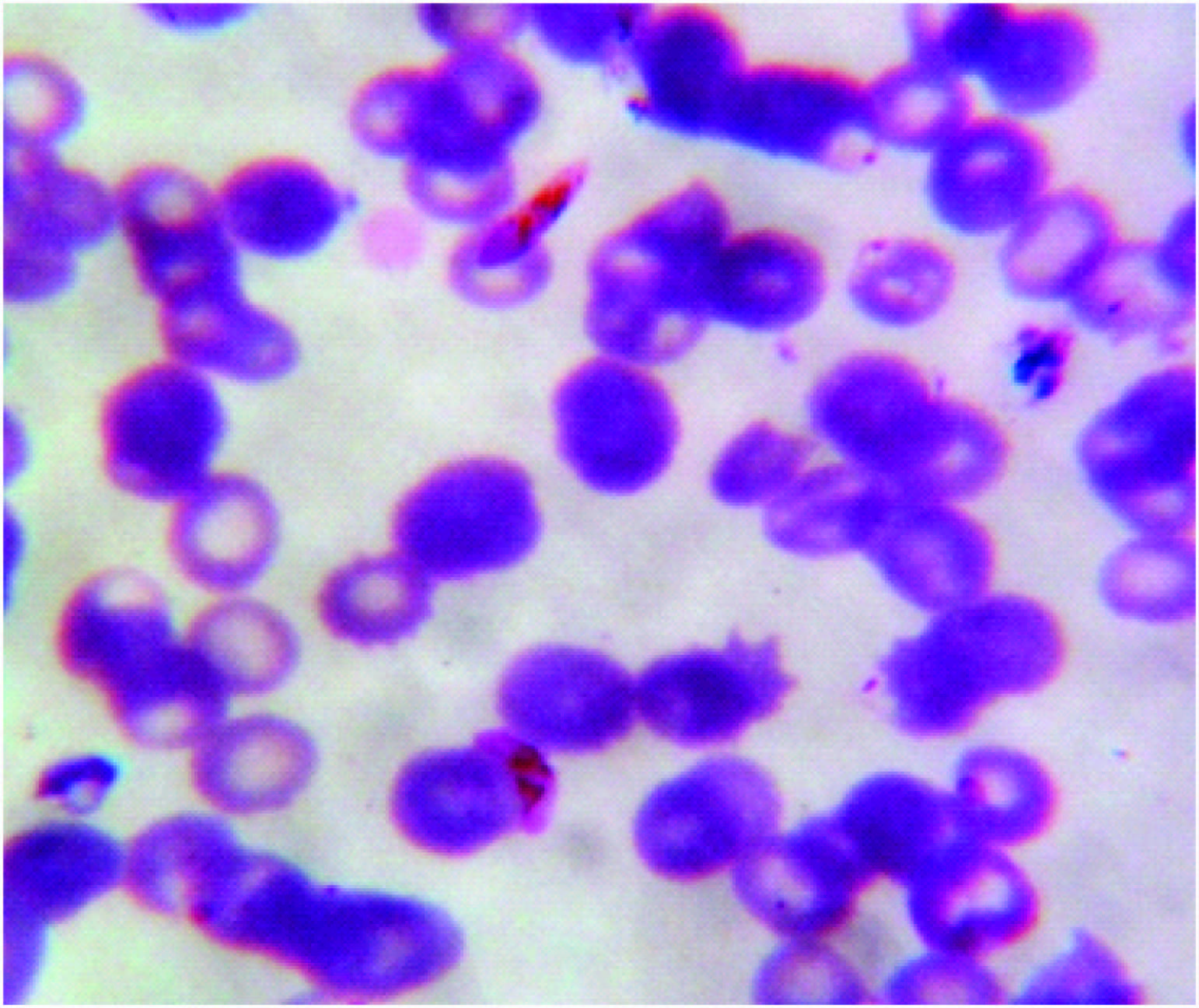
Field stain, 40x field, MIPS

Wet mount, 40x field, box type digital camera
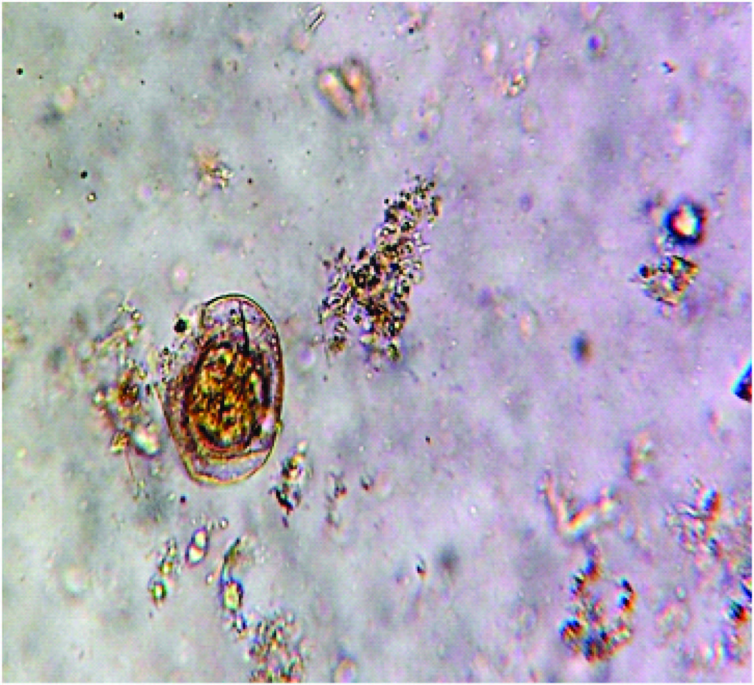
Wet mount, 40x field, MIPS

H & E Stain
Photomicrographyby box type digital camera vs Photomicrography by MIPS
[Table/Fig-6,7](10x fields) & [Table/Fig-8,9](40x fields)
PAS Stain
Photomicrography by box type digital camera vs Photomicrography by MIPS
[Table/Fig-10,11] (10x fields) & [Table/Fig-12,13](40x fields)
Peripheral smear (Fields Stain)
Photomicrography by box type digital camera vs Photomicrography by MIPS
[Table/Fig-14,15](10x fields) & [Table/Fig-16,17] (40x fields)
Stool examination (wet mount slide)
Photomicrography by box type digital camera vs Photomicrography by MIPS [Table/Fig-18,19].
Fluorescent microscopy
We had also tried to capture images of histopathology slides stained by acridine orange stain under a fluorescent microscope by box type digital camera, but the results were not satisfactory as with light microscopy method [Table/Fig-20,21].
Acridine orange, fluorescent microscopy, box type digital camera
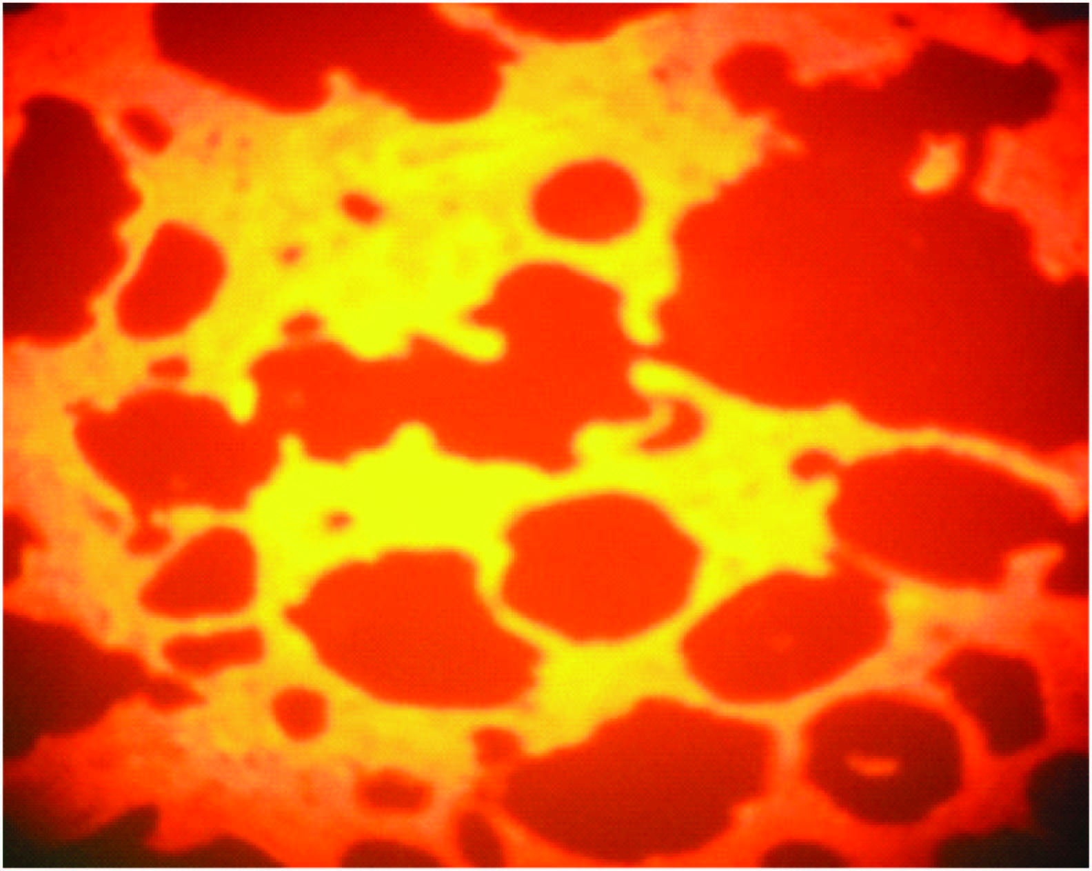
Acridine orange, fluorescent microscopy, MIPS
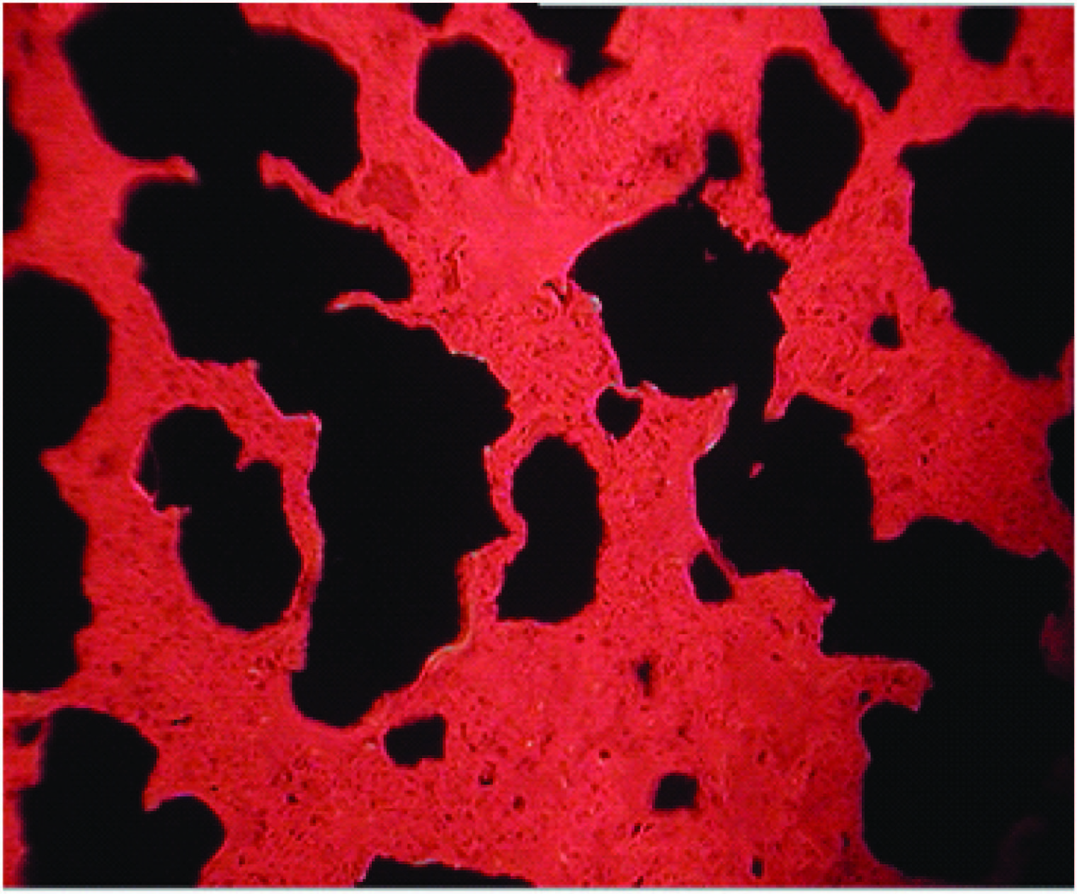
Photomicrography by box type digital camera vs Photomicrography by MIPS.
Discussion
The presented method of capturing microscopic images by a low cost box type digital camera is not a new one. It is already evolved by various authors [1–7]. But, it is still in its developing stage. Hereby, we have confirmed that it is a simple, economical, and highly practical technique. We have mentioned details of both, the box type digital camera, and standard microscopic photography camera unit (MIPS)to compare which is not given in certain reference studies.
Earlier workers have used different tools for photomicrography, like use of paper sleeve between eyepiece of microscope and camera lens [2]. However, we did not require this kind of paper sleeve or adaptor tube and the results were also good [Table/Fig-3&4]. Similarly, Bellina L et al., [5] took images by simply approaching the lens of the mobile-phone camera to the ocular of the microscope without using any sleeve or adaptor.
Most models of light microscope and box type digital camera, and even some camera-phones, can be used in this technique. An advantage of digital camera or mobile phone camera is its affordability and availability which makes it feasible to use as and when required. At the same time it is handy to carry at different sites [1,3].
The technique is quick to learn and can easily be performed. Graduation students who are pursuing research, and for whom the resources are usually meagre,as well as post-graduate students can also use such an easy and affordable technique for their field of interest. The images captured thus can be reproduced in their dissertations or theses [1]. It also enables the accumulation of a library of locally relevant clinical images for use in teaching laboratory staff and clinicians and for documentation in research [3]. Many research workers have found this method a very useful diagnostic technique for malaria and tuberculosis at the peripheral or remote areas [6,7].
We may say that the method which we have used has certain limitations; as it has not yielded useful digital images with fluorescent microscopy.
However, this simple cost effective spot image capture technique is quite useful and must be encouraged in a resource-poor setting or remote areas, where it can be used to assist with diagnosis and can be extremely useful for teaching to improve the quality of diagnostics and academics.