Ovarian Pregnancy: Uncommon Mode of Presentation
Saubhagya Kumar Jena1, Gayatri Kar2, Soumya Samal3, Basanta Kumar Behera4
1 Assistant Professor, Department of Obstetrics and Gynaecology, AIIMS, Bhubaneswar, Odisha, India.
2 Retired Professor and HOD, Department of Obstetrics and Gynaecology, MKCG Medical College, Berhampur and GSL Medical College and Gen. Hospital, Rajahmundry, India.
3 Assistant Professor, Department of Anaesthesiology, IMS and SUM Hospital, Bhubaneswar, Odisha, India.
4 Professor, Department of Community Medicine, KIMS, Bhubaneswar, Odisha, India.
NAME, ADDRESS, E-MAIL ID OF THE CORRESPONDING AUTHOR: Dr. Saubhagya Kumar Jena, Assistant Professor, Department of Obstetrics and Gynaecology, AIIMS, Bhubaneswar, At: Sijua, Po: Dumduma, Bhubaneswar, Odisha-751019, India. Phone : 9438884133, E-mail : drsaubhagya@gmail.com
Ovarian pregnancy is very rare, and its incidence is 1 in 3000 live births. In this condition, common risk factors for ectopic pregnancy not usually found. It usually occurs in fertile women and more commonly with in-situ intrauterine device (IUD). Preoperative diagnosis is always not possible although the patient commonly presents with abdomen-pelvic pain, per vaginal bleeding and hypovolemic shock. High degree of suspicion with estimation of serum beta HCG, transvaginal ultrasonography by an experienced sonologist and laparoscopy is required for confirming the diagnosis. Though, the usual treatment is surgery, it can be managed by medical methods only in hemodynamically stable patients. In this case report, we describe the unusual mode of clinical presentation in an elderly woman with ovarian pregnancy.
Ectopic, Extra-uterine pregnancy, Pregnancy
Case Report
A 45-year-old female presented with pain in lower abdomen for six months, excessive vaginal discharge for one year and scanty menstruation for five years. Her previous menstrual cycles were regular, with 3 to 4 days bleeding occurring every 28 to 30 days and average flow. For last 5 years, she had irregular cycles with scanty flow for 1 to 2 days. She was para 3 with 2 living children and last childbirth was 19 years back. Her husband vasectomized 17 years back. She was under treatment for major depressive disorder.
On examination her height was 146cm, weight 50kg without any clinical evidence of pallor. Both breasts were normal on examination. Her pulse rate was 80 beats per minute; blood pressure was 120/80 mm of Hg and temperature was 98.8oF. On examination of cardiovascular and respiratory system, no abnormality detected. Abdomen was soft, non-tender, and no mass was palpable. Per speculum examination revealed parous cervical os, erosion of both lips of the cervix with minimal mucoid discharge. In bimanual examination uterus was bulky, mobile, tender and fornices were free. A provisional diagnosis of adenomyosis made, and investigations started.
In investigations, blood group was A+ve, haemoglobin 9.1 gm%. Total count and differential count was normal. Bleeding time and clotting time was 1min 55s and 3min 20s respectively. Antibody tests for VDRL, HBSAg and HIV, were non-reactive. Urine examination was normal. Pap smear was inflammatory. Pelvic ultrasonography revealed a bulky, anteverted uterus with heterogeneous myometrium and thin endometrium. Both the ovaries were not visualized. Abdominal ultrasonography was unremarkable.
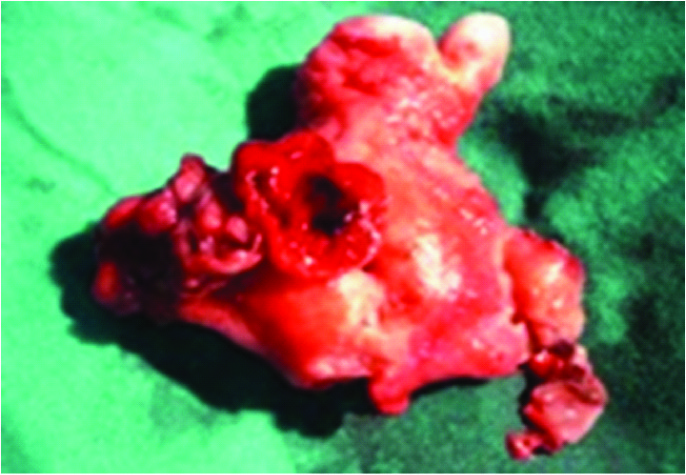
Laparotomy done under epidural anaesthesia. Abdomen opened by pfannensteil incision. Intraoperative findings were bulky uterus with normal, intact and healthy tubes on both sides. Left ovary was healthy and atrophic. Right ovary was enlarged in size (4cm x 3.5cm x 4cm) with the presence of old blood clots. It was attached to the posterior surface of uterus [Table/Fig-1].Total abdominal hysterectomy with bilateral salpingo-oophorectomy done. Postoperative period was uneventful.
Specimen of uterus, tube and ovaries showing blood clots and attached right ovary over posterior surface of uterus
On histopathology examination,there was secretory endometrium and normal myometrium. Both tubes and left ovary was normal. Trophoblastic villi and corpus luteum embedded in the right ovarian tissue seen, which was confirmatory of ovarian pregnancy [Table/Fig-2].
Histopathology slide showing trophoblastic villi embedded in ovarian tissue

Discussion
Our case fulfilled all the four criteria of Spiegelberg (1878) for the diagnosis of ovarian pregnancy: (a). The tube has to be entirely normal (b). The gestational sac has to be anatomically situated in the ovary (c). The ovary and the gestational sac have to be connected to the uterus by the utero-ovarian ligament (d). Placental tissues to be mixed with ovarian cortex [1–4]. Though there was abdominal pain, history of amenorrhoea and vaginal bleeding was not present. She had not noticed it due to the irregular menstrual cycles. We also not suspected it preoperatively as her husband undergone vasectomy. The possibility of ovarian pregnancy was suspected intraoperatively and confirmed by histopathology.
Ovarian pregnancy occurs in the fertile patient in contrast to tubal ectopic pregnancy, which is more frequently associated with impaired fertility [5]. The cause of ovarian pregnancy remains obscure. Though, there are different hypotheses like interference in the release of the ovum from the ruptured follicle, malfunction of the tubes and inflammatory thickening of the tunica albuginea. Usually, it is secondary to reflux of the fertilized oocyte to the ovary [6]. The cases of ovarian pregnancy after IVF reported in the literature support the theory of reflux [3]. Intrauterine contraceptive devices may also be a cause [7].
In our case, the cause of pregnancy was failed vasectomy. The cause of ovarian pregnancy may be due to unruptured follicle leading to intrafollicular pregnancy or reflux of the fertilized oocyte. Vasectomy failure might not be responsible for ovarian pregnancy. There might be some pathology in the ovaries that lead to ovarian pregnancy.
Patients usually present with abdominal pain (100%), vaginal bleeding (33%) and hypovolemic shock (8%) [8]. In suspected cases, serum beta-HCG estimation is valuable for diagnosis [9]. Abdomen-pelvic ultrasonography can not always differentiate ovarian pregnancy from other forms of ectopic pregnancy [3]. The sonographic diagnosis of ovarian pregnancy is difficult. Only an experienced sonographer, who is aware of the symptoms of this type of ectopic pregnancy that is a rare event, can demonstrate a gestational sac adjacent to the ovary or as some people described a double ring hyperechoic within a hypoechoic adnexal mass. Culdocentesis has no clinical diagnostic benefits. Laparoscopy is invaluable, as diagnosis and treatment can be carried out in a single setting. Laparoscopic wedge resection is the treatment of choice [1]. However, successful management of hemodynamically stable patients with methotrexate has been reported.
Conclusion
The diagnosis of ovarian pregnancy continues to challenge clinicians. Whenever suspicion arises serum beta HCG estimation and transvaginal ultrasound by an experienced sonologist can help in the diagnosis. Laparoscopy is very much needed in those cases both for diagnosis and treatment.
[1]. Raziel A, Schachter M, Mordechai E, Friedler S, Panski M, Ron-El R, Ovarian pregnancy- a 12-year experience of 19 cases in one institutionEur J Obstet Gynecol Reprod Biol 2004 114(1):92-96. [Google Scholar]
[2]. Ercal T, Cinar O, Mumcu A, Lacin S, Ozer E, Ovarian pregnancy;relationship to an intrauterine deviceAust N Z J ObstetGynaecol 1997 37:362-64. [Google Scholar]
[3]. Sergent F, Mauger-Tinlot F, Gravier A, Verspyck E, Marpeau L, Ovarian pregnancy: reappraisal of diagnostic criteriaJ Gynecol Obstet Biol Reprod 2002 31:741-46. [Google Scholar]
[4]. Spigelberg O, Casusistik der ovarialschwangerschaftArch Gynecol 1878 13:73 [Google Scholar]
[5]. Piero Seinera, Alessandro di Gregorio, Riccardo Arisio, Andrej Decko, Francesco Crana, Ovarian Pregancy and operative laparoscopy : Report of eight casesHuman Reporduction 1998 12(3):608-10. [Google Scholar]
[6]. Kraemer B, Kraemer E, Guengoer E, Juhasz-Boess I, Solomayer EF, Wallwiener D, Ovarian ectopicpregnancy: diagnosis, treatment, correlation to Carnegie stage 16and review based on a clinical caseFertil Steril 2009 92(1):392 [Google Scholar]
[7]. Bouyer J, Rachou E, Germain E, Fernandez K, Coste J, Pouly J, Risk factors for extrauterinepregnancy in women using an intrauterine deviceFertil Steril 2000 74(5):899-908. [Google Scholar]
[8]. Odejinmi F, Rizzuto MI, Macrae R, Olowu O, Hussain M, Diagnosis and Laparoscopic Management of 12 Consecutive Cases of Ovarian Pregnancy and Review of LiteratureJournal of Minimally Invasive Gynaecology 2009 16(3):354-59. [Google Scholar]
[9]. Royal College of Obstetricians and GynaecologistsManagementof Tubal Pregnancy. Green Top Guidelines May 2004 LondonRCOGGuideline No. 21 [Google Scholar]