Case Report
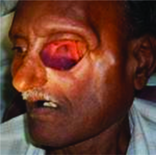
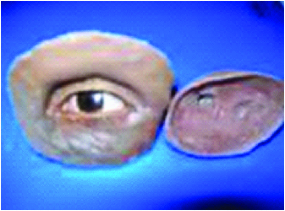
A 62-year-old male patient was referred to the institution with a chief complaint of facial disfigurement on left side, following surgery due to carcinoma one year back. The patient reported after one year for rehabilitation. The patient history revealed of surgical exenteration of the left eye and left side hemi maxillectomy was done to treat Rhino cerebral mucormycosis had been carried out one year before. On examination the socket was thoroughly healed and partial loss of zygomatic bone with a dip in surrounding structure [Table/Fig-1]. Extra orally, tissue was healthy and sufficient anatomical soft tissue undercuts for retention of the prosthesis present.
Intraoral examination shows a through and through connection between orbit and the oral cavity as total maxillectomy on the left side was done. To rehabilitate, a custom made extra-oral orbital prosthesis and a definitive intra-oral obturator was planned.
Procedure
A definitive hollow bulb obturator replacing missing teeth was done in a conventional manner and delivered to the patient after finishing and polishing.
Recording the Orbital Impression
• After evaluation and inspection of the anophthalmic socket and defect region, the diameter of the iris and pupil on the intact side was measured using a pair of Boley Gauge callipers [1,2].
• The patient was draped for impression procedures and patient’s eyebrows and eyelashes were lubricated with petroleum jelly in order to facilitate removal of the impression material and minimize discomfort to the patient.
• Direct impression was made according to Mathew’s [1] classification. Irreversible hydrocolloid (Zelgan, Dentsply India Ltd.) along with reinforcement by dental plaster (Goldstone, Dental stone plaster class II, Asian chemicals, Rajkot, India) was applied. Subsequently, a cast was poured in type III dental stone (Goldstone, Dental stone plaster class III, Asian chemicals, Rajkot, India).
• This cast was duplicated with laboratory silicone and three facial moulage / working cast are prepared from it for the following three different techniques.
TECHNIQUE I {Conventional Orbital Silicone Prosthesis}
-In the first working cast, the floor of the orbit and all the sensitive and non required undercut were blocked out and sealed with dental plaster(Goldstone,Dental stone plaster class II, Asian chemicals,Rajkot,India)
• One base plate thickness of modeling wax(The Hindustan Dental Products,Hyderabad,India) was adapted over the cast.
• A suitable prefabricated ocular stock shell selected according to the colour of the right eye and placed on the cast. Orientation was done with pupils of the unaffected side taken as reference. The patient was made to sit in an erect position and asked to focus his vision on a point directly ahead.
• Another base plate wax (The Hindustan Dental Products, Hyderabad, India) is adapted over the eye shell and necessary opening of the eye given. The upper and lower eyelids & associated structures were then sculptured. The prepared wax pattern was tried on the patient’s face to check the orientation of pupil, colour, size and volume of sclera visible as compared to the contra lateral eye [Table/Fig-2].
• Overall scrapping of the stone cast along the outer margin done for approximately 0.5 mm. Once the trial was found satisfactory, flasking, dewaxing and packing were done as per Prosthodontic protocols and processed in RTV (room temperature vulcanizing) {Cosmesil HC, Principally medical, Newport, UK} silicone material. Silicone-specific colours were used to match the colour of the patient’s skin.
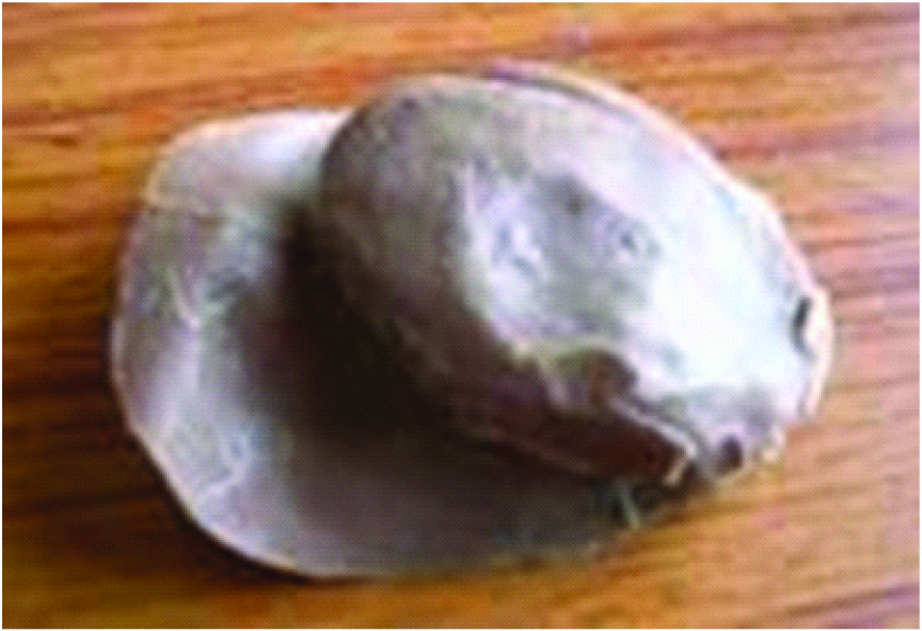
• Eye prosthesis was retrieved, characterized and delivered to the patient. Eyelashes are attached on upper and lower eyelids with cyanoacrylate adhesives {LOCTITE R}. Prosthesis was fitted to the patient and retained by undercuts [Table/Fig-3,4].
TECHNIQUE 2: {Hollow bulb Orbital Silicone Prosthesis}
• A suitable stock ocular prosthesis was positioned with base plate wax (The Hindustan Dental Products, Hyderabad, India) in the defect of the second working cast.
• All the undesirable undercuts were blocked. Here the wax up of the orbital contents are done separately as two parts i.e. the orbital and the periorbital part which were latter fused to form a hollow orbital prosthesis.
• Hence the periorbital tissue was sculptured to mimic the right eye, taking care that the line of junction was feathered. After sculpturing the prosthesis pattern, the final surface contour and skin texture were established by carving in lines and wrinkles found around the normal eye. Pressing a wet piece of gauze square into softened wax would produce a texture similar to normal skin.
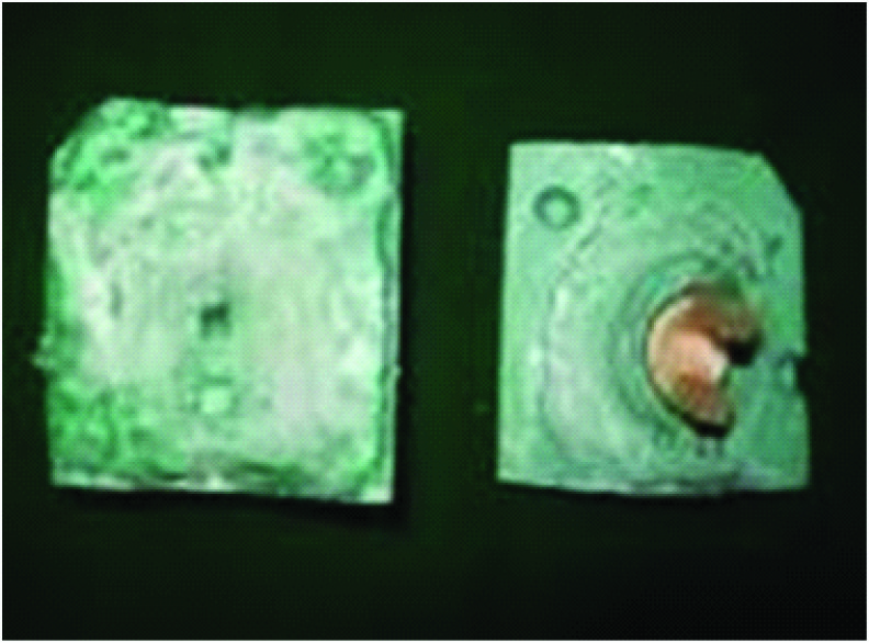
• Two orientation stents of autopolymerising resin (DPI cold cure, Dental Product of India, Mumbai, India) were prepared with the dimension of 0.5cmx0.5cmx4cm & were fixed to the posterior part of the stock ocular prosthesis. These were placed to help in orientation of orbital and periorbital prosthesis after processing [Table/Fig-5].
• Two separate moulds were then fabricated, one with the orbital part and another with the periorbital part and dewaxing done.
• Shade matching was done in natural daylight by mixing different intrinsic colours according to the different periorbital area. Orbital part was packed with the base colour silicone and both the moulds were left overnight for bench curing. After the polymerization was complete, the residual flush were trimmed back with a scalpel and finished with an abrasive stone. Then the hair of the patient was weaved to make the eyelashes and extrinsic colouring done.
• While placing the prosthesis the orbital silicon prosthesis was placed first followed by the periorbital prosthesis which were oriented by the orientation stents [Table/Fig-6]. Both parts were snuggly fitted into each other like a matrix and patrix part of a semi precision attachment and adjusted to attain a path of insertion and removal without interference. The patient was instructed in the use and care of the prosthesis
TECHNIQUE 3: {Magnetic Attached Orbital Silicone prosthesis}
• A thin layer of base plate modeling wax was adapted to the orbital defect of the third working cast. Over that, one layer thickness of self cure acrylic resin (DPI cold cure, Dental Product of India, Mumbai,
• The self-cure resin orbital prosthesis was invested separately in a dental flask and replaced by heat-cure resin (high impact heat cure acrylic, Travelon). On the tissue surface of the heat-cure resin orbital prosthesis, tissue conditioner (GC Tissue conditioners, GC Corporation, Europe) applied and placed in the patient orbit.
• Along with the acrylic orbital prosthesis in the patient orbit, a facial impression was made of irreversible hydrocolloid (Zelgan, Dentsply India Ltd.) and poured with die stone (Silky Rock, whipmix U.S.A).
• Stock eye mimicking the adjacent eye was selected and aligned in the new cast. Wax sculpting done along the periorbital areas and the wax pattern sealed, flasked and dewaxed. Colour matching done and the silicon material packed and cured as described earlier.
• The retrieved prosthesis was checked for fit and magnets {MAGFIT DX400}are placed on the stock acrylic eye and the acrylic side of the hollow orbital obturator and sealed by cynoacrylate and self-cure resin(DPI-RR Cold Cure, The Bombay Burmah trading corporation Ltd, Mumbai, India).
• As said in the above technique, initially the tissue conditioned acrylic orbital prosthesis with magnet was placed & then the periorbital silicone prosthesis with the counter-magnet positioned [Table/Fig-7,8].
Discussion
Prosthetic rehabilitation is the treatment of choice for patients with large facial defect of maxillary-orbital complex following surgical resection of the tumour. The tolerance and retention of the prosthesis is improved if the defect is lined by split thickness graft. However this is not recommended if there is a likelihood of recurrence. In the treatment of a patient requiring a custom ocular prosthesis many successful techniques are available to the practitioner. Although implant-retained ocular prosthesis [3] plays an important role in the success of treatment, conventionally retained orbital prostheses are practical, trouble-free, cost-effective, and successful. The retention of the orbital prosthesis can be achieved by using adhesive, attachment to eye glasses or engagement to hard and soft tissue undercut [4].
In all of these prosthesis, the anatomic undercut were utilized for retention. Compared to other two techniques fabrication of the conventional silicon retained by adhesive was quite simple. But studies have proven that the patients skin are allergic even to these medically grade adhesives. Hence, scrapping of the cast along the outer circumference of the orbital rim gives a tight fit to the silicone prosthesis on the patient face, there by adding to its retention. Advantages of using this particular method include adequate retention, economical, comfortable, and noninvasive unlike implants. Because of this non rigid attachment, slight movement produced in the eye prosthesis with the movement of obturator gave a slight life like appearance to the orbital prosthesis. The silicone eye prosthesis provided all the advantages of being light weight, better esthetics than acrylic prosthesis and a life like appearance. But, use of adhesive was not required as the prosthesis gains the retention through anatomic undercut. So the chance of allergic reaction caused by adhesives was overcome in this case as more of tissue was in contact with the prosthesis.
Use of spectacle was optional for the patient but it was not used as the method of retention for the present patient as he was comfortable with the prosthesis.
Through undoubtedly implants are the best choice for retention of an orbital prosthesis, but for a patient undergoing radiotherapy and high financial cost involved were the limitations. And the bar and clip system used for retention in an implant have the disadvantage of being rigid and providing a hindrance for proper cleansing of that area. The alternative for all these are the use of magnets or buttons which are less technique sensitive, improves hygiene and retention and was user friendly for the patient. Besides being an expensive option, there may be loss of magnetism with time or corrosion of magnets may occur.
Older reports have described the fabrication of acrylic resin orbital prosthesis but are outdated by the use of medical grade silicone elastomer. Significant advances in the field of material science have led to the production of new silicones with improved characteristics & colouration [5,6]. Hence, the appearance & mechanical strength are not affected. Therefore, selection of a reasonable maxillofacial prosthetic material and economically feasible retentive aid should be the goal of rehabilitating such patients. Since silicone has better marginal adaptation and life-like appearance, it has been used for the fabrication of orbital prostheses [7,8]. A limitation of silicone orbital prostheses is its lack of chemical/mechanical bonding with the eyeglass frame, making it difficult to retain the prosthesis[9]. Use of spectacle frame is an easy and economical treatment option but the method is not user friendly as the frame becomes heavy and patient has to wear the spectacle unconditionally. Modern prosthetic replacements are secured with adhesives that are readily available, easily applied, and provide satisfactory retention, but for a limited period of time. However, continual use of adhesives may cause allergic response or irritation [10].
Orbital prosthesis is subjected to greater scrutiny due to the prominent position on the face. They demand a high degree of accuracy in colour & surface characterization to blend with adjacent skin. Orbital prosthesis presents an attractive and viable alternative when esthetic and functional demands are beyond the capacity of local reconstructive efforts [11]. Hence, the minimum thickness considered for the prosthesis is 1 cm. It is difficult to achieve acceptable esthetics & prosthesis durability with less than 1 cm in thickness. A thin prosthesis is likely to tear during application, removal & cleaning. However, careful margin planning & execution has done to produce a visually well-integrated prosthesis.
Intelligible speech is achievable with both the closed bulb orbital prosthesis. Speech was assured by asking the patient to repeatedly utter his name. The degree of articulation is improved when the acrylic resin bulb is used for the orbital content. As it is rigid, it makes a snuggy fit, tight and high contact with the tissues to produce the necessary vacuum and improve the resonance & voice quality. The patient’s ability to handle the mucous secretions was significantly improved with the Hollow-bulb silicone prosthesis {Technique 2}. This produces less irritation to the mucous tissues than the acrylic bulb. Advantages of using the silicone for three of these prosthesis include improved adaptation and increased mobility to the underlying tissues, decreased irritation and less mucous secretion, reduced weight of the prosthesis and enhanced aesthetics.
Frontal & side view of the defect – pre operative view
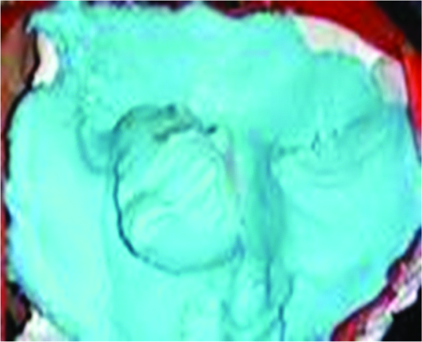
Facial view of the conventional silicone prosthesis

Tissue surface of the conventional silicone prosthesis
Mouldspace for the hollow bulb silicone prosthesis with two orientation stents
Facial surface of the hollow bulb silicone prosthesis
Facial view of the magnetic attached orbital silicone prosthesis


Conclusion
Fabrication of the maxillofacial prosthesis is a time consuming, labour intensive, artistic job. A well-retained, user-friendly, removable maxillofacial prosthesis is the key to successful prosthetic rehabilitation. Three different techniques of fabrication of orbital prosthesis are described elaborately along with the pros and cons of each procedure. The different ways of retention of the prosthesis were also described. The advantages of these methods are its better retention, restoration of function, cost-effectiveness, tissue tolerance, aesthetics, and comfort for use and wear.
Thus, the accumulation of positive effect as a result the use of the orbital prosthesis for a disfigured face has undoubtedly improved the quality of life of the patient.
[1]. MM Hanasono, JC Lee, JS Yang, JS Roman, R Gregory, B Esmaeli, An Algorithmic Approach to Reconstructive Surgery and Prosthetic Rehabilitation after Orbital Exenteration.Plast Reconstr Surg. 2009 123:98-105. [Google Scholar]
[2]. Hafezeqoran Ali, Koodaryan Rodabeh, A Technique for Fabrication of an Orbital Prosthesis: A Case Report.J Dent Res Dent Clin Dent Prospect. 2010 4(2):69-73. [Google Scholar]
[3]. D Shome, SG Honavar, BSK Raizada, BSD Raizada, Implant and prosthesis movement after enucleation: a randomized controlled trial.Ophthalmology 2010 117:1638-44. [Google Scholar]
[4]. J Beumer, TA Curtis, DN Firtill, JJ Hefferren, Maxillofacial Rehabilitation: Prosthodontic & surgical considerations.CV Mosby:st Louis 1979 :364-71. [Google Scholar]
[5]. T Aiz, M Waters, R Jagger, Surface modification of an experimental silicone rubber maxillofacial material to improve wettability.J Prosthet Dent. 2003 31:213-16. [Google Scholar]
[6]. GL Polyzois, Colour stability of facial silicone prosthetic polymers after outdoor weathering.J Prosthet Dent. 1999 82:447-50. [Google Scholar]
[7]. VK Shenoy, K Shenoy, Prosthetic rehabilitation of a patient after partial rhinectomy: a clinical report.J Prosthet Dent. 2005 93:125-18. [Google Scholar]
[8]. CKA Avinash, R Nadiger, SS Guttal, K Lekha, Orbital Prosthesis: A Novel Treatment Approach.Int J Prosthodont Restor Dent. 2012 2(1):19-23. [Google Scholar]
[9]. NP Patil, RK Nadiger, Use of acrylic resin base as an aid in retaining silicone orbital prosthesis.J Indian Prosthodont Soc. 2008 8:112-15. [Google Scholar]
[10]. SM Parel, Diminishing dependence on adhesives for retention of facial prosthesis.J Prosthet Dent. 1980 43:552-60. [Google Scholar]
[11]. P Scolozzi, M Hamedani, Rehabilitation of orbital cavity after total orbital exenteration using oculofacial prostheses anchored by osseointegrated dental implants posed as a one step surgical procedure.Klin Monbl Augenheilkd. 2006 223:400-04. [Google Scholar]