Cancer is the third leading cause of death worldwide. Among that oral cancer is an important health issue. Oral Squamous cell carcinoma (OSCC) is one of the most widely prevalent cancer types in developing countries, including India, and is associated with tobacco and alcohol abuse [1].
Cancer arises by means of two prominent alterations namely genetic alterations and epigenetic alterations. The genetic alterations along with epigenetic events like promoter hypermethylation affect the tumour suppressor genes causing loss of function [2].
Hypermethylation of the tumour suppressor genes were found to be associated with several types of cancers [3]. In the present study, we have analysed promoter hypermethylation pattern of five genes such as p16, p15, hMLH, MGMT and E-cadherin in patients who presented with oral precancer and cancer to evaluate if aberrant methylation occurs earlier in carcinogenesis (premalignant lesions).The results were compared between oral precancerous lesion and oral cancer to find methylation markers in oral cancer susceptible patients.
We included five tumour-suppressor genes in our study. p16, it is a cyclin dependent kinase inhibitor, involved in the regulation of the cell cycle, control of which is lost in virtually all types of tumour [4]. p15 another CDK inhibitor involved in regulation of cell cycle, which is frequently altered in human tumours [5]. hMLH, it is the human homolog of bacterial MutL, involved in mismatch repair [6]. MGMT - DNA repair gene, this gene involves in repair of methylated guanosine residues formed due to alkylated carcinogens.E-cadherin, it involves in homotypic epithelial cell-cell adhesion. It’s regarded as an invasion-suppressor gene [7]. All of these genes are known to be inactivated by methylation in various cancers [8].
Materials and Methods
Study design: The present study was a case control study conducted in the Department of Oral Medicine and Radiology, Saveetha Dental College, chennai, India. The study was approved by institutional ethical committee of Saveetha Dental College and hospital. The study subjects were divided into 3 groups. Group I – Control Group of five individuals - They are normal healthy individuals came to hospital for dental extraction. Group II consists of 10 patients suffering from oral leukoplakia. Group III – Consists of 10 patients suffering from OSCC. Informed consent was obtained from the subjects who were included in the study.
Inclusion criteria
Patients who were willing to participate in the study,
Patients who were attending dental OPD and who were diagnosed as oral leukoplakia and oral cancer,
Patients who were free from systemic diseases,
Healthy subjects were included as control.
Exclusion criteria
Patients who were reluctant to participate in the study.
Collection of samples and DNA isolation
Biopsies of oral mucosa for control study were taken among the normal healthy individuals. Incisional biopsy was done in patients suffering from leukoplakia and OSCC. Part of the tissue sent for histological examination and part of tissue sent for hypermethylation study of certain tumour suppressor genes. The samples were transported on ice in phosphate buffered saline (PBS) to the laboratory, where it was freed of surrounding normal tissue, rinsed in PBS and immediately processed for DNA isolation.
Restriction digestion of genomic DNA
Genomic DNA (3 μg) was digested initially with 5U of Hap II restriction enzyme in 1X supplied buffer L for 2h at 37°C. The reaction was refreshed with 10 U of Hap II and incubated again for 2h. DNA was purified and the restriction enzyme inactivated by extraction with phenol chloroform immediately after digestion.
Methylation-specific polymerasechain reaction
DNA was amplified by methylation-specific PCR (MSP) [9] using two sets of primers, one specific for methylated and other for unmethylated sequence of each gene.
Agarose gel electrophoresis
The PCR product was mixed with appropriate volume of 6X loading dye (0.25% bromophenol blue, 0.25% xylene cyanol and 30% glycerol in water) and electrophoresed through 0.75-3.0% agarose gel and visualized under ultraviolet illumination after staining with ethidium bromide. The promoter hypermethylation status of the five genes p16, p15, MGMT, hMLH and E-cadherin were analysed.
Results
Hypermethylation status of five tumour suppressor genes were carried out for the control group, oral leukoplakia and OSCC patients. Positive and negative promoter hypermethylation were recorded for each tumour suppressor gene separately and results were recorded. Appearance of two bands using enzyme treated genomic DNA has template would indicate hypermethylation of the CpG sight in each tumour suppressor gene. A single band amplified by the nested primer would indicate unmethylated DNA.
It was recorded that no methylation was found among the 5 normal individuals for all the 5 genes. This shows that hypermethylation process do not occur for these genes in normal individuals [Table/Fig-1].
Promoter hypermethylation patterns of various genes in control group
| Genes | No. of Cases | Promoter hypermethylation positive | Promoter hypermethylation negative |
|---|
| No. of cases | Percentage | No. of cases | Percentage |
|---|
| p16 | 5 | Nil | 0 | 5 | 100 |
| p15 | 5 | Nil | 0 | 5 | 100 |
| hMLH | 5 | Nil | 0 | 5 | 100 |
| MGMT | 5 | Nil | 0 | 5 | 100 |
| E-Cadherin | 5 | Nil | 0 | 5 | 100 |
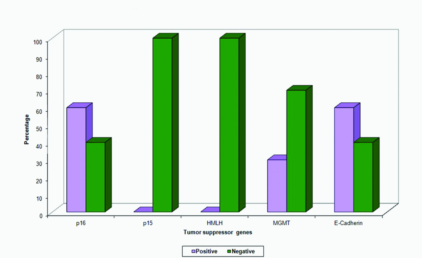
It was recorded that in leukoplakia patients, 60% of methylation in the case of p16, 30% of methylation in the case of MGMT and 60% of methylation in the case of E-cadherin gene [Table/Fig-2]. The other two genes namely p15 and hMLH clearly showed no methylation in all leukoplakia samples. In the case of leukoplakia 70% of samples showed methylation in any one of the genes, remaining 30% of the samples showed nil methylation in all the genes analysed. Further 30% of the samples showed methylation in two genes whereas 10% of the sample showed methylation in three genes (p16, MGMT and E-cadherin).
Promoter hypermethylation patterns of various genes in oral leukoplakia
| Genes | No. of Cases | Promoter hypermethylation positive | Promoter hypermethylation negative |
|---|
| No. of cases | Percentage | No. of cases | Percentage |
|---|
| p16 | 10 | 6 | 60 | 4 | 40 |
| p15 | 10 | Nil | 0 | 10 | 100 |
| hMLH | 10 | Nil | 0 | 10 | 100 |
| MGMT | 10 | 3 | 30 | 7 | 70 |
| E-Cadherin | 10 | 6 | 60 | 4 | 40 |
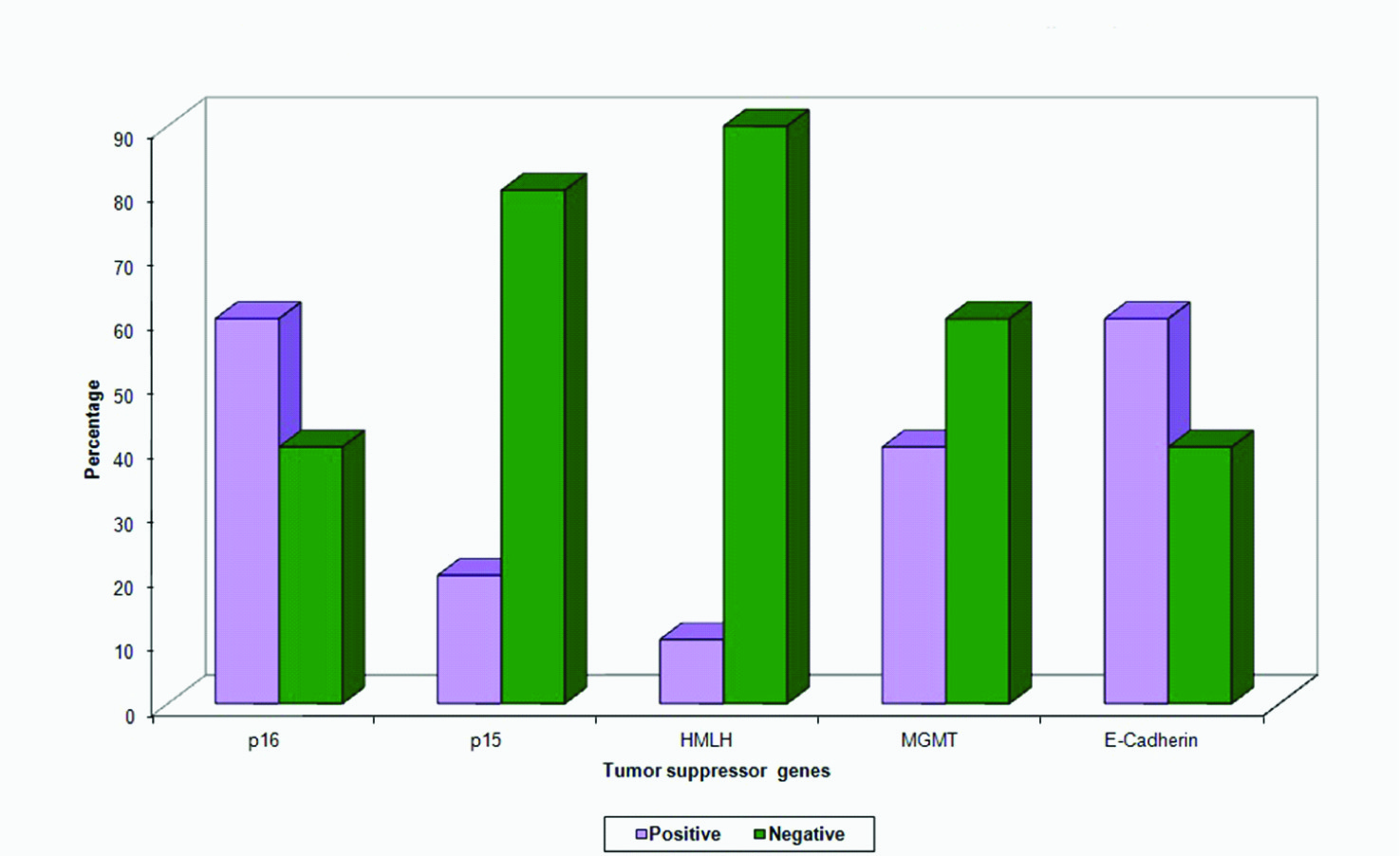
The results in OSCC showed, 60% of methylation in the case of p16, 40% of methylation in the case of MGMT, 60% of methylation in the case of E-cadherin, 20% in case of p15, 10% in case of hMLH gene [Table/Fig-3]. In the case of oral cancers, 80% of the samples showed methylation in any one of the genes analysed, remaining 20% of samples were not shown any methylation. Further 40% of the samples showed methylation in two genes where as 10% of the samples showed methylation in three genes (p16, MGMT and E-cadherin).
Promoter hypermethylation patterns of various genes in oral Squamous cell carcinoma
| Genes | No. of Cases | Promoter hypermethylation positive | Promoter hypermethylation negative |
|---|
| No. of cases | Percentage | No. of cases | Percentage |
|---|
| p16 | 10 | 6 | 60 | 4 | 40 |
| p15 | 10 | 2 | 20 | 8 | 80 |
| hMLH | 10 | 1 | 10 | 9 | 90 |
| MGMT | 10 | 4 | 40 | 6 | 60 |
| E-Cadherin | 10 | 6 | 60 | 4 | 40 |
Discussion
Recently, epigenetic disruptions, such as methylation of CpG island in promoter region of tumour suppressor genes and microRNA methylation have been recognized as key events in squamous cell carcinoma development [10]. Aberrant promoter hypermethylation can interfere with the binding of transcription factors to the DNA of tumour suppressor genes, resulting in transcriptional silencing of key genes involved in protection of cells from the process of unregulated growth [11]. These methylation changes serve as potential and sensitive molecular markers in defining risk states, achieving early diagnosis and tracking the prognosis of cancer [12].
The significance of environmental contaminants such as heavy metals have known to be related with multiple diseases, such as cancer, cardiovascular diseases, neurological disorders and autoimmune diseases [13]. Metal ions induce reactive oxygen species (ROS), and thus, lead to the generation of free radicals [14,15]. ROS accumulation can affect epigenetic factors [16].
Several groups investigated the methylation profile of various genes like p16, p15, MGMT, hMLH, Ecadherin and DAP-K in oral squamous cell carcinoma [17,18]. Though currently, multiple reports have identified the correlation between some tumour-related genes such as CDKN2A/p16, DAP-K and MGMT in endometrial, endobronchial and cervical premalignant lesions [19,20] yet, no such significant correlation have been studied in oral premalignant lesion. In this study, we investigated the methylation status of p16, p15, MGMT, hMLH and E-cadherin genes in leukoplakia and squamous cell carcinoma patients, to study the significance of epigenetic alterations in head and neck carcinogenesis.
As part of the study with respect to oral leukoplakia samples, the hypermethylation analysis showed 60% of methylation in E-cadherin and p16 gene [Table/Fig-4]. There are already reports of p16 gene methylation marker linked to leukoplakia [21] populations who are at high risk for susceptibility to oral squamous cell carcinoma. MGMT gene also showed 30% methylation frequency in leukoplakia. The fact that patients affected with leukoplakia have an increased susceptibility to oral squamous cell carcinoma has been supported by reports revealing the presence of MGMT gene methylation markers [22]. However, no methylation was found in the Cyclin dependent kinase inhibitor p15 and mismatch repair gene hMLH.
Shows promoter hypermethylation patterns of various genes in oral leukoplakia
Next with respect to OSCC samples, the methylation analysis showed 60% methylation in p16 and E-cadherin genes [Table/Fig-5]. Our studies indicate that the p16 and E-cadherin are the most preferentially targeted genes for methylation in oral squamous cell carcinoma of Indians. There are already reports of p16 and E-cadherin genes suitable for diagnosis of OSCC in individuals at high risk for this disease [23]. The same high percentage methylation in leukoplakia samples suggests that the methylation of these two genes [Table/Fig-6] may be an early event in the genesis or progression of oral squamous cell carcinoma. MGMT gene hypermethylation showed only a marginal difference between leukoplakia and oral cancer patients. This may also be taken as a premalignant early event suggesting its prognostic significance [24].
Shows promoter hypermethylation patterns of various genes in oral squamous cell carcinoma
Shows promoter hypermethylation patterns of various genes in oral leukoplakia and oral squamous cell carcinoma
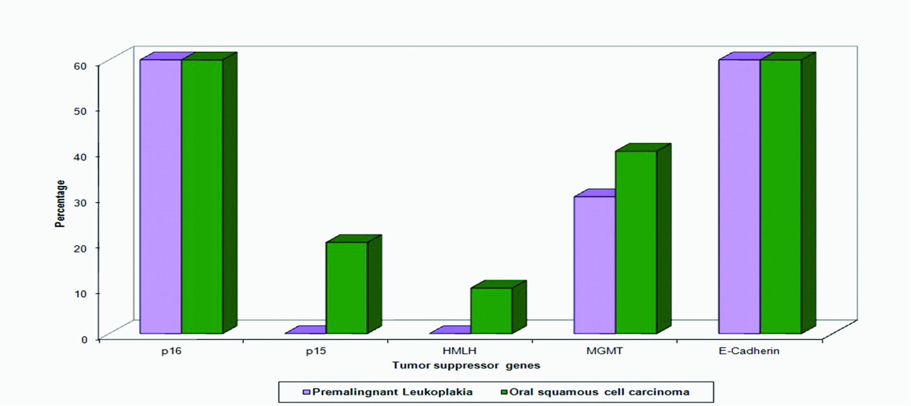
Contrary to leukoplakia samples p15 and hMLH genes in oral cancers showed 20% and 10% methylation respectively. This suggests that it may be a delayed event in the Oral squamous cell carcinoma progression. Low levels of methylation inactivation of hMLH1 indicated in our study are consistent with the observation of very low incidence of microsatellite instability in OSCC [25]. Hence, our study suggests the possible usage of Hypermethylation of two genes E-cadherin along with p16 as a diagnostic marker for OSCC susceptibility patients.
Conclusion
In conclusion, the observations with high percentage (60%) of hypermethylation in p16 and E-cadherin gene in leukoplakia can be taken as a diagnostic tool for oral squamous cell carcinoma susceptible patients.This shows the importance of methylation as a crucial event in the progression of oral squamous cell carcinoma, which offers a possible therapeutic target. Thus Methylation changes constitute potentially sensitive molecular markers to define risk states, achieve early diagnosis and track the prognosis of the cancer.
Hypermethylation profiling is a relatively expensive procedure which though currently is seldom practiced yet has excellent future prospective with technical sensitivity requiring a lot of standardization.