Oral squamous cell carcinoma (OSCC) is the eighth most common malignant epithelial neoplasm. Tobacco habit and excessive alcohol consumption have been estimated to account for about 90% of oral cancers [1]. OSCC is recognised as a disease ensuing from genetic damage, leading to unrestrained cell proliferation of damaged cells [2]. There are multiple steps that are associated with tumour progression, of them one is neoangiogenesis, which is one of the essential factor for tumour growth and metastasis [3]. An important predictor of tumour behavior is intratumour micro vessel density (MVD). The intensity of angiogenesis has been associated with poor prognosis in several cancers. Angiogenesis is determined by immunohistochemical assessment and with several endothelial cell markers, in which Endoglin CD105 is a potent pleiotropic angiogenic factor expressed on activated endothelial cells during angiogenesis and considered to be a specific marker for the detection of tumour angiogenesis [4]. Endoglin, CD105 is Type I, 180-KDa homodimeric transmembrane glycoprotein, a receptor for two types of transforming growth factors (TGF-1 and TGF-3) [4]. It has been established that endothelial cells of tumour-associated neovasculature proliferate 20 to 2,000 times more rapidly than endothelial cells of normal tissue [5].
Therefore, the present study attempts to assess the expression of Endoglin CD-105 and MVD in clinically normal mucosa of non-tobacco and tobacco habituated patients and also histopathologically confirmed cases of OSCC, with other important clinico-pathological characteristics.
Materials and Methods
The present study is a comparative study and sampling was done by convenient sampling method. The patients for the present study were drawn from the outpatient department of H.K.E.S’s S. Nijalingappa Institute of Dental Sciences and Research, Gulbarga, Karnataka, India. The patients were selected with the following parameters. A formal written consent was taken from the volunteers who actively participated in the study and the ethical clearance was obtained from the institutional ethical committee.
Group I - Twelve patients who do not use tobacco in any form, and with clinically normal oral mucosa served as control group.
Group II - Twenty patients with tobacco chewing or smoking habits but with clinically normal oral mucosa. Oral biopsy’s were done to ascertain the status of epithelial cells. Dysplasia present if any was recorded as mild, moderate or severe [6].
Group III - Twenty histopathologically confirmed cases of OSCC.
The tissue biopsies taken were stained with haematoxylin and eosin and subsequently were subjected to immunohistochemical staining for evaluation with Endoglin CD 105.
Procedure
Formalin fixed paraffin embedded tissues were sectioned at 3-4 μm, using a semi automated microtome. Consecutive sections were placed on slides pre-coated with egg albumin for routine Haematoxylin & Eosin staining and on positively charged microscope slides (25*75*10 mm Fisher Brand, Super frost plus. Biogenex) for immunohistochemical staining for Endoglin CD 105.
Immunohistochemical staining-procedure:
Sections of 3-4 microns thick were taken on positively charged microscope slides, these slides were baked on hot plate at 60° for 2h. Then dewaxing was done using xylene for 5min (2 times). Then slides were rehydrated in two baths of 100% propanol, subsequently the slides were transferred to one bath of 95% and 80% propanol for 3min each. Then the slides were rinsed with distilled water for 5min.
Antigen Retrieval: The antigen retrieval solution was Tri sodium citrate buffer solution (1000 ml distilled water with 2.94 grams of Tri sodium citrate buffer) at PH 6 – 6.2.
For antigen retrieval, two liters capacity stainless steel pressure cooker was used. The antigen retrieval was done by boiling in pressure cooker, after that, the pressure cooker was left to cool for 20min prior to opening. Then all slides were transferred to distilled water for two dips, of 1min each. Then the slides were dipped into TBS (Tris Bufferd Saline) once for 10min.
Immunostaining
Blocking: The slides were covered with 3% hydrogen peroxide block (HK111) reagent. The sections were incubated for 10min at room temperature in humid chamber and rinsed well with buffer wash for 5min twice. The tissues were then covered with power block (HK083) reagent and incubated for 5-10min at room temperature in humid chamber. The tissues were then gently blotted.
Labeling: Tissues were covered with primary antibody (Anti CD 105, Rabbit polyclonal BioGenex, USA).Similarly rabbit negative control serum (HK408) was added to the negative control slide. The slides were incubated for 1h at room temperature in humid chamber and rinsed well with wash buffer for 5min twice. The recommended positive control tissue was thyroid carcinoma tissue. Tissues were covered with super enhancer (HK518) and incubated for 20 min at room temperature in humid chamber. The slides were then rinsed well with wash buffer for 5 minute twice. Sections were covered with polymer Horse Radish Peroxidase (HRP) (HK519) reagent and incubated for 30min, at room temperature in humid chamber. The slides were then rinsed well with wash buffer for 5min twice.
Tissues were covered with Subsrate Solution [One drop of Diaminobenzidine (DAB) chromogen] DAB- 3-3 (HK124) mixed with 1 ml of substrate buffer DAB (HK520) and incubated for 10min, at room temperature in humid chamber. The slides were rinsed well with wash buffer. The sections were counterstained with haematoxylin, dehydrated, cleared and mounted.
The evaluation of the stained slides was carried out using a binocular research microscope (Lawrence & Mayo) under 100X and 400X magnification by two oral pathologists to overcome observer bias.
The expression of CD - 105 was determined with respect to Localisation, Intensity and Area (percentage) of stained cells as specified by Sappayatosok K et al., [7], and Weidner N et al., [8,9]. The immunopositivity of CD - 105 was mainly confirmed by the presence of brown stained cells in the tissue sections.
The Intensity/ Mean Microvessel Density (MVD) of the stain was defined on the following scale:
0 = No stained cells in any microscopic field
1 = Less than 25% of endothelial cells stained positively
2 = Between 25 and 50% of endothelial cells stained positively
3 = Between 50 and 75% of endothelial cells stained positively
4 = Greater than 75% of endothelial cells stained positively.
For the purpose of statistical analysis, MVD of the tissue specimen for all three groups was calculated.
Statistical Analysis
Chi - square analysis was used to test the immunohistochemical expression of Endoglin CD 105 with different parameters. Statistical significance was set at p ≤ 0.05.
Results and Observations
Tobacco use:
Moderate - Chewing tobacco less than 10 packets daily for less than 10yrs or smoking less than twenty bidis/cigarettes for less than 10yrs.
Heavy - Chewing tobacco more than 10 packets daily for more than 10yrs or more than 20 bidis /cigarettes for more than 10yrs.
Dysplasia Grade: Mild, Moderate, Severe, No Dysplasia (ND)
Endoglin CD 105 expression in various Groups of patients and intensity of staining:
In Group I, (Controls) out of twelve patients, two were negative for CD 105 expression, two specimens showed mean microvessel density of 5 – 25%, four specimens showed mean microvessel density of 25 – 50%, three specimens showed mean microvessel density of 50 – 70%, and one specimen showed mean microvessel density upto 80% [Table/Fig-1].
Endoglin CD 105 expression in oral biopsies of normal oral mucosa of individuals without habit of tobacco chewing and smoking
| Sl No. | Age/sex | Mean MVD | Scale |
|---|
| 1. | 47/M | 70 | 3 |
| 2. | 38/M | 25 | 2 |
| 3. | 28/F | 0 | 0 |
| 4. | 34/M | 70 | 3 |
| 5. | 48/M | 60 | 3 |
| 6. | 45/M | 30 | 2 |
| 7. | 50/F | 50 | 2 |
| 8. | 28/M | 80 | 4 |
| 9. | 35/M | 25 | 1 |
| 10. | 28/M | 10 | 1 |
| 11. | 47/M | 0 | 0 |
| 12. | 42/M | 50 | 2 |
In Group II i.e., tobacco users with clinically normal mucosa patients, out of twenty patients, seven were negative for CD 105 expression, eight specimens showed mean microvessel density of 5 – 25%, three specimens showed mean microvessel density of 25 – 50%, one specimen showed mean microvessel density of 50 – 70%, and one specimen showed mean microvessel density above 80% [Table/Fig-2].
CD 105 expressions in oral biopsies of tobacco users with clinically normal oral mucosa
| Sl No. | Age/sex | Tobacco Habit [Smoking (S) Chewing (C)] | Dysplasia Grade | Mean MVD | Scale |
|---|
| 1. | 42/M | Heavy (S+C) | Moderate | 67.5 | 3 |
| 2. | 49/M | Heavy (S+C) | Moderate | 30 | 2 |
| 3. | 23/M | Moderate(C) | ND | 0 | 0 |
| 4. | 33/M | Heavy (c) | ND | 20 | 1 |
| 5. | 52/M | Heavy (S+C) | Mild | 0 | 0 |
| 6. | 28/M | Heavy (c) | Moderate | 35 | 2 |
| 7. | 27/M | Moderate(C) | ND | 0 | 0 |
| 8. | 27/M | Heavy (S+C) | Mild | 0 | 0 |
| 9. | 19/M | Moderate(C) | Moderate | 10 | 1 |
| 10. | 28/M | Moderate(S+C) | Moderate | 40 | 2 |
| 11. | 40/F | Heavy (C) | ND | 25 | 1 |
| 12. | 31/M | Moderate(C) | Mild | 10 | 1 |
| 13. | 32/M | Moderate(S+C) | ND | 0 | 0 |
| 14. | 47/M | Heavy (S+C) | Moderate | 25 | 1 |
| 15. | 35/M | Heavy (S+C) | Moderate | 100 | 4 |
| 16. | 18/M | Heavy (S+C) | Mild | 25 | 1 |
| 17. | 44/M | Heavy (C) | Mild | 0 | 0 |
| 18. | 32/M | Heavy (C) | ND | 20 | 1 |
| 19. | 24/M | Heavy (C) | Mild | 0 | 0 |
| 20. | 23/M | Moderate(C) | ND | 15 | 1 |
In Group III i.e., OSCC patients, out of twenty patients, six were negative for CD 105 Expression, one specimen showed mean microvessel density 5 – 25%, three specimens showed mean microvessel density of 25 – 50%, four specimens showed mean microvessel density of 50 – 70%, and six specimens showed mean microvessel density above 80% [Table/Fig-3].
Endoglin CD 105 expressions in oral mucosal biopsies of oral squamous cell carcinoma
| Sl No. | Age/Sex | HPR* | TNM** | H1*** | H2**** | Mean MVD | Scale |
|---|
| 1. | 54/M | WD | T2N0M0 | 25 | 40 | 32.5 | 2 |
| 2. | 50/F | WD | T2N0M0 | 40 | 45 | 42.5 | 2 |
| 3. | 50/M | WD | T3N0M0 | 10 | 0 | 10 | 1 |
| 4. | 50/F | WD | T2N0M0 | 0 | 0 | 0 | 0 |
| 5. | 39/M | WD | T4N0M0 | 80 | 90 | 85 | 4 |
| 6. | 60/F | WD | T2N0M0 | 50 | 45 | 47.5 | 2 |
| 7. | 32/F | WD | T2N0M0 | 0 | 0 | 0 | 0 |
| 8. | 45/F | PD | T2N0M0 | 85 | 75 | 80 | 4 |
| 9. | 50/M | WD | T2N0M0 | 100 | 100 | 100 | 4 |
| 10. | 60/M | WD | T1N0M0 | 75 | 45 | 60 | 3 |
| 11. | 30/F | MD | T2N0M0 | 50 | 50 | 50 | 3 |
| 12. | 37/M | WD | T3N0M0 | 70 | 65 | 67.5 | 3 |
| 13. | 45/M | WD | T2N1M0 | 0 | 0 | 0 | 0 |
| 14. | 58/F | MD | T2N1M0 | 0 | 0 | 0 | 0 |
| 15. | 62/M | MD | T1N0M0 | 100 | 70 | 85 | 4 |
| 16. | 45/M | WD | T1N0M0 | 68 | 75 | 71.5 | 3 |
| 17. | 58/M | WD | T2N0M0 | 75 | 75 | 75 | 4 |
| 18. | 70/F | MD | T4N0M0 | 90 | 75 | 82.5 | 4 |
| 19. | 38/M | WD | T2N0M0 | 0 | 0 | 0 | 0 |
| 20. | 65/M | WD | T4N0M0 | 0 | 0 | 0 | 0 |
* - Histopathological Report. ** - T describes the size of the original (primary) tumour and whether it has invaded nearby tissue, N describes nearby (regional) lymph nodes that are involved, M describes distant metastasis (spread of cancer from one part of the body to another). *** - Hot Spot 1. **** - Hot Spot 2
The Comparison of the expression of CD 105 positivity & CD 105 negativity in Group I with Group II, Yates corrected Chi square value i.e., t-value (<1.86 for p=0.05), and comparison between Group I and Group III, Yates corrected Chi square value i.e., t-value (< 0.43 for p=0.05), shows no significant difference [Table/Fig-4].
Comparsion of MVD between various groups
| Between | t-value |
|---|
| Group I and Group II | <1.86 |
| Group I and Group III | <0.43 |
| Group II and Group III | >1.86 |
The Comparison of the expression of CD 105 positivity & CD 105 negativity in Group II with Group III, Yates corrected Chi-square value i.e., t-value (>1.86 for p=0.05) shows significant difference between group II and group III [Table/Fig-4].
Discussion
Angiogenic phenotype serves the development of malignant tumour at multiple stages. In addition to forming microvascular networks via angiogenesis, endothelial cells may also affect tumour growth by releasing plethora of cytokines and proteases, which act to alter the proliferative and invasive behavior of adjacent tumour cells. The proposition that CD105 may be associated with tumour angiogenesis developed from observations that CD105 was strongly up-regulated in the endothelium of various tumour tissues compared with that in normal tissue [10,11,12].
Several reports had shown that CD105 expression was related to metastasis, whereas there were only few studies investigating CD105 expression in head and neck regions [5].
Several neoplasm have been investigated for Endoglin CD 105 expression including colorectal cancer [13]; Breast Carcinomas [14]; clear cell renal cell carcinoma [15]; endometrial carcinoma [16]; esophageal adenocarcinoma [17]; gastric carcinoma [18]; laryngeal squamous cell carcinoma [19]; advanced Epithelial Ovarian Cancer [20]; Pancreatic carcinoma [21].
Endoglin CD 105 expression was studied by many workers in various oral lesions like, oral submucous fibrosis [22], angiogenic squamous dysplasia-like phenomenon in oral epithelial precursor lesions [23].
Endoglin CD 105 expression in OSCC was also studied by these researchers and are considered to be significant [24,25,26,27].
In, the present study expression of Endoglin (CD-105) and Microvessel Density was studied by immunohistochemical staining method using Anti CD 105 in normal oral mucosal tissue, in oral dysplastic epithelium and OSCC.
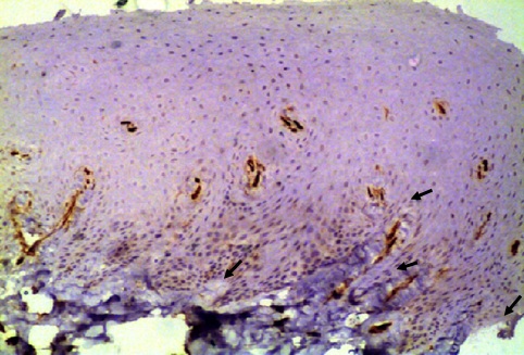
In our study, Group I consisted of 12 cases of normal healthy oral mucosa, where the mean MVD ranged from 0-4 [Table/Fig-5], which is consistent with previous studies [24], who reported CD 105 expression in normal oral mucosa and microvessel density which ranged from 1-3. Similar studies were done on normal mucosa [23,28], and found mean MVD scores significantly higher in dysplastic epithelium than in normal oral mucosal epithelium. The results of the present study found 66.67% of normal healthy oral mucosal cases positive for CD 105 and the microvessel density ranged between 0 – 49% [Table/Fig-2].
Low endoglin expression in normal healthy oral mucosa (X400)
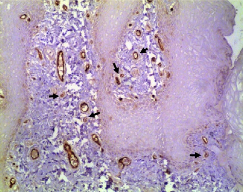
Expression of Endoglin CD 105 in normal oral mucosa was completely absent [22]. In the present study CD105 expression was found to be positive in 65% of the 20 cases of tobacco users. Mild to Moderate degree of epithelial dysplasia was observed in 13 cases of tobacco users, of these only 9 cases showed positive for CD105 expression [Table/Fig-6]. The expression of CD 105 in a few of them shows the susceptibility of these patients for further changes. The expression of CD 105 in epithelial dysplasia, in premalignant lesions, though variable, but is expressed faintly in oral submucous fibrosis without dysplasia, it became remarkable in dysplastic conditions, with maximum presence in severe oral submucous fibrosis condition, which indicates that CD105 expression, a marker for tumour vasculature, was upregulated in dysplastic conditions [22]. Our result of 65% of very small sample is quite consistent.
Moderate expression of CD105 in dysplastic epithelial (X400)
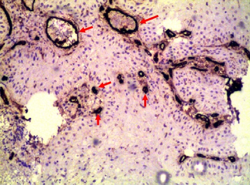
The expression of CD105 observed by different researchers in OSCC was significantly higher i.e., to the extent of 75% of cases [25,27–28]. Expression of CD 105, in OSCC patients habituated to tobacco, in this present study was observed in 13 of 20 cases (65%) [Table/Fig-7]. CD 105 expression in this study is consistent with the previous study results [27–28]. However, the higher expression of CD 105 can be attributed to the intensity, duration and form of tobacco used and the peculiar Indian habit of smokeless tobacco use. All the patients in this study were tobacco users of smoking and smokeless tobacco along with betel leaf and areca nut, of not less than 10yrs duration. Thus, the high incidence could be explained due to combined effect of tobacco smoke, chewing tobacco and arecanut which is proved to be genotoxic.
High expression of CD105 in oral squamous cell carcinoma (X400)
The statistical analysis in our study did not show any significant association of MVD with age, sex, clinical stage or differentiation grade. Since, four cases, in this study belonged to moderately differentiated squamous cell carcinoma, 15 belonged to well differentiated squamous cell carcinoma and only one belonged to poorly differentiated squamous cell carcinoma [Table/Fig-7]. Association between CD 105 expression and histological grades is not found statistically. This fact is owned mainly to small number of investigated cases.
Quantification of MVD with CD105 in our study reveals that the highest density was present in tumour specimens, than in normal healthy mucosa and dysplastic epithelium.
Though, there have been many studies of CD105 expression on oral biopsies of squamous cell carcinoma, the expression of CD105 in clinically normal oral mucosa of tobacco habituated individuals has not been studied.
Conclusion
The immunohistochemical technique undertaken in our study was reliable, as the expression of CD 105 was found in control and study group, is consistent with the findings of other workers in normal mucosa and OSCC.
The finding of 65% CD 105 positive MVD in tobacco users in clinically normal mucosa indicates, neo angeogenisis or angiogenic squamous dysplasia like phenomenon occurring as important intermediate pathological biomarker preceding oral cancer development, and may therefore be useful as a predictive marker of malignancy.