Prevelance of Haller’s Cells: A Panoramic Radiographic Study
Jitender Solanki1, Sarika Gupta2, Neelkant Patil3, Venkatesh V Kulkarni4, Meenakshi Singh5, Sanjeev Laller6
1Assistant Professor, Department of Public Health Dentistry, Vyas Dental College & Hospital, Jodhpur, Rajasthan, India.
2Post Graduate Student, Department of Oral Medicine and Radiology, Vyas Dental College & Hospital, Jodhpur, Rajasthan, India.
3Reader, Department of Oral Medicine and Radiology, Vyas Dental College & Hospital, Jodhpur, Rajasthan, India.
4Associate Professor, Department of Oral Pathology, Bharati Vidyapeeth Dental College & Hospital, Pune, Maharashtra, India.
5Assistant Professor, Department of Prosthodontics, Jodhpur Dental College & Hospital, Jodhpur, Rajasthan, India.
6Assistant Professor, Department of Oral medicine and Radiology, PDM Dental College & Hospital, Bahadurgarh, Haryana, India.
NAME, ADDRESS, E-MAIL ID OF THE CORRESPONDING AUTHOR:
Dr. Jitender Solanki, Assistant Professor, Department of Public Health Dentistry, Vyas Dental College & Hospital, Jodhpur, Rajasthan, India. Phone : 91-9571580558,
E-mail: solankijitender@gmail.com
Background: Infraorbital ethmoid cells, also known as Haller’s cells can be seen on panoramic radiographs. These help in identification of various pathologies and patient symptoms.
Objective: To determine the prevelance and characteristic of Haller’s cells on panoramic radiographs. Infraorbital ethmoid cells are extensions of ethmoid air cells into areas of orbit and maxillary sinus.
Materials and Methods: This study comprised of 1000 panoramic radiographs of healthy adults of the age 18-80 years. Each radiograph was interpreted for the presence of haller’s cells. The data collected were then tabulated and subjected to descriptive statistics and chi-square test.
Results: Haller’s cells were observed in 19.2% patients. Majority of cells were present unilaterally (176 cells) while only 15 were seen bilaterally. Maximum cells were oval in shape, unilocular and single in number.
Conclusion: Presence of haller’s cells helps in enumerating the differential diagnosis for orofacial pain and in avoiding surgical complications in endonasal procedures.
Endonasal procedures, Ethmoid air cells, Maxillary sinus, Orthopantomograph, Orofacial pain
Introduction
Infraorbital ethmoid cells were first described in 1765 by an anatomist Albert yon Haller, and have thereby been designated as Haller’s cells [1]. Haller cells are the extensions of anterior ethmoid sinus into the floor of the orbit and superior aspect of the maxillary sinus [2]. On imaging these cells may be seen below the ethmoid bulla, along the line of maxillary sinus and the most inferior portion of the lamina orbitalis [3]. Haller cells are often seen on panoramic radiograph and many authors have proven there prevelance in their studies. The importance of identifying haller cells on panaromic radiograph is to rule out the patient’s symptoms associated with this anatomical variation. These symptoms include orofacial pain and sinusitis, nasal obstruction, impaired nasal breathing, headache, chronic cough and mucoceles [2,4,5] Haller’s cells may appear from small to considerably large in size, single or multiple, round, oval or teardrop-shaped radiolucency [6]. These cells may or may not appear to be corticated and are medial to the infraorbital foramen on a panaromic radiograph [2]. Due to the clinical significance of these cells a study was carried out with the aim to determine the prevelance of infraorbital cells on panaromic radiograph and to identify the patterns, variations in shape and size of Haller’s cells with respect to the gender, age and side of the individual.
Materials and Methods
The present study was carried out in the Department of Oral Medicine and Radiology, Vyas Dental College and Hospital, Jodhpur city, India for a period of one year, from January 2013 to December 2013. The patients who were more than 18 y in age and required panaromic radiograph for dental treatment were randomly included in the study. Patients with history of trauma, fractures or surgery of the oral and maxillofacial region were excluded from the study. Patients with any systemic diseases affecting growth of the maxillofacial complex and with clinical or radiographic evidence of developmental anomalies or pathologies of the maxillofacial region were also excluded. The institutional ethical clearance was obtained prior to the conduct of the study. After carrying out complete clinical examination, a digital panoramic radiograph was taken for each patient, requiring the same. Kodak 8000C extraoral imaging system was used which was operated at 8-12 mA and 70-80 kVP, depending on the subject’s jaw size. The digital images obtained were then saved on the computer and interpreted for the presence of Haller’s cells. According to the selection criteria, a total of 1000 panoramic radiographs of the patients in the age range of 18–80 y were divided into six age groups and studied. The presence of Haller’s cells was confirmed by Ahmad et al’s criteria [2].
1) Well-defined round, oval, or tear-drop shaped radiolucency, single or multiple, unilocular or multilocular, with a smooth border, which may or may not appear corticated.
2) Located medial to infraorbital foramen.
3) All or most of the border of the entity in the panoramic section is visible.
4) The inferior border of the orbit lacks cortication or remains indistinguishable in areas superimposed by this entity.
The cells were recognized only when they fulfilled these criteria. The various observations related to the Haller’s cells were entered in the study proformas which consisted of data pertaining to date, patient name, age, gender, presence or absence of haller’s cell, side, shape, number of cells and number of loculae (unilocular or multilocular). Two examiners examined the radiographs. Training of the examiners for radiographic interpretation was done by a senior staff in the department. Each examiner viewed the radiograph twice for the presence of cells in a time interval of one week. The presence of haller’s cell was confirmed only when both intra and inter examiner results matched. In case of result which did not match, a mutual decision was obtained by both the examiners under the guidance of a senior staff. Inter examiner reliability test was done by using kappa statistics and score was found to be 0.80 (good). The data collected were then tabulated and subjected to descriptive statistics and chi square test using SPSS version 18.
Results
The study was done to assess the prevalence of haller’s cels in 1000 Orthopantamograph (OPG). In the total study group of 1000 patients 593 were males and 407 females. The study subjects were divided into 6 different age groups. Various variables associated with haller cells such as presence, shape, loculae, number and side were studied. After interpretation of 1000 OPG, Haller’s cells were observed in 192 radiographs, out of which 104 (54.2%) were female and 88 (45.8%) were male subjects [Table/Fig-1]. Maximum numbers of cells were seen in the age range of 18-28 years (33.3%)[Table/Fig-2]. Also, it was seen that 47.4% of cells were oval in shape with very few cases showing more than one shape of cells (6.8%) [Table/Fig-3,4,5,6,7,Table/Fig-8].
Chi-square test was used to compare the correlation between the various variables and a significant correlation p-value (0.034) was observed between the shape of the haller cells and the number i.e. whether it is single or multiple [Table/Fig-9,10], However, no significant correlation was observed between the gender and shape of the cells (p=0.820) [Table/Fig-11]. Also, no significant correlation was observed when the shape of the cells and loculae were correlated (p=0.296) [Table/Fig-12,13]. A significant correlation was observed when the shape of the cells and side of cells were correlated p-value (0.001) [Table/Fig-14,15]. No positive correlation was observed between any other two variables.
Discussion
Various studies have reported the appearance of haller’s cells in their literature, both in OPG and in CT imaging. The prevelance has varied hugely in various studies, ranging from 4.7-45.1% [2,7,8]. OPG studies have shown the prevelance to be as high as 38.2% [2] which is much higher than the prevelance seen in this study (19.2%). This difference could be due to the variation in population, larger sample size and observation bias. In the present study analyses have been performed to determine the variations of Haller’s cells with respect to gender, age, number, type and shape which were remotely been reported in the past. A study conducted by A Raina et al., [9] have reported the prevelance of hallers cells of 16% which lies in the same range as that of this study. Maximum number of cellcells was seen in the age range of 18-28 years. These findings are consistent with the study done by M Kantarci et al., [7]. No significant difference was reported among prevelance of haller’s cells between males and females which was consistent with the findings of N Basic et al., [10].
Unilateral occurrence of Haller’s cells was found to be statistically significant. These findings were in corelation with other studies who have seen side distribution [2,7,8]. In the present study the unilateral occurance of the cells was found out to be 74%. No statistically significant differences were noted in the occurrence of Haller’s cells on the right and left side, which were 69 and 73 cells respectively. Similar findings were seen in study conducted by A Raina et al.,[9].
Of the, 192 cells seen 91.7% were unilocular while only 8.3% were multilocular. Also, the majorities of the Haller’s cells were oval or round in shape with very few cases depicting a teardrop shape. Two new shapes have been seen in our study of pyramidal and heart shape cells, which has not been reported by any other studies previously.
Distribution of study subjects with Haller’s cells according to gender and side
| Unilateral | Bilateral | Both | Total |
| Male | 80 | 08 | 00 | 88 |
| Female | 96 | 07 | 01 | 104 |
| Total | 176 | 15 | 01 | 192 |
Distribution of study subjects with haller’s cells according to different age groups
| Age Groups | Percentage % |
| 18-28 years | 64 (33.3) |
| 29-38 years | 37 (19.3) |
| 39-48 years | 38 (19.8) |
| 49-58 years | 34 (17.7) |
| 59-58 years | 15 (7.8) |
| 69-80 years | 4 (2.1) |
Distribution of study subjects with Haller’s cells according to Shape of the Cells
| Shape of the Cells | Percentage % |
| Combination of more than 1 shape | 13 (6.8) |
| Heart | 2 (1.0) |
| Oval | 91 (47.4) |
| Pyramidal | 11 (5.7) |
| Round | 41 (21.4) |
| Teardrop | 34 (17.7) |
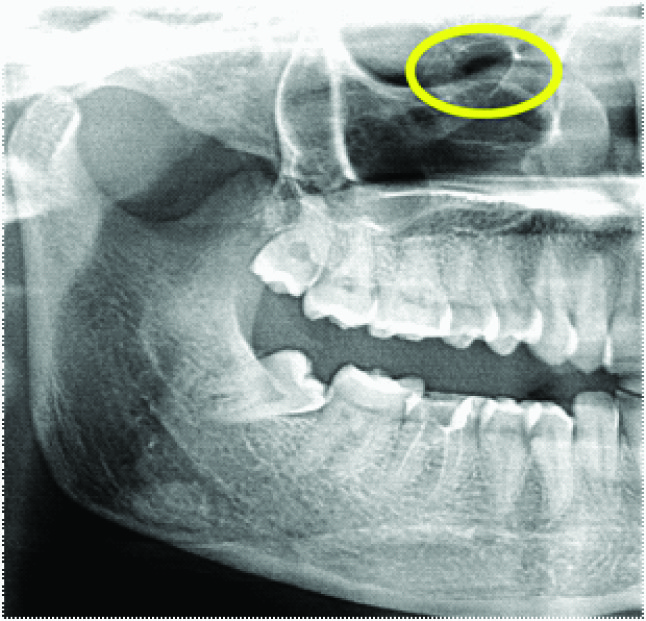
Round shape haller’s cell

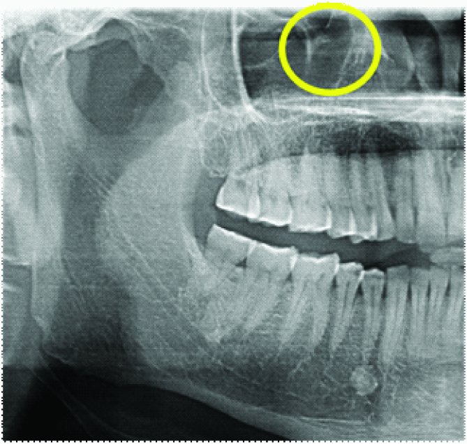
Heart shape haller’s cell
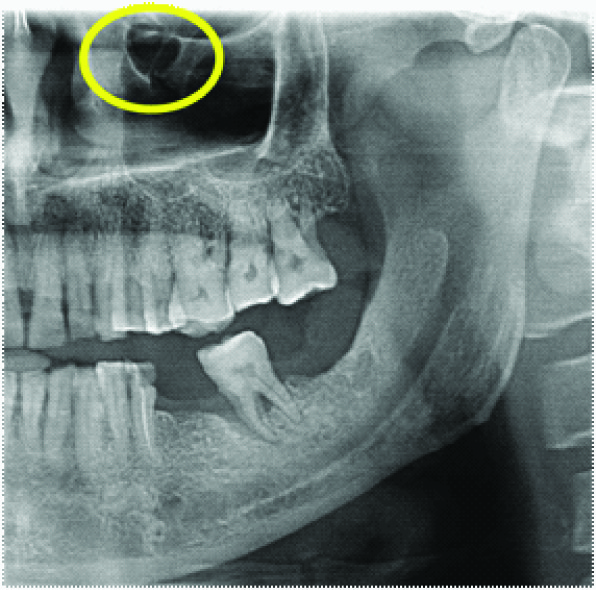
Tear drop shape haller’s cell

Pyramidal shape haller’s cell
Correlation between shape of cells and number (chi square test) (p value<0.05) significant
| | Number | Chi - Square | p- Value | Ns/S |
| Multiple | Single |
| Shape of cells | Combination of more than 1 shape | 4 | 9 | 12.031 | 0.034 | S |
| Heart | 0 | 2 |
| Oval | 22 | 69 |
| Pyramidal | 0 | 11 |
| Round | 7 | 34 |
| Tear drop | 1 | 33 |
oval shape cell on left side and tear drop shape haller’s cell on right side

Correlation between gender and shape of cells (chi-square test) (p-value<0.05) significant
| | Gender | Chi - Square | p- Value | Ns/S |
| Male | Female |
| Shape of cells | Combination of more than 1 shape | 6 | 7 | 2.205 | .820 | NS |
| Heart | 0 | 2 |
| Oval | 43 | 48 |
| Pyramidal | 6 | 6 |
| Round | 18 | 18 |
| Tear drop | 15 | 15 |
Correlation between shape of cells and locular (chi square test) (p-value<0.05) significant
| | Locular | Chi - Square | p- Value | Ns/S |
| Multilocular | Unilocular |
| Shape of cells | Combination of more than 1 shape | 0 | 13 | 6.103 | 0.296 | NS |
| Heart | 0 | 2 |
| Oval | 10 | 81 |
| Pyramidal | 1 | 10 |
| Round | 5 | 3 |
| Tear drop | 0 | 34 |
Multilocular haller’s cell

Correlation between shape of cells and side (chi-square test) (p value<0.05) significant
| | Side | Chi - Square | p- Value | Ns/S |
| Right | Both | Left |
| Shape of cells | Combination of more than 1 shape | 0 | 10 | 2 | 29.229 | 0.001 | S |
| Heart | 1 | 1 | 0 |
| Oval | 33 | 26 | 32 |
| Pyramidal | 2 | 1 | 8 |
| Round | 18 | 7 | 16 |
| Tear drop | 14 | 5 | 15 |
Oval shaped haller’s cells seen bilaterally

Conclusion
In the present study it has been noted that the presence of haller’s cells can be clearly appreciated on OPG. Our study highlights two new shapes of haller’s cells other than the various parameters recorded to distinguish these cells. These shapes have not been reported in the previous studies. Knowledge of different shapes can help dentists in easy identification of these cells. The ability to identify these cells on routine panoramic radiographs can help come to diagnosis of many conditions. Detection of these cells may also help overlook any untoward intraoperative complications prior to endonasal procedures. Use of advanced imaging modalities would further help in confirming the presence of cells and formulating a definative diagnosis related to patients symptoms.
[1]. A Von Hailer, W Cullen, First lines of physiology.Teratology18031stAmerican ed. Edinburgh Obrabran, Penniman:224 [Google Scholar]
[2]. M Ahmed, N khurana, J Jaberi, C Sampair, KR Kuba, Prevalence of infraorbital ethmoid (Haller’s) cells on panoramic radiographs.Oral Surgery Oral Medicine Oral Pathology Oral Radiology and Endodontology. 2006 101(1):658-61. [Google Scholar]
[3]. J Earwaker, Anatomicvariants in sinonasal CT.Radiographics 1993 13:381-415. [Google Scholar]
[4]. MM Tatli, I San, M Karaoglanoglu, Paranasal sinus computed tomographic findings of children with chronic cough.Int J Pediatr Otorhinolaryngol. 2001 60:213-17. [Google Scholar]
[5]. M Kantarci, RM Karasen, F Alper, O Onbas, A Okur, A Karaman, Remarkable anatomic variations in paranasal sinus region and their clinical importance.Eur J Radiol. 2004 50:296-302. [Google Scholar]
[6]. HH Wanamaker, Role of Haller’s cell in headache and sinus disease: A case report.Otolaryngology - Head and Neck Surgery. 1996 114(2):244-47. [Google Scholar]
[7]. M Kantarci, RM Karasen, F Alper, O Onbas, A Okur, A Karaman, Remarkable anatomic variations in paranasal sinus region and their clinical importance.Eur J Radiol. 2004 50:296-302. [Google Scholar]
[8]. HH Wanamaker, Role of Haller’s cell in headache and sinus disease: a case report. Otolaryngol Head Neck Surg. 1996 114:324-27. [Google Scholar]
[9]. A Raina, MV Guledgud, K Patil, Infraorbital ethmoid (Haller’s) cells: a panoramic radiographic study.Dentomaxillofac Radiol. 2012 41(4):305-08. [Google Scholar]
[10]. N Basic, V Basic, T Jukic, M Basic, M Jelic, J Hat, Computed tomographic imaging to determine the frequency of anatomical variations in pneumatization of the ethmoid bone.Eur Arch Otorhinolaryngol. 1999 256:69-71. [Google Scholar]