Giant Inguinoscrotal Hernia Repaired by Lichtensteins Technique Without Loss of Domain -A Case Report
Dinesh HN1, Jagadish Kumar CD2, Shreyas N3
1 Associate Professor, Department of Surgery, Mysore Medical College and Research Institute, Karnataka, India.
2 Postgraduate Trainee, Department of Surgery, Mysore Medical College and Research Institute, Karnataka, India.
3 Postgraduate Trainee, Department of Surgery, Mysore Medical College and Research Institute, Karnataka, India.
NAME, ADDRESS, E-MAIL ID OF THE CORRESPONDING AUTHOR: Dr. Jagadish Kumar CD, #1188,1st Floor, 3rd Cross, 4th Main, Aravindanagar, Mysore, Karnataka-570023, India. Phone : +917259759549, E-mail : drjagadishkumarcd@gmail.com
Giant inguinal hernia is a formidable surgical problem. It is defined as inguinal hernia extending up to mid thigh or below in standing position. Giant inguinal hernia is usually associated with compromised quality of life due to sexual discomfort and constant weight bearing. It is a challenge for the operating surgeon since it is rare. It may require multistage repair with recurrence being common. A 45-year-old male patient presented with Giant inguinal hernia and compromised quality of life due to pain and sexual discomfort. Lichtenstein’s polypropylene mesh repair was done after reducing the sac contents (omentum and transverse colon) with partial omentectomy. There was no loss of intra-abdominal domain. Postoperative period was uneventful. In literature many techniques are available to increase the intra-abdominal cavity (a) Creating progressive preoperative pneumoperitoneum (b) Creation of ventral wall defect (c) surgical debulking of hernia contents. Recurrence is prevented by reconstruction of the abdominal wall using Marlex mesh and a Tensor fasciae lata flap. Laparoscopic repair is associated with more recurrence. Lichtenstein’s technique is one of the preferred treatments.
Abdominal compartment syndrome, Formidable surgical problem, Giant, Liechtenstein, Polypropylene mesh, Tension free repair
Case Presentation
A 45-year-old male patient came with complaint of left Inguinoscrotal swelling since 25 yrs, insidious onset, gradually progressive and recently reached below level of mid thigh, associated with dragging type of pain. Patient’s quality of life has been compromised because of interference with sexual activity and dribbling of urine to the scrotum. Patient was not able to perform his daily routine activities due to constant weight bearing and pain in his scrotum. No history of Chronic Cough / previous surgery / constipation and vomiting.
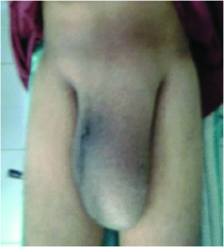
On examination Pulse 78/min, BP 130/80 mm Hg, an oval shaped swelling 35x15x10 cms in left Inguinoscrotal region, extending from left inguinal region up to left mid thigh, surface smooth, borders well made out, only partially reducible, doughy in consistency, no cough impulse, non tender, no local raise in temperature, not able to get above the swelling, scrotal skin was thickened and penis is buried in scrotum [Table/Fig-1].
Giant Left inguinal hernia with penis buried in scrotum
Chest X-ray normal, USG abdomen-Left indirect inguinal hernia with enterocele and omentocele as its contents with good vascularity.
Patient posted for elective hernia repair and bladder catheterized after anaesthesia before incision. Hernia approached with Left Inguinal incision, indirect hernial sac noticed and sac was carefully dissected separating it from cord structures. The sac opened and contents found to be omentum and transverse colon with part of ascending and descending colon [Table/Fig-2a,b]. Adhesions were lysed to make colon free from omentum and hernial sac without any bowel resection. Partial omentectomy was done. The colon and remaining omentum were successfully reduced back to abdominal cavity through wide deep ring without any change in intraoperative airway pressures. Direct hernia component also noted through Hasselbach’s triangle. Direct hernial contents were reduced. A 6cm x 11cm polypropylene monofilament mesh was placed over the posterior wall of inguinal canal and fixed to it (LICHENSTEINS TENSION FREE ONLAY MESH REPAIR). Drain was placed in the scrotum but not in inguinal site and wound closed in layers. Post operatively after 24 hours drain was 30 ml (bloody) without cardio respiratory distress. Post operative day 2 and 3 drain decreased to 15 ml and 5ml (serosangous) respectively. Drain was removed and patient discharged on postoperative day 4. Patient was followed up weekly for the first month and every15 days from 2nd month. Patient developed mild scrotal hematoma in his first follow up visit and subsequently after 3 months scrotal oedema reduced significantly.
Giant inguinal hernia sac containing omentum and transverse colon

Discussion
Giant inguinal hernia is a chronic condition and rare in recent practice. It is defined as inguinal hernia extending till mid thigh or below in the standing position [1]. Penis is usually buried inside the scrotum causing dribbling of urine over an already edematous scrotal skin (lymphatic and venous oedema) leading to excoriation, ulceration and secondary infection. It is a challenge for the operating surgeon. Reducing large amount of abdominal viscera of giant hernial content to abdomen cavity may cause abdominal compartment syndrome which push the diaphragm towards thoracic cavity resulting in respiratory difficulty and compromised venous return leading to circulatory collapse [2]. Replacement of intestine into the abdominal cavity can also cause intestinal obstruction and wound dehiscence postoperatively.
There are few surgical techniques described in the literature for repairing giant Inguinoscrotal hernias. Most common one is creation of progressive preoperative pneumoperitoneum for two weeks by insufflating air into abdominal cavity [3]. Another technique describes de-bulking the contents of the hernia sac by performing resection of the bowel in the hernia sac, and reconstruction of the abdominal wall using Marlex mesh and a tensor fasciae lata flap to prevent recurrence [4]. In 2001, El-Dessouki described a new way to achieve this by creating a midline abdominal wall defect to increase the intra-abdominal capacity to accommodate the hernia contents. The hernia sac is then pulled up to the abdomen and fashioned as a rotation flap to augment and close the peritoneum over the replaced contents [5]. Merret et al., described another similar technique which includes reduction of sac, hernial orifice repair with merlex mesh and creation of a midline abdominal wall defect followed by marlex Mesh with rotation flap of inguinoscrotal skin covering the defect [6]. Lastly, a giant polypropylene mesh is inserted in the preperitoneal space to cover the midline defect created and to buttress both inguinal regions [7]. Laparoscopic repair of giant hernia can also be done but recurrence rate is high. Current guidelines suggest Lichtenstein repair as preferred surgical technique for large irreducible inguinal hernia [8].
Conclusion
Giant inguinal hernia is a formidable and challenging surgical disease associated with compromised quality of life. We treated it by simple Lichtenstein’s mesh repair without loss of intra-abdominal domain. Postoperative period was uneventful.
[1]. Hodgkinson DJ, Mcllrath DC, Scrotal reconstruction for giant inguinal herniasSurg clin North Am 1984 64:301-13. [Google Scholar]
[2]. Serpell JW, Polglase AL, Anstee EJ, Giant inguinal herniaANZ Journal of surgery 1988 58:831-34. [Google Scholar]
[3]. Piskinv T, Aydin C, Barut B, Dirican A, Kayaalp C, Preoperative progressive pneumoperitonium for giant inguinal herniaA Saudi Medicine 2010 30:317-20. [Google Scholar]
[4]. Mehendal FV, Taams KO, Kingsnorth AN, Repair of a giant inguinoscrotal herniaBr J Plast Surg 2000 53:525-29. [Google Scholar]
[5]. El-Dessouki NI, Preperitoneal mesh hernioplasty in giant inguinoscrotal hernia: a new technique with dual benefit in repair and abdominal roomingHernia 2001 5:177-81. [Google Scholar]
[6]. Merrett ND, Waterworth MW, Green MF, Repair of giant inguinoscrotal hernia using marlex mesh and scrotal skin flapsAust N Z J Surg 1994 64:380-83. [Google Scholar]
[7]. Bernhardt GA, Gruber K, Gruber G, TAPP repair in giant bilateral scrotal hernia-limits of a methodAZN J Surg 2010 80:947-48. [Google Scholar]
[8]. Simsons MP, Aufenacker T, Bay-Nielsen M, European Hernia Society guidelines on the treatment of inguinal hernia in adult patients 2009 13:343-403. [Google Scholar]