Aesthetic demands have been climbing ever higher in dentistry driven by an enhanced awareness of beauty. A concept of creating something tangibly better, “Dental aesthetics” is fueled by fascination, generating compliments and popularity. A successful aesthetic dental treatment helps regain the patient’s self-image, revive social skills and experience professional success. Modern aesthetic dentistry involves not only the restoration of lost teeth and their associated hard tissues, but increasingly the management and reconstruction of the encasing gingiva [1]. In the past, periodontal treatment has been aimed more at the preservation and restoration of periodontal health than at the aesthetic outcome of treatment. However, recent advances have enhanced the periodontist’s proficiency to address the aesthetic concerns [2]. Periodontal plastic surgery consists of a broad range of procedures aiming at correcting or eliminating anatomical, developmental and/or traumatic deformities of the gingiva or alveolar mucosa [3]. One among such problems is open interproximal spaces or the black triangles.
“Black triangles” or the interproximal spaces are one of the most troubling dilemmas in dentistry, can cause aesthetic concerns, phonetic difficulties, and food impaction. Several reasons contribute to the loss or absence of interdental papilla and establishment of ’Black Triangle’, including gingival inflammation, attachment loss, and interproximal bone resorption. The most common reason for black triangle in the adult population is plaque associated loss of periodontal support as well abnormal tooth shape or traumatic oral hygiene [4]. While Kandaswamy et al., reported that black holes (dark triangles) are more likely to develop following labial movement of overlapping or palatally placed incisors and diastema closure [5]. An interproximal contact point and an adequate level of bone support are essential for maintenance of a healthy papilla that completely fills the interproximal space [6]. From a biological point of view, the presence or absence of the papilla primarily depends on the distance between the interdental contact point and the interproximal crest of bone. Tarnow et al., stated that the distance of 5 mm is critical for this purpose [7]. Various periodontal plastic surgical procedures like soft tissue sculpturing, use of connective tissue / free gingival grafts, use of enhanced conservative new mucoperiosteal flap designs, and methods to improve soft tissue topography with/without GTR/GBR, all are invented to enhance regeneration of lost interdental hard and soft tissue. The most difficult and elusive goals for periodontists is reconstruction, regeneration of lost interdental papilla and to achieve the Aaesthetics which is lost because of open interproximal spaces [8]. Several non-surgical and surgical procedures have been presented to treat the soft tissue deformities in the interproximal areas such as prosthetic covering, perio surgeries, orthodontic teeth alignment or combination of above [4]. Inoceincio et al., observed that combined periodontal surgery and orthodontic repositioning tooth helps in achieving periodontium with better aesthetic results and proper formation of interdental papilla [9]. The non-surgical approaches modify the interproximal space whereas the surgical approaches aim to recontour, preserve, regenerate and reconstruct the soft tissue between the teeth and implants [10]. Surgical techniques aiming at correcting the “black hole problem” have been used mainly with free epithelialized gingival grafts, repeated interproximal curettage, or displacement of the interproximal palatal tissue in the buccal direction [2,11], but limited success has been achieved with these procedures. The major limiting factor for the complete and predictable survival of the graft tissue is the lack of a minimal source of blood supply [12]. The healing principle on which the subepithelial connective tissue graft for root coverage and ridge augumentation are based have been applied to the reconstruction of the interdental papilla, thus increasing both the success rate and predictability [13].
So, in the present study an attempt has been made to clinically evaluate the surgical reconstruction of interdental papilla by using an advanced papillary flap with interposed subepithelial connective tissue graft.
Materials and Methods
The present study was carried out on patients selected amongst the outpatient Department of Periodontology & Oral Implantology, M.M. College of Dental Sciences and Research, Maharishi Markandeshwar University Mullana, Ambala, India and were explained the whole study protocol and were asked to submit a duly signed written informed consent. The study was carried out as per Helsinki declarations (1964) with the ethical clearance from the M.M. University.
Selection Criteria Inclusion criteria
Patients between the age group of 18-55 y, of either sex (male/female).
Patients with presence of “Black triangles” / papilla recession in the maxillary anterior teeth having contact points.
Patients having adequate zone of attached gingiva with minimal probing depth adjacent to the open embrasure.
Patients willing to follow recommended plaque control and follow up regimen.
Exclusion criteria
Unaesthetic open embrasures in the mandibular anterior region.
Patients who are unable to undergo minor surgical procedure.
Patients exhibiting allergy / systemic disease/ treatment that contraindicate surgical procedure.
Patients exhibiting trauma from occlusion.
Patients having gingival recession on the labial surface of the teeth adjacent to the open embrasure.
Patients with habit of tobacco chewing, smoking and alcohol consumption.
History of previous periodontal surgical treatment and untreated non vital teeth.
Study design
A total of 15 sites from ten patients having black triangles /papilla papilla recession in the maxillary anterior region were selected and subjected to presurgical evaluation. Bone assessment can be done clinically or radiogarphically. For standaridisation, clinically we have to split the papilla prior to surgery and at the follow up visits (surgical rentry) which will jeoparadise the results and will affect the technique to be used. Radiographically there was difficulty in standardization in measuring the distance from contact point to bone crest hence, bone assessment was not done. Distance between contact point and crest of alveolar bone was not taken in the present study as it was difficult to standardise the results. There are studies done for reconstruction of interdental papilla where the distance was not measured from contact point to bone crest as no bone augumentation technique was used and obtained fairly good result [14,15].
The following clinical parameters were recorded at baseline (preoperative) and postoperatively at 1st month, 3rd month and at 6th month interval.
Plaque Index Loe, [16].
Gingival Index Loe And Sillness, [16].
Papillary Bleeding Index Saxers and Muhlemann H.R. [17].
Papilla Presence Index Cardaropoli D, Re S and Corrente G [18].
Distance from Contact Point to Gingival Margin.
Width Of Keratinized Gingiva [19].
Surgical Procedure
After the assessment of pre-treatment records and clinical examination, patients who demonstrated satisfactory response to phase I therapy were considered and subjected to surgical procedure.
1.a – Pre-operative (presence of “black triangle” between maxillary central incisors), 1.b –crevicular incison followed by semilunar incison, 1.c – coronal displacement of lingivopapillary unit, 1.d - void created by displacement of gingivo papillary unit

The selected operative sites [Table/Fig-1a] were anesthetized with 0.2% Xylocaine with 2% adrenaline (1:200000). A 3-5mm semilunar incision was given with No. 11 blade 2 mm coronal to the mucogingival junction, just over the papillary region followed by intercrevicular incision [Table/Fig-1b] over the teeth neighbouring the defect extending from the buccal aspect to the palatal aspect keeping the existing papilla preserved. Through the semilunar incision the gingivopapillary unit was freed from the underlying bone using an orban knife extending toward the palate [Table/Fig-1c]. Taking care to avoid perforating the palatal tissue or damaging the interproximal papilla, the tissue was completely released from the root as well as bone, so that flap became mobile, which allow for the coronal displacement of the gingivopapillary unit. A buccal/palatal void (dead space) could be established between the soft tissue and the bone structure [Table/Fig-1d]. To maintain the whole gingivopapillary unit coronally, the dead space was filled with the connective tissue graft.
2.a – Trap door incison on donor site (palate), 2.b - partial thickness flap elevation, 2c – harvested subepithelial connective tissue graft, 2.d - interposed subepithelial connective tissue graft at the recipient site

The desired length and width of the subepithelial connective tissue graft was obtained using trap door technique by giving a horizontal incision in the area of molar and premolars followed by two vertical incisions at each end of the horizontal incision, the partial thickness flap was raised and separated from the underlying connective tissue with the help of surgical blade no.11 and tissue holding forcep [Table/Fig-2a,b]. The subepithelial connective tissue graft was harvested and preserved in the normal saline [Table/Fig-2c]. The donor site was covered by repositioning the partial thickness flap, secured in place by interrupted sutures using 3-0 black braided silk sutures to obtain primary closure.
Placement and Suturing of the Graft in the Recipient Site
The subepithelial connective tissue graft was trimmed to the desired size and shape and placed under the flaps to fill the dead space [Table/Fig-2d] and to maintain the gingivopapillary unit coronally and stabilized over the recipient site using 6-0 vicryl sutures [Table/Fig-3a]. The area was irrigated and covered with sterilized tin foil followed by periodontal dressing. Patients were prescribed with oral antibiotic, amoxicillin 500 mg thrice a day and combiflam (Iboprufen 400 mg and paracetamol 325 mg) thrice a day for five days. The patients were instructed to rinse with 0.2% chlorhexidine digluconate twice daily for two weeks and were discharged from the hospital with post surgical instructions. It was difficult to use and manipulate 6-0 vicryl suture but its usage causes minimal tissue trauma and enhance the final aesthetic outcome.
3a - Securing subepithelial connective tissue graft with 6-0 vicryl suture , 3.b- healing after 1 month, 3c – healing after 6 months
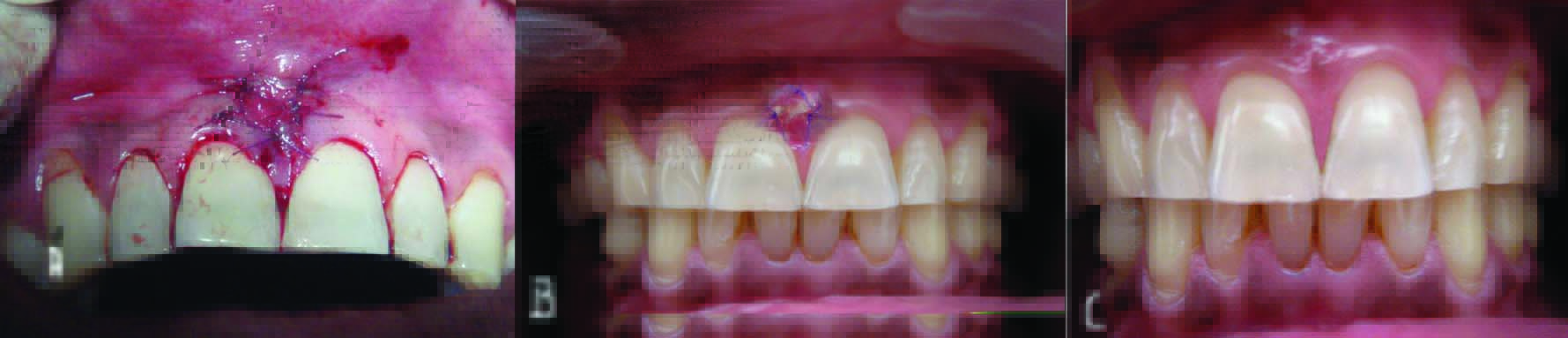
Post surgical follow up
All the patients were recalled after 24 h to assess any postoperative complication such as bleeding, pain, swelling and hematoma etc. After an interval of 10 d, patients were recalled for the removal of the periodontal dressing, sutures from the donor site and appraisal of the healing response. The area was irrigated and patients were kept on a postsurgical follow up after every 15 d. Clinical parameters were evaluated during follow up visits at 1 month, 3 months and at 6 months interval [Table/Fig-3b,c].
Results and Observations
In the present study, ten subjects with 15 sites having black triangle/papilla recession in the maxillary anterior region were selected, on the basis of inclusion and exclusion criteria. Clinical parameters like plaque index, gingival index, papillary bleeding index, papilla presence index, distance from contact point to gingival margin, width of keratinized gingiva were recorded at different time intervals from baseline to 1st, 3rd, and at 6th months. All the clinical parameter values obtained at different intervals were entered in the standard performa drawn for the study and were subjected to statistical analysis. The scores were statistically analysed by calculating their mean values and standard deviation. The mean difference between the intervals was calculated by using Paired Sample t-test to calculate the p-values.
Clinical Observations
Mean and mean difference of plaque index
| Assessment interval | Mean ± SD | Mean difference | t value | p-value |
|---|
| Baseline | 1.24±0.18 | Baseline to | | |
| 1 month | 1.02±0.18 | 0.22±0.10 | 8.37 | <0.001 |
| 3 month | 0.87±0.23 | 0.37±0.13 | 10.46 | <0.001 |
| 6 month | 0.80±0.27 | 0.44±0.19 | 9.04 | <0.001 |
The mean plaque index score at baseline was 1.24±0.18. At 1st, 3rd and 6th month intervals the scores were 1.02±0.18, 0.87±0.23, 0.80±0.27 with the mean difference of 0.22±0.10, 0.37±0.13, 0.44±0.19 and t value 8.37, 10.46, 9.04 respectively which were statistically highly significant.(p-value <0.001).
Mean and mean difference of gingival index
| Assessment interval | Mean ± SD | Mean difference | t value | p-value |
|---|
| Baseline | 1.20 ±0.18 | Baseline to | | |
| 1 month | 1.02±0.16 | 0.18±0.09 | 7.87 | <0.001 |
| 3 month | 0.91±0.17 | 0.29±0.14 | 8.30 | <0.001 |
| 6 month | 0.85±0.17 | 0.35±0.13 | 10.82 | <0.001 |
The mean gingival index score at baseline was 1.20 ±0.18. At 1st, 3rd, and at 6th month intervals the scores were 1.02±0.16, 0.91±0.17, 0.85±0.17 with the mean difference of 0.18±0.09,0.29±0.14, 0.35±0.13 and t-value 7.87, 8.30, 10.82 respectively which were statistically highly significant (p-value <0.001).
Mean and mean difference of papilla presence index
| Assessment interval | Mean ± SD | Mean difference | t value | p-value |
|---|
| Baseline | 2.80±0.94 | Baseline to | | |
| 1 month | 2.40 ±0.91 | 0.40±0.51 | 2.06 | 0.009 |
| 3 month | 2.40 ±0.91 | 0.40±0.51 | 2.06 | 0.009 |
| 6 month | 2.40 ±0.91 | 0.40±0.51 | 2.06 | 0.009 |
The mean papilla presence index score at baseline was 2.80±0.94. At 1st, 3rd and at 6th month the score was 2.40 ±0.91 with the mean difference of 0.40±0.51 and t value 2.06.
Mean and mean difference of papillary bleeding index
| Assessment interval | Mean ± SD | Mean difference | t value | p-value |
|---|
| Baseline | 2.20±0.67 | Baseline to | | |
| 3 month | 0.26±0.45 | 1.93±0.45 | 16.35 | <0.001 |
| 6 month | 0.33±0.61 | 1.86±1.06 | 6.82 | <0.001 |
The mean papillary bleeding index score at baseline was 2.20±0.67. At 3rd and at 6th months the scores were 0.26±0.45, and 0.33±0.61 with the mean difference of 1.93±0.45, and 1.86±1.06, and t value 16.35 and 6.82 respectively which were statistically highly significant. (p-value <0.001)
Distance from Contact Point to Gingival Margin [
Table/Fig-8]
Mean and mean difference of distance from contact point to gingival margin
| Assessment interval | Mean ± SD | Mean difference | t value | p-value |
|---|
| Baseline | 2.60±0.98 | Baseline to | | |
| 1 month | 1.87±1.13 | 0.80±0.94 | 3.29 | 0.005 |
| 3 month | 1.87±1.13 | 0.80±0.94 | 3.29 | 0.005 |
| 6 month | 1.87±1.13 | 0.80±0.94 | 3.29 | 0.005 |
The mean score of distance from contact point to gingival margin at baseline was 2.60±0.98. At 1st, 3rd and at 6th month the score was 1.87±1.13, with the mean difference of 0.80±0.94 and t-value 3.29 which was statistically significant (p-value 0.005).
Mean and mean difference of width of keratinized gingiva
| Assessment interval | Mean ± SD | Mean difference | t value | p-value |
|---|
| Baseline | 6.60±0.91 | Baseline to | | |
| 1 month | 7.46± 0.91 | 0.86±0.51 | -6.50 | <0.001 |
| 3 month | 7.46± 0.91 | 0.86±0.51 | -6.50 | <0.001 |
| 6 month | 7.46± 0.91 | 0.86±0.51 | -6.50 | <0.001 |
The mean score of width of keratinized gingiva at baseline was 6.60±0.91 and at 1st 3rd and at 6th month the score was 7.46 ±0.91, with the mean difference of 0.86±0.51 and t-value of -6.50 which was statistically highly significant (p-value <0.001).
Discussion
A good and healthy aesthetic smile improves the self confidence of a person [14]. Reconstruction of lost interdental papilla is one of the major aesthetic challenges in periodontal plastic surgery [4]. Black triangles generally seen in the anterior teeth are a point of aesthetic concern. Different nonsurgical and surgical approaches are proposed in the periodontal literature to provide satisfactory interdental papilla reconstruction. The non surgical approaches modify the interproximal spaces whereas the surgical approaches aim to recontour, preserve or reconstruct the soft tissue between the teeth [10].
The predictable creation of the lost papilla by surgical means must follow the principle of using the most advantageous pattern of blood supply to the newly created tissue. Therefore, a form of pedicle grafting using the semilunar incision and the coronal displacement of entire gingivopapillary unit, held in place with interposed subepithelial connective tissue graft may be a method that could be useful in reconstructing a lost gingival papilla. The technique used in this study to reconstruct the interdental papilla offers the predicatble results [13,20]. The healing principle on which the subepithelial connective tissue for root coverage and ridge augmentation are based on double blood supply have been applied to the reconstruction of the interdental papilla too. Caranza stated that reconstruction of interdental papilla with an underlying subepithelial connective tissue graft is successful and this technique minimizes surgical trauma and blockage of blood supply to the existing papilla by accessories to papillary area through vertical incision [15]. Sawai et al., observed fairly successful results and improvement in interdental papilla with reconstruction of interdental papilla in maxillary anterior region [14]. Jaiswal et al., concluded from their clinical study that, SCTG procedure is successful in papilla reconstruction [4].
The rationale of using semilunar incision is to allow the coronal displacement without creating the tension and preventing gingiva from rebounding back to its original position while the intra sulcular incision free the connective tissue from the root surface to allow the coronal displacement of the gingivopapillary unit keeping the existing papilla fully preserved. To eliminate the dead space and to maintain whole unit coronally, the dead space was filled with interposed subepithelial connective tissue graft. The graft was harvested just before the surgical detachment of the papilla to prevent the development of blood clot between the bone and connective tissue because blood clot might compromise the immediate blood supply to the graft and therefore can induce partial necrosis of the transplanted tissue as suggested by Carnio J [20]. The graft was harvested from palate using trap door technique by Edel A, [21] indicated when there is concern for underlying anatomy and need for larger amount of tissue. Trap door technique has advantage over the other techniques of having greater visibility, easiest to execute, and graft size is similar to the incision design (Liu CL and Weisgold AS) [22].
The graft was trimmed to the desired size and shape and fit under the flaps to fill the dead space and to maintain the gingivopapillary unit coronally and stabilized over the recipient site using 6-0 absorbable suture as done in the similar study by Carnio J [20] Since the graft receives nourishment from all direction, flow of plasma and ingrowth of capillaries from surrounding tissue can result in aaesthetic achievement [23]. Therefore, both the maximized blood supply and maintenance of papillary integrity by the flap design were essential in avoiding flap necrosis and enhancing the graft tissue “take”.
Limitations
The limitations of the present study includes the lack of radiographic parameter assessment, the presence of small sample size as well as presence of short span of the follow up visits. Therefore, future studies with clinical, radiographical parameter along with histological and longitudinal analysis on large sample size to evaluate the predictability and stability of the papilla reconstruction are required.
Conclusion
The advantage of this technique is that semilunar incision allows the coronal displacement without creating the tension and prevents the gingiva from rebounding back to its original position. To maintain this new coronal position the measured amount of the subepithelial connective tissue obtained from the palate is interposed under the flap. Also this surgical technique involves the maintenance of integrity of interdental papilla.
So, within the limitations of the study it may be concluded that although there was decrease in the distance from contact point to gingival margin from baseline to 1st month and it was statistically significant. Also, if papilla loss occurs solely due to soft-tissue damage, reconstructive techniques can completely restore it; but if due to periodontal disease reconstruction is generally incomplete.