Increasing incidence of oral cancer and precancer in Indian population necessitates in depth probing of various premalignant lesions and conditions [1]. Oral sub mucous fibrosis (OSMF) is a pre malignant condition with malignant potential of 7% to 13% [2]. It is a disease which mainly occurs due to guthka and quid chewing [3]. The prevalence is increasing in India, in 1980 250,000 cases were estimated and it rises in 1993 affecting 2 million people [4]. Worldwide estimate in 1996 indicate that 2.5 million people were affected by the disease. In 2002 the statistic report for OSMF from the Indian population alone was about 5 million people [5].
OSMF manifests variety of clinical features that varies from reduced mouth opening and burning sensation to hearing loss and dysphagia in advanced cases [6]. One of the clinical features not to be missed out is masseter muscle hypertrophy (MMH), seen on both right and left side of face in some patients of OSMF [Table/Fig-1]. Prolonged high activity of the muscles results in increased thicknesses of the masseter muscle [7]. The probable cause of this hypertrophy is increased demand on the muscle due to the habit of guthka chewing [8].
To measure the MMH various soft tissue imaging modalities are Magnetic Resonance Imaging (MRI) and Ultrasound (US). USG is the technique that is reliable, easily available and less cost effective then MRI [9].
Very few studies have been carried out to measure the thickness of masseter muscle in OSMF. Hence this study was carried out to measure and compare the thickness of masseter muscle through USG in OSF patients, who were chronic gutka chewers with a clinically appreciable MMH and in normal patients. It was also planned to establish the difference in the thickness of masseter muscle during contracted and relaxed state and to check the specificity of the ultrasonography for measuring thickness of masseter muscle.
Materials and Methods
The study was conducted on a total of 50 subjects. The study was carried out during the time period of January 2011 to October 2011. A total of 25 patients with OSMF of age group 25-50 y of age, with clinically evident masseteric hypertrophy and habit of chewing from more than 5 y, who consistently attended the Department of Oral Medicine and Radiology of Institute of Dental Sciences Bareilly (U.P), India were included in study. Diagnosis of OSMF was made on basis of history and characteristic clinical features including mucosal blanching, burning, hardening and presence of characteristic fibrous bands. Clinical diagnosis was confirmed histopathologically following incisional biopsy from most affected area of buccal mucosa. Staging was done according to Haider et al., [10]. Stage I,II and stage III patients were included in present study.
Equal number of age and sex match patients who presented at the department for various other complaints and no adverse habits were included as control group. Information was given to all the participants regarding the need and design of the study, and the need for undergoing a clinical examination, radiological and histopathological examination. Informed consent was taken from all the cases and the control patients. The study was approved by the Ethical Committee, Institute of Dental Sciences, Bareilly. The patients having the habit of bruxism and patients having temporomandibular joint disorders were excluded.
Subjects of the study were following.
Group 1- OSMF patients with clinically evident (MMH) - 25 Patients
Group 2- control patients- 25 Patients.
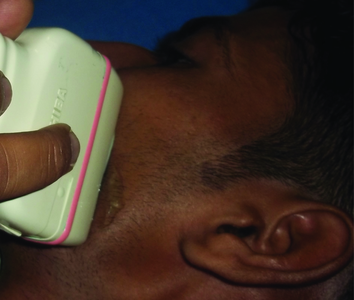
For ultrasonographic examination of MM, a water-based gel was applied to the probe before the imaging procedure [Table/Fig-2]. During imaging, the transducer was held perpendicular to the surface of the skin and special care was taken to avoid excessive pressure. The measurement site was at the thickest part of the masseter, close to the level of the occlusal plane, approximately in the middle of the mediolateral distance of the ramus (a line drawn on the skin parallel to and two centimeter above the inferior border of the mandible, approximately corresponding to the most bulky part of the superficial portion of the MM). Ultrasound examinations were performed with patient in supine position by single examiner using Toshiba power vision 6000 ultrasound imaging system with linear transducer (5-11 MHz).
Examination done with Ultrasonography
The real time imaging of MM was performed bilaterally both in relaxed and contracted state [Table/Fig-3]. Images were recorded at three sites that were one centimeter apart from each other (bulkiest part of MM). The measuring sites were taken as PMA where P is posterior, one centimeter from the posterior border of the ramus of the mandible, M is middle, over the midpoint, and A is anterior near the anterior border of the ramus of the mandible. The data was coded on Microsoft excel sheet and analysed using SPSS statistical package version 17. Statistical analysis was done by Student t-test.
Ultrasonographic measurements of masseter muscle in OSMF patient, Contracted state (A) and Relaxed state (B)

Results
Ultrasonographic measurement of MM thickness in control group and study group was done. [Table/Fig-1,2] shows both mean relaxed and contracted thickness, and also relaxed versus contracted thickness on right and left side respectively.
Right Side
In the study group, the mean relaxed thickness of right masseter muscle was 10.952 ± 1.356 mm, whereas the mean contracted thickness of right masseter muscle was 13.672 ± 1.946 mm. In the control group, the mean relaxed thickness of right masseter muscle was 7.752 ± 1.104 mm, whereas the mean contracted thickness of right masseter muscle was 9.168 ± 1.440 mm [Table/Fig-4].
Thickness of masseter muscle on right side in osmf patients and control group * p < 0.001; Highly significant
| Thickness of masseter muscle on right side | Study Group | Control Group | Study vs Control |
|---|
| Diffe-rence | t-value | p-value |
|---|
| Relaxed | 10.952 ± 1.356 | 7.752 ± 1.104 | 3.200 | 9.151 | <0.001* |
| Contracted | 13.672 ± 1.946 | 9.168 ± 1.440 | 4.504 | 9.302 | <0.001* |
| Relaxed vs Contr-acted | Diff. | 2.720 | 1.416 | | | |
| t-value | 13.056 | 11.527 | | | |
| p-value | <0.001* | <0.001* | | | |
Left Side
In the study group, the mean relaxed thickness of left masseter muscle was 11.876 ± 1.198, whereas the mean contracted thickness of left masseter muscle was 14.536 ± 1.861 mm. In the control group, the mean relaxed thickness of left masseter muscle was 8.104 ± 0.972 mm, mean contracted thickness of left masseter muscle was 9.544 ± 1.159 mm [Table/Fig-5].
Thickness of masseter muscle on left side in OSMF patients and control group
| Thickness of masseter muscle on left side | Study Group | Control Group | Study vs Control |
|---|
| Diffe-rence | t-value | p-value |
|---|
| Relaxed | 11.876 ± 1.198 | 8.104 ± 0.972 | 3.772 | 12.228 | <0.001* |
| Contracted | 14.536 ± 1.861 | 9.544 ± 1.159 | 4.992 | 11.386 | <0.001* |
| Relaxed vs Contr-acted | Diff. | 2.660 | 1.440 | | | |
| t-value | 12.705 | 11.850 | | | |
Right Vs Left
The comparison of right vs left MM thickness was not significant in control group both in relaxed and contracted state. In study group, the relaxed vs contracted thickness of MM was significant, p = 0.001. In study group, the relaxed vs contracted thickness of MM was highly significant, p < 0.001.
On comparing the ultrasonographic measurements of the right and left MM thickness in study and control groups, both in relaxed and contracted state, revealed that [Table/Fig-1,2], the mean of the relaxed and contracted thickness of muscles on both right and left side was highly significant, p < 0.001 in the study as well as in the control groups. Relaxed verses contracted muscle thickness in study and control group was also highly significant with p < 0.001 [Table/Fig-6].
Comparison of relaxed & contracted states according to stages of OSMF * p < 0.05; Significant; ** < 0.001; Highly Significant
| Stage | N | Relaxed | Contracted |
|---|
| II | 12 | 10.692 ± 0.928 | 12.642 ± 1.029 |
| III | 13 | 12.081 ± 0.869 | 15.454 ± 0.968 |
| Difference | 1.389 | 2.812 |
| t-value | 3.865 | 7.040 |
| p-value | 0.001* | <0.001** |
The study group showed higher thickness, both on the right and left side in relaxed as well as in the contracted state when compared to the controls. The thickness of MM was more in contracted state than relaxed state both in study as well as control group, which was highly significant. The comparison of USG measurements with the clinical staging showed significant results, p < 0.01 [Table/Fig-7].
Comparison between thickness of masseter muscle on right and left side in OSMF patients and control group * p < 0.05; Significant
| Group | Thickness of masseter muscle on right side versus left side | Right Side | Left Side | Differ-ence | t- value | p- value |
|---|
| Study | Relaxed | 10.952 ± 1.356 | 11.876 ± 1.198 | 0.924 | 3.841 | 0.001* |
| Contracted | 13.672 ± 1.946 | 14.536 ± 1.861 | 0.864 | 2.755 | 0.011* |
| Control | Relaxed | 7.752 ± 1.104 | 8.104 ± 0.972 | 0.352 | 2.183 | 0.039* |
| Contracted | 9.168 ± 1.440 | 9.544 ± 1.159 | 0.376 | 2.417 | 0.024* |
Discussion
Masseter muscle is the bulkiest and strongest muscle of the face and maximum force is applied to it during mastication. Muscle thickness has been considered as one of the indicators of jaw muscle function [11]. Intensive use of any skeletal muscle may cause changes in the muscle fibre size and composition, which in turn will increase the strength of the muscle and the resistance to fatigue. This is also true of the masticatory muscles. Prolonged high activity of these muscles resulted in increased ultrasonographic thickness of the masseter muscle and increased maximal bite force values [7].
Ultrasonography is based on the transformation of sound waves into visible light waves. Clinicians use these images to identify differences in anatomical structures examined. It is an accurate method, convenient, easy, and inexpensive to apply [12]. It has been used as an instant, non-invasive method for the observation of relatively deep areas, recently, however high frequency echography has been developed that can provide detail investigation of more superficial regions [9]. Ultrasonography is an appropriate method for measuring the thickness of Masseter Muscle (MM). It reveals a large variation in thickness of the masseter muscle between and among individuals during both relaxed and contracted conditions [13].
OSMF is a premalignant condition and a topic of interest now-a –days as of the rise in its incidence and its malignant potential. Earlier it was seen in south East Asia only but now this disease is found worldwide, may be due to increase in habit of quid chewing, which is the most common aetiological factor for OSMF [14]. It is very well documented in literature that fibrosis of buccal mucosa in OSMF produces the sunken cheek appearance; due to which masseter seems to be enlarged [8]. Other conditions like scleroderma can be included as differential diagnosis. This can be ruled out on basis of history of chewing areca nut and burning sensation, which is seen in OSMF. The present study was done to measure the thickness of masseter muscle in 50 patients out of which 25 were of OSMF patients and 25 were normal individuals. According to literature search only one study has been conducted so far to measure the thickness of masseter muscle in OSMF, by Kamla KA et al.,[8] the results of that study is coinciding with our results.
In our study, the mean thickness of right masseter muscle during contraction in OSMF patients was 13.672 ± 1.946 mm and mean thickness in relaxed position is 10.952 ± 1.356 mm. These results were consistent with the results of study conducted by Kamla KA et al., [8] So the difference between relaxed and contracted state is about 3mm, which means thickness increased during contraction. Similar results have been shown on left side during contraction it is 14.536 ± 1.861mm and during relaxation it is 11.8 ± 1.198mm.
On right side, in control group mean of muscle during contraction is 9.168 ± 1.440 mm and relaxation is 7.752 ± 1.104mm, on left side according to us it is 8.104 ± 0.972 mm for relaxation, mean contracted thickness of left masseter muscle was 9.544 ± 1.159 mm. Again the results were coinciding with the results of study conducted by Kamla KA. So there is a clear association between massetric hypertrophy and occurrence of OSMF.
Main drawback of our study is that the sample size was small. Further ultrasonographic studies are advised with larger samples size of OSF patients, so that the incidence of MMH in OSF patients could be strongly established in future.
Conclusion
Masseter Muscle thickness was increased as the duration and frequency of the habit was increased and also as the disease progressed clinically and histologically. Similarly our study also showed association of MMH with OSF, and ultrasound measurements of MM thickness was statistically significant. It is also well documented that if a muscle is not used for a large interval of time then atrophy of the muscle occurs, but according to our study it was found that hypertrophy of masseter muscle occurs when there is increased demand over muscle, due to chewing.