Parry-Romberg Syndrome: A Case of Late Onset with Rapid Progression
Senthil Kumar1, Dinesh Kumar2, Ramesh Kumar3, Rajkumar4, Ramachandra Prabhakar5
1 Reader, Department of Oral Surgery, Thai Moogambigai Dental College, Chennai, India.
2 Senior Lecturer, Department of Oral Pathology, S.R.M Dental College, Chennai, India.
3 Professor, Department of Oral Pathology, S.R.M Dental college, Chennai, India.
4 Professor and Head, Department of Oral Pathology, S.R.M Dental College, Chennai, India.
5 Dean and Head, Department of Orthodontics, Thai Moogambigai Dental College, Chennai, India.
NAME, ADDRESS, E-MAIL ID OF THE CORRESPONDING AUTHOR: Dr. Senthil Kumar, Reader, Department of Oral Surgery, Thai Moogambigai Dental College, Chennai-600 0095, India. Phone : 9444468399, E-mail : dr.ramachandra.prabhakar@gmail.com
Parry–Romberg syndrome (PRS) or progressive hemifacial atrophy is rare, poorly understood condition with an unclear aetiology and characterized by slow and progressive atrophy affecting one side of the face. PRS is a syndrome with diverse presentation and the most common early sign is a painless cleft, the “coup de sabre” near the midline of the face which marks the boundary between the normal and atrophic tissues. Characteristically, the atrophy starts in the first decade of life and progresses slowly for several years before it becomes quiescent. This article describes a case of PRS in a 19-year-old female patient affecting the right side of the face which is unique in the fact that it had a late onset with rapid progression.
Facial hemiatrophy, Parry-romberg syndrome, Progressive hemifacial atrophy
Case Report
A19-year-old female patient reported to the clinic with a chief complaint of facial asymmetry and abnormally textured skin on the right side of the face. She had first observed skin discolouration at 16 years of age. There were no associated symptoms. The patient’s medical and family history was non-contributory.
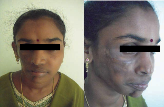
On examination, obvious facial asymmetry was noticed due to the loss of subcutaneous tissue on the right side of the face. Loss of orbital fat resulted in decreased scleral show with enophthalmos on the affected side. The facial skin was pigmented, more pronounced in the infra-orbital and malar region. The nose was deviated to the right side. A clear linear demarcation was seen on the skin of the chin between normal and abnormal side is termed as coup de saber [Table/Fig-1a,b]. On palpation, generalized muscular atrophy was marked on the right face, more pronounced in the lower third of the face. On smiling, there is increased exposure of the teeth on the right side than the left [Table/Fig-2]. Examination of relevant cranial nerves revealed no obvious anomaly with normal vision, smell and skin sensation on the affected side of the face. Intraoral examination did not reveal any dental or soft tissue defects [Table/Fig-3]. With the presenting features, a diagnosis of Parry-Romberg syndrome was given. Her routine haemogram report was within normal limits.


CT scan of the brain and facial bones [Table/Fig-4] revealed reduction in the size on the right side of skull bones and thickness of soft tissues. The right orbit and right maxillary sinus appeared smaller in size. There was mild deviation of the nasal septum to the right. Mandibular bone was normal. Brain showed no focal lesions. A serum antinuclear antibody (ANA) was ordered, which was negative. The patient was planned for facial augmentation after confirmation of cessation of the disease process completely.
Computed tomography views

Discussion
Parry-Romberg syndrome (PRS) also commonly referred to as “progressive hemifacial atrophy” [1,2] was first described by Caleb Parry in 1815 and by Moritz Romberg in 1846. It is a rare neurocutaneous disorder that is characterized by slow and progressive atrophy of the skin and subcutaneous tissues and bones of one or occasionally both sides of the face [3,4]. Sometimes extending to the neck and even to the entire body [5]. The end result of these progressive changes is facial asymmetry that may be accompanied by ocular, neurological, cutaneous, and dental abnormalities [6].
PRS has a prevalence rate of 1 in 70,000 of general population [1, 3] affecting mainly females and usually involves the left side of the face. In our case it involved the right side of the face. The onset is insidious and the condition usually manifests as an atrophy which starts in the first decade of life and progresses slowly for several years before it becomes quiescent. Progression of the disease is rapid in the two to ten years following onset and then stabilizes [7–9]. The complications of early-onset PRS include severe facial deformity, and other co-morbidities when compared to late-onset PRS. In our case, unlike the common cases the deformity involved the right side of the face and developed with a rapid progression within 2 years of onset.
The etiology of this disturbance is unknown and remains unclear, and many possible factors have been proposed including disturbance in fat metabolism, trauma, viral infection, and auto-immunity. Other proposed theories include trigeminal neuritis, increased sympathetic nerve activity triggering facial atrophy, myelopathies or peripheral neuropathies [10].
The most important presenting features of this condition include enophthalmos, deviation of the mouth and nose to the affected side and increased exposure of the teeth on the affected side. All of these features were present in the reported cases. The osseous lesions described in progressive hemifacial atrophy are variable and are strictly related to the age at which the condition appears. When the condition commences after the age of 15 years, the lesions are considered to appear exclusively in soft tissues [11, 12]. And when the onset occurs in children younger than 5 years of age, the fronto-orbito-zygomatic area is commonly affected, whereas the onset is late, the skeletal changes take place dominantly in the lower third of the face [11]. Our patient reported the onset of the facial discoloration at the age of 16 years with no facial deformity, which rapidly progressed to the presented facial deformity. As the onset of the disease was in the later part of the second decade of life, patient had very minimal bony deficit. Uniqueness of our case is being late in onset it affected the upper facial skeleton rather than the lower one third, but cutaneous involvement was more on the lower half of the face. PRS is commonly associated with alopecia, blanched hair, degenerative brain lesions, intracranial calcifications or vascular malformations, sensory impairment, excessive sweating, tear duct dysfunction. The neurological association with 15% PRS patients may include trigeminal neuralgia, epilepsy, facial paraesthesia, and headaches [10]. None of these manifestations were present in our patient.
PRS usually overlaps with a condition called linear scleroderma [13]. There still exists a controversy regarding whether these two entities are different disorders or belong to the same spectrum. Differentiating clinical features between linear scleroderma “en coup de sabre” and PRS include paramedian atrophy in Romberg syndrome without induration of the skin overlying the scalp and with atrophy extending down to one side of the face. In linear scleroderma there is induration of the skin in the region of the scalp and usually does not extend below the forehead. Serum antinuclear antibodies (ANA) are usually raised in linear scleroderma and not so in Romberg syndrome. Although, the differentiating features between these two entities are mainly clinical, there are reports which suggest their coexistence and common cutaneous manifestations [14].
Treatment of Parry-Romberg syndrome aims at cosmetic correction of the atrophied face, after cessation of the pathological process. Aesthetic treatment and facial augmentation is advised to be undertake after the recommended pause of 1 to 2 years after stabilization of the disease process. Immunosuppressive drugs might be considered in some patients especially in the case of cerebral involvement to halt the disease and prevent further cerebral damage and control its systemic manifestations.
[1]. Deshingkae SA, Barpande SR, Bhavthankar JD, Hume JE, Progressive hemifacial atrophy (Parry- Romberg Syndrome)Contemporary Clinical Dentistry 2012 3(5):78-81. [Google Scholar]
[2]. Saude A, Risbud M, Kshar A, Paranjpe AO, Progressive hemifacial atrophyDent Res J(Isfahan) 2013 vol 10(1):108-11. [Google Scholar]
[3]. Stone J, Parry Romberg syndromePractical Neurology 2006 6:185-88. [Google Scholar]
[4]. Gulati S, Jain V, Gar G, Parry Romberg syndromeIndian J Pediatr 2006 73(5):448-49. [Google Scholar]
[5]. Rangare RA, Babu GS, Thomas PS, Shetty RS, Parry-Romberg Syndrome: a Rare Case ReportJ Oral Maxillofac Res 2011 2(2):e5 [Google Scholar]
[6]. Asher SW, Berg BO, Progressive hemifacial atrophy: report of three cases, including one observed over 43 years and computed tomographic findingsArch Neurol 1982 39(1):44-46. [Google Scholar]
[7]. Miller MT, Spencer MA, Progressive hemifacial atrophy. A natural history studyTrans Am Ophthalmol Soc 1995 93:203-15. [Google Scholar]
[8]. Mendonca J, Viana SL, Freitas F, Lima G, Late-onset progressive facial hemiatrophy (Parry-Romberg syndrome)J Postgrad Med 2005 51(2):135-36. [Google Scholar]
[9]. Gomez-Diez SG, López LG, Escobar ML, Gutiérrez LJ, Oliva NP, Progressive facial hemiatrophy with associated osseous lesionsMed Oral Patol Oral Cir Bucal 2007 12(8):E602-04. [Google Scholar]
[10]. Patel H, Thakkar C, Patel K, Parry–Romberg Syndrome: A Rare Entity. J. MaxillofacOral Surg 2010 9(3):247-50. [Google Scholar]
[11]. Moore MH, Wong KS, Proudman TW, David DJ, Progressive hemifacial atrophy (Romberg’s disease): skeletal involvement and treatmentBr J Plast Surg 1993 46(1):39-44. [Google Scholar]
[12]. Urban J, Toruniowa B, Chibowska M, Progressive hemifacial atrophy: ten-year observation of a caseCutis 1996 58(2):165-68. [Google Scholar]
[13]. Demir Y, Karaaslan T, Aktepe F, Yucel A, Denir S, Linear scleroderma “en coup de sabre” of the cheekJ Oral Maxillofac Surg 2003 61:1091-94. [Google Scholar]
[14]. Patel Hiren, Thakkar Chintan, Patel Kajal, Parry–Romberg Syndrome: A Rare EntityJ. Maxillofac. Oral Surg 2010 9(3):247-50. [Google Scholar]