Electrocautery Assisted Harvesting of Free Gingival Graft to Increase the Width of Attached Gingiva - An Uncommon Case Report
Priyanka K. Cholan1, P. Harinath2, Mangaiyarkarasi Subramanian3, Niroshini Rajaram4, Aravindhan T Ranganathan5
1 Senior Lecturer, Department of Periodontics, SRM Dental College, Ramapuram, SRM university, Chennai, Tamilnadu, India.
2 Professor, Department of Periodontics, SRM Dental College, Ramapuram, SRM university, Chennai, Tamilnadu, India.
3 Senior Lecturer, Department of Pedodontics, SRM Dental College, Ramapuram, SRM university, Chennai, Tamilnadu, India.
4 Senior Lecturer, Department of Oral Pathology, SRM Dental College, Ramapuram, SRM university, Chennai, Tamilnadu, India.
5 Reader, Department of Periodontics, Tagore Dental College, Ramapuram, SRM university, Chennai, Tamilnadu, India.
NAME, ADDRESS, E-MAIL ID OF THE CORRESPONDING AUTHOR: Dr. Priyanka.K, Senior Lecturer, Department of Periodontics, SRM Dental College, Ramapuram, Chennai-6000089, Tamilnadu, India. Phone : 09840771507, E-mail : priyankacholan@gmail.com
Procuring a free gingival autograft for the purpose of gingival augmentation has been advocated in areas of inadequate width of attached gingiva that result in gingival recession and/or accumulation of local factors. As obtaining the graft from the palatal donor site with conventional scalpel techniques can result in problems such as prolonged bleeding, increased surgical time and patient discomfort, alternative methods have been advocated to procure such grafts using lasers and electrocautery. This case report elaborates, a free gingival graft harvested for the purpose of increasing the width of attached gingiva using electrocautery principles. The parameters assessed included the extent of patient reported discomfort at the donor site and clinical gain of keratinized and attached gingival width.
Attached gingiva, Electrocautery, Free gingival graft, Keratinised gingiva
Case Report
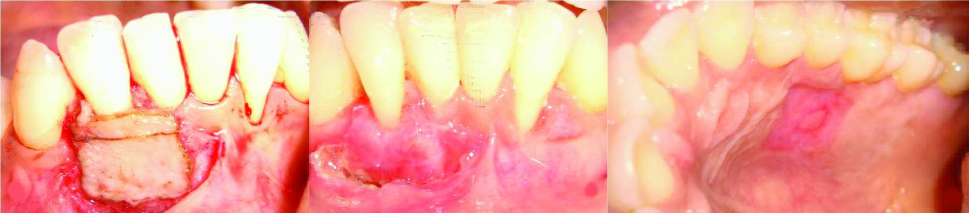
A 32-year-old female patient reported to the Department of Periodontics with the chief complaint of receeding gums in the lower front teeth region and poor oral hygiene maintenance. Intra-oral examination of the area revealed gingival recession in 32 and 42 with inadequate keratinized gingival width and nil width of attached gingiva (WAG) [Table/Fig-1a]. Further evaluation showed shallow vestibular depth with multiple frenal attachments. To correct these mucogingival problems a free gingival autograft (FGG) was planned in 32 and 42. However, the management of the mucogingival defects in 42 with the FGG harvested using electrocautery alone is emphasized in this current case report. Routine blood investigations were done and a written informed consent was obtained from the patient.
a- Pre-operative clinical view., b- De epithelialisation of the recipient site., c&d- Free gingival graft harvested from the palatal donor site using electrocautery

Following phase 1 therapy the donor (Palatal mucosa in relation to 24, 25, 26) and recipient sites (42) were adequately anaesthetized and de-epethelialisation of the recipient bed was done with a no.15 scalpel [Table/Fig-1b]. A tin foil with appropriate measures of the recipient bed was used as a template. The dimensions of the FGG taken were greater than the tin foil measurements, hence compensating for the soft tissue shrinkage as a result of thermal tissue ablation and soft tissue remodeling.
The incisions on the palate are made using the needle (incision-blue) electrodes and the under surface of the graft was dissected using the same [Table/Fig-1c&d]. A 2mm thickness of graft was harvested from the donor site and thinned down to a uniform thickness of 1.5 mm with a surgical scissors once it was harvested, thus maintaining the graft thickness throughout. The thinned graft is then stabilized at the recipient site using a cyanoacrylate tissue adhesive [Table/Fig-2a] and a periodontal dressing was given. A palatal stent was provided and adequate post op instructions and medications were given.
FGG stabilised with a cyanoacrylate tissue adhesive., b- 1 week healing of the graft., c- 1 week healing of the donor site
The patient was monitored at 1, 2, 4 and 24 weeks after surgery [Table/Fig-2b&c]. The parameters recorded included immediate bleeding at the donor site, delayed bleeding at the donor site, complete wound epithelialisation and the level of discomfort/pain at the palatal site. Discomfort was assessed as the level of pain experienced by the patient at the palatal donor site. The patient was asked to rate the discomfort as none, mild/ moderate or severe.
As complete haemostasis was attained with the electrosurgery there was no immediate or delayed bleeding at the donor site. Patient experienced a mild pain at the palatal donor region one week following surgery which reduced considerably and was absent two weeks post surgery [Table/Fig-3].
Comparative values of various variables pre and post surgery
| PARAMETERS | BASELINE (mm) | POST-SURGERY-after 6 months (mm) |
|---|
| Width of keratinized gingival | 1 | 4 |
| Width of attached gingival | 0 | 3 |
| Probing depth | 1 | 1 |
| Recession depth | 5 | 4 |
Discussion
An adequate WAG has been associated with, easy maintenance, increased long term survival of teeth, ease of prosthetic fabrication, resistance to mechanical and bacterial insults, improved smile aesthetics and soft tissue stability around dental implants [1,2]. Beyond the controversies revolving around the adequate WAG, In individuals with inadequate WAG, the possibility of progressive gingival recession and persistent inflammatory changes are substantially increased forming a vicious cycle. The need for gingival augmentation is thus primarily indicated in cases of esthetic requirement and to facilitate oral hygiene maintenance [3].
Whilst various mucogingival techniques been reported to increase the WAG, the free gingival autograft (FGG) has remained the most predictable technique. Yet, the FGG comes with a few hitches including, open bleeding palatal wound associated with discomfort and difficulty in haemostasis that affects operator visibility and consequently graft dimensions [4,5]. To overcome these disadvantages associated with graft procurement using routine scalpel technique, alternatives such as lasers and electrocautery have been suggested. Considering the clinical uses of these two modalities overlap considerably, electrocautery seemed to be a cost effective alternative to the pricey lasers and hence executed in this case report.
Electrosurgery is a controlled, precise application of heat to the soft-tissue site to be cut, achieved by means of carefully designed electrodes. The result is a controlled, irreversible thermal alteration of the soft tissue. With electrocautery, the clinician can control the inherent variables such as waveform, frequency, size of the electrode, time of contact and cooling periods [6].
Though the scalpel technique is commonly advocated for obvious reasons of control and uniformity, its main drawbacks include that of maintaining haemostasis at the palatal donor site and subsequent patient discomfort. On the contrary, electrocautery alleviated this problem and has been advocated for several reasons including immediate haemostasis, consistent cutting, reduced operative time and increased operator efficiency due to a bloodless field of surgery (coagulating property). In addition the electrode which cuts on its sides as well as on its tip, may be bent to meet the clinical needs and the cuts are made with comparative ease. Also, patient compliance and motivation for future surgeries are easier due to the reduced discomfort and uneventful healing [6]. However, the procedure comes with few drawbacks such as objectionable odour, low tactile sensitivity and need for Anaesthetic, in contrast to the lasers which requires no or minimal anaesthesia [7].
This case report elaborates a FGG harvested by electrocautery for the purpose of increasing the width of attached gingiva. The electrocautery unit (Servotome-Acteon/Satalec) used in this case uses fully rectified filtered alternating current in monopolar mode with its main socket in its earthed connection. In monopolar electrosurgery units, the current begins with the electrosurgery device and travels along a wire to the oral site, then to an indifferent plate placed behind the patient’s back. As the surgical electrode contacts the patient’s oral soft tissues, heat is produced and controlled cutting is achieved. Smoke and pain also are produced as the tissue is cut, necessitating the use of anaesthetic [5].
This electrocautery assisted harvesting of FGG showed a 3mm increase in width of both the keratinized and attached gingiva with an increase of 1mm of root coverage despite no change in the probing depth after 24 weeks [Table/Fig-4a,b]. The experience as reported by the patient, was least traumatic, with a very mild discomfort in the 1st week and no discomfort thereafter, though the “burning flesh” odour, during the procedure, which is an important shortcoming of electrocautery, was obvious. The healing of the palatal wound was assessed and complete healing of the palatal site occurred only four weeks after surgery [Table/Fig-4c,d]. The results of this case report showed similar results in clinical parameters when compared to the FGG harvested by conventional scalpel technique done by Del pizzo et al., and thus validating its use [8]. Cyanoacrylate was used to stabilize the graft, instead of the standard sutures thus, making it a traumatic, which may have also contributed to the uneventful healing process.
a-Healing of palatal donor site after 1month., b-Healing of palatal donor site after 3 months., c-Healing of the recipient bed after 3 months., d-Healing of the recipient bed after 6 months

In dentistry though many cases of soft tissue incisions and excisions have been done with electrocautery, no studies have been reported in the literature so far for graft procurement in FGG, owing to the fear of graft necrosis due to insufficient vascularity. The possible reasons for the success of this case could be attributed to the thickness of the graft, the ample vascularity at the donor and recipient sites, the intrinsic vascular channels in the harvested graft and the bigger graft dimensions, hence making electrosurgery a simple and efficient alternative to scalpel or laser surgeries.
Conclusion
Within the limits of this case report, it can be reported that electrosurgery assisted FGG harvesting has evident advantages and hence can be promoted as a viable alternative to the traditional scalpel techniques. Further randomized controlled studies with large sample sizes are required to form a strong scientific basis for its continued usage.
[1]. Krygier G, Glick PL, Versman KJ, Dahlin CJ, Cochran DL, To minimize complications, it it essential that implant abutments be surrounded by keratinized tissue?Int J Oral Maxillofac Implants 1997 12:127 [Google Scholar]
[2]. Bader HI, Soft-tissue considerations in esthetic dentistryCompendium 1991 12(8):534:536-8.:540-42. [Google Scholar]
[3]. Mehta P, Peng LL, The width of the attached gingival-Much ado about nothing?J Dent 2010 38(7):517-25. [Google Scholar]
[4]. Saglam M, Köseoglu S, Treatment of localized gingival recessions with free gingival graftEur J Gen Dent 2012 1:10-14. [Google Scholar]
[5]. Christensen GJ, Soft-tissue cutting with laser versus electrosurgeryJ Am Dent Assoc 2008 139:981-84. [Google Scholar]
[6]. Raghavan Rohit, Shajahan PA, Koruthu Anil, Sukumar B, Nair Anoop, Divakar KP, Second stage surgery: A clinical case report comparing efficacy of laser and electrocauteryInt J of Dent Res 2014 2(1):26-28. [Google Scholar]
[7]. Babuz SK, Agila S, Root coverage with free gingival autograft using a diode LaserJournal of dent lasers 2012 2(6):72-75. [Google Scholar]
[8]. Del Pizzo M, Modica F, Bethaz N, Pritto P, Romagnoli R, The connective tissue graft: a comparative clinical evaluation of wound healing at the palatal donor site. A preliminary studyJ Clin Periodontol 2002 29(9):848-54. [Google Scholar]