Introduction: Arterial compliance will result in stabilizing the fluctuations in arterial pressure and blood flow. So arterial stiffness can be a good indicator for monitoring the cardiovascular system. Arterial stiffness can be measured using indices like reflection index (RI), stiffness index (SI) and Brachial Finger Pulse Wave Velocity (BFPWV).
Objectives: Aim of our study was to evaluate the changes in RI, SI and BFPWV during different phases of the menstrual cycle and to correlate RI with SI in healthy female subjects between the age group of 18-30 years from Bangalore, India.
Materials and Methods: Basal recordings of RI and SI were determined by Photo Pulse Plethysmography (PPG) picked up from the fingertip using BIOPAC system and BFPWV was obtained using Doppler. Recordings were obtained at three different time points during the menstrual cycle. Analysis was done using repeated measures ANOVA with Bonferroni correction.
Result: There was a significant decrease in above parameters p <0.05 during the mid-cycle. Correlation between RI and SI was also significant p<0.05.
Conclusion: These findings suggests that the menstrual cycle affects the arterial stiffness and one of the factor is oestrogen. Hence, women are less prone to the incidence of cardiovascular diseases before menopause. Screening for arterial stiffness in a general population, using these indices is valid, economical and reliable.
Arterial stiffness, Hormone, Non-Invasive method, Pulse wave velocity
Introduction
Elasticity of an arterial wall will help it to expand and recoil during cardiac contraction and relaxation respectively, thus buffering the fluctuations in arterial pressure and blood flow [1] . Reductions in central arterial compliance impair this buffering function, contributing to elevations in systolic blood pressure, development of left ventricular hypertrophy, and reductions in arterial baroreflex sensitivity [2,3]. Recently, need of simple, economical, convenient and noninvasive cardiovascular assessment techniques have reinstated the interest to investigate Photo pulse plethysmography (PPG). It is
Physiology Sectionan instrument used to determine and register the variations in blood volume or blood flow in the body, which occurs with each heartbeat [4]. So using this instrument we can monitor the behaviour of pulse volume and many indices are there for the same. Like reflection index (RI) is an index which indicates peripheral arterial stiffness and vascular tone, where as stiffness index (SI) and pulse wave velocity (PWV) indicates the large arterial stiffness [5].
Increased arterial stiffness is a risk factor for progression of hypertension, cardiovascular disease, stroke and dementia. Number of lifestyle changes can reduce the arterial stiffness like exercise, reduce intake of coffee, lowering the salt intake, etc. By measuring these indices, we can identify the diseases and reduce the level of progression by changing their life styles [6]. Also, studies done to look at the effect of hormonal changes (Estrogen and Progesterone) on arterial elasticity during various phases of the menstrual cycle, which are still controversial [7,8]. However, the postmenopausal use of hormonal therapy is associated with lower risk of coronary heart disease can’t be neglected [9].The present study was aimed to look at the effect of different phases of menstrual cycle on peripheral arterial stiffness with help of indices like RI, SI and BFPWV by using a noninvasive method like PPG.
Materials and Methods
Subjects- A pilot study was carried out in our institute from August 2008 to October 2008. A total of 20 subjects were selected based on the inclusion and exclusion criteria.
Inclusion criteria: Young healthy female volunteers, ranging in the age from 18-30 years who were not regularly involved in extreme physical activity, having regular menstrual cycles (according to menstrual history).
Exclusion criteria: Subjects suffering from cardiovascular disease, diabetes mellitus, hypertension and any other chronic diseases & those who are on any treatment or drugs and oral contraceptive therapy were excluded.
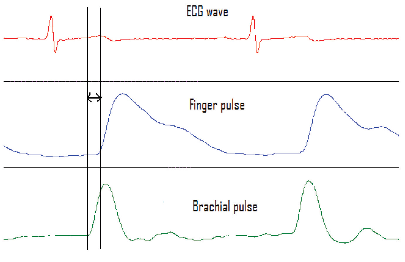
Study protocol: After explaining the procedure to the subjects in their own language, written informed consent was obtained from the individual. Subjects were asked to come to the lab between 6:30 to 7:00 am in fasting state. Height, weight and forearm length (from left brachial artery to the left index finger was measured with the help of measuring tape) were recorded. After 5-10 minutes of rest to the subject, blood pressure was recorded. Basal recordings of RI, SI and BFPWV were measured using PPG (which is used to pick up the finger pulse), Electrocardiogram (ECG) lead-II and Doppler (which is used to pick up the brachial artery wave forms) [Table/Fig-1] . Recordings (in sitting position) were taken at three time points i.e., 1st recording on 2nd or 3rd day of menstruation, 2nd recording during mid-cycle (around 12th-15th day) & 3rd is on 22nd-24th day of menstrual cycle.
Determination of arterial stiffness: Finger PPG, lead II ECG and Doppler recording was done and the waves obtained were acquired by the BIOPAC PRO software. A pulse transducer was wrapped around the left index finger to record the finger pulse wave by PPG. The PPG measures the density of blood in the fingertip. Doppler probe was used to record the left brachial artery wave. Both waves were recorded simultaneously for five minutes.
Statistical Analysis
In reference to earlier studies, a difference of 10% between the mean and Standard deviation (SD) of PWV, with 90% power and significance of 0.05, the sample size was calculated as 20. Paired t-test was used to compare between baseline values. Results expressed as mean ± SD. Repeated measures ANOVA was used to look at the effect of different phases of the menstrual cycle on measurement of RI, SI and BFPWV.
To evaluate the Coefficient of variance (CV) for biological variability, eight subjects were assessed for BFPWV, SI and RI on three different days, consecutively. CV was calculated using RMS (Root means square equation) [Table/Fig-2]. The coefficient of variance (within subject variability) for the measurements of BFPWV, SI and RI was 4.2%, 3.5% and 2.4 % respectively.
Results
The basal recordings of different parameters of 20 healthy female subjects are given in the [Table/Fig-3].
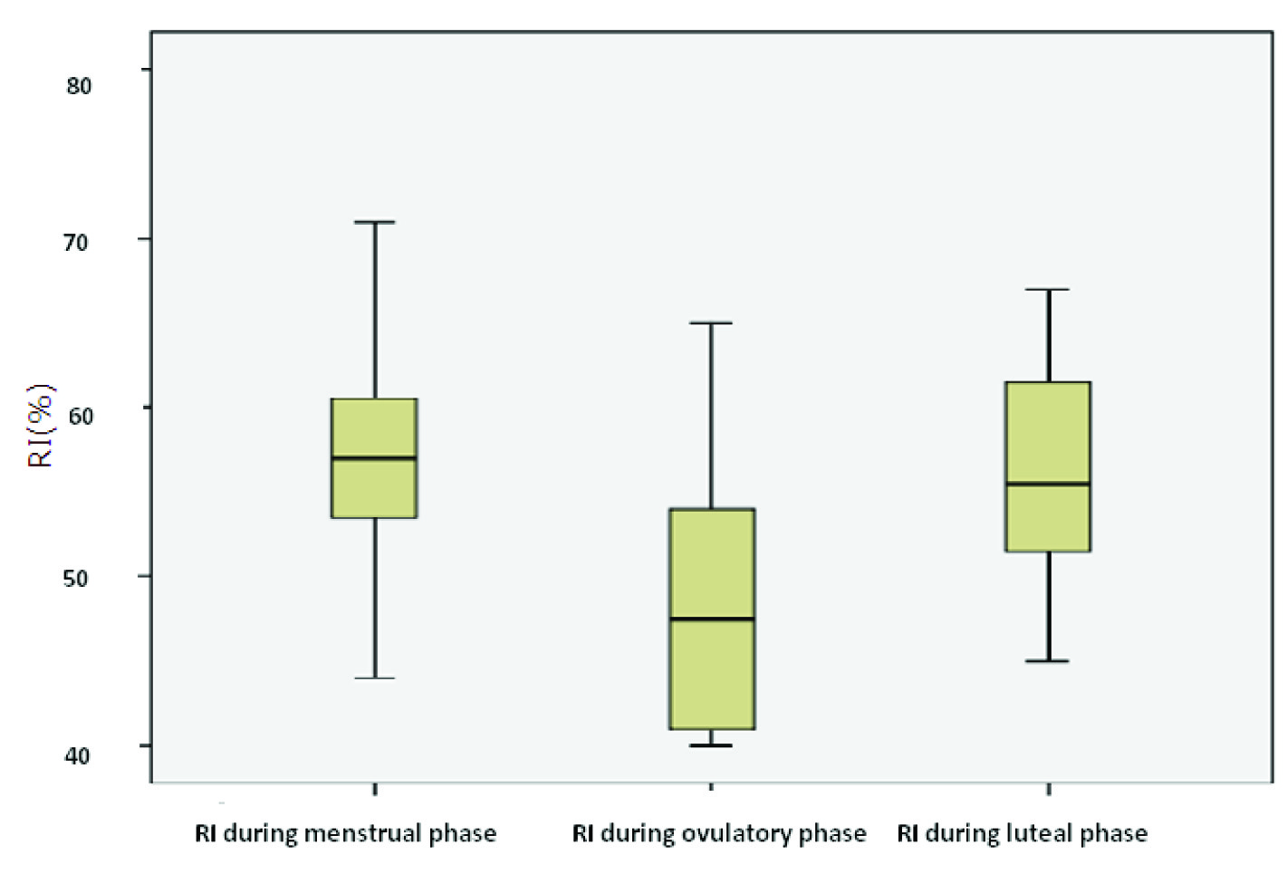
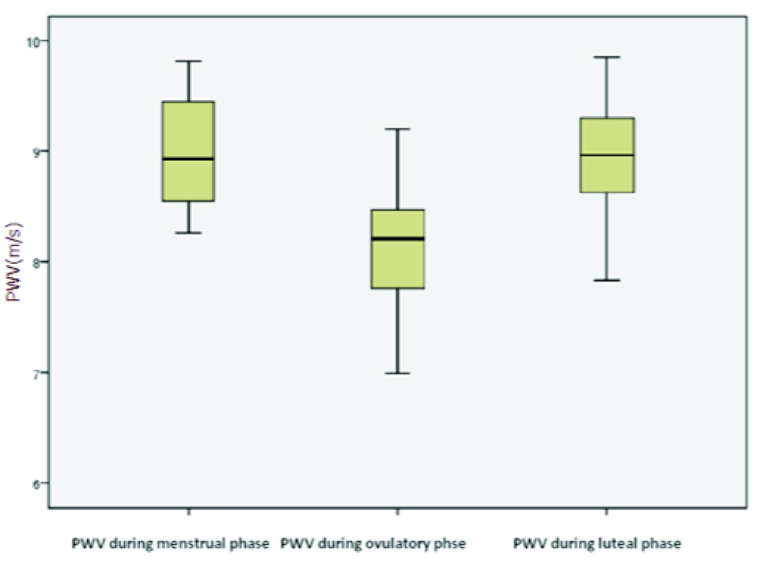
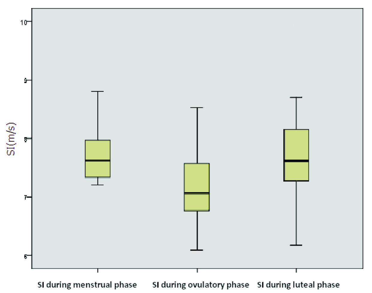
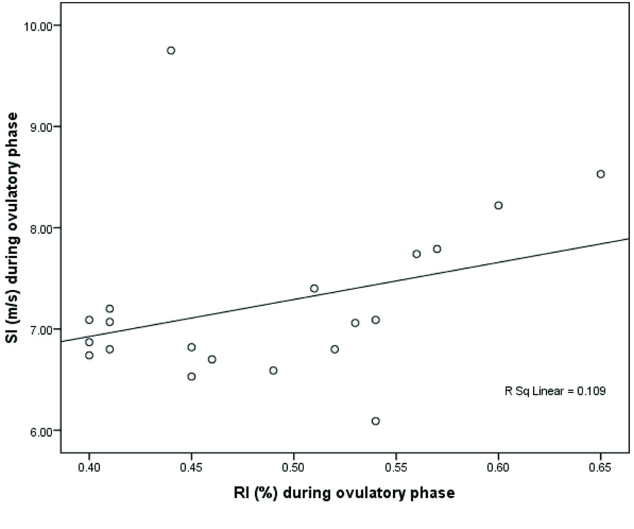
Observations: The salient findings of our study were as follows: There was a significant decrease in all the parameters during the mid-cycle (p= 0.00) [Table/Fig-4,5,6]. The correlation between the RI and SI was also significant [Table/Fig-7,8].
Discussion
In this study, we explored the complex relationship between variations in ovarian hormones and the elastic properties of peripheral arteries at three distinct time points throughout the menstrual cycle. Also, this study is unique as for the first time an attempt was made to demonstrate a correlation between peripheral arterial stiffness with menstrual phases using a noninvasive technique. We will discuss this under two subheadings,
Changes in arterial stiffness during different phases of the menstrual cycle: Our study showed a significant reduction in arterial stiffness during midcycle, indicating an estrogen dependent reduction in vascular smooth muscle tone. This is comparable with the other studies showing similar changes using different indices like distensibility index [12], Flow mediated dilatation (FMD) and whole body arterial compliance (WBAC) [8]. There are many studies showing similar results using central artery compliance by invasive procedures (mean arterial pressure, aortic augmentation index (AIx) and central systolic blood pressure (SBP) [7,13] and non invasive techniques (MRI and USG) [14,15]. But contrary to our study some of the studies have not shown change in peripheral arterial compliance during different phases of menstrual cycle [7,16].
Use of Pulse wave plethysmography: We have used indices like SI, RI and PWV for measuring peripheral arterial stiffness. There are many studies showing effectiveness of SI and PWV for measuring 3peripheral arterial compliance under different clinical scenarios like diabetic, hypertensive patients, postmenopausal women and women on oral contraceptive pills [10,11,13,17,18]. Regarding these indices they provide information on mechanical properties and endothelial function of arteries. The method is simple, valid, and reliable. Most importantly they are non-invasive. Large number of the general population can be screened for detecting and also the severity of CAD. Instruments like PPG are easily available, and high level of technical expertise is not required as needed in Ultrasound and Magnetic resonance imaging (MRI) [11,19,20]. As our second objective was correlation between RI and SI, which is found to be significant. Study performed by Brilliante et al., showed a significant correlation of RI and SI with age, heart rate, systolic blood pressure and diastolic blood pressure [21].
Recording of ECG, Pulse wave and Brachial Wave forms
BFPWV (m/s)[10] = Distance (forearm length) (meters)
Delta t (Seconds)
SI (m/s) [11] = Subjects Height (meters)
Time T (Seconds)
RI (%)[11] = Height of reflected wave (2nd wave)
Height of forwarded wave (1st wave)n
Within subject variability for vascular measurements
| Parameter | Within subject variability |
| BFPWV | 4.2% |
| SI | 3.5% |
| RI | 2.4% |
Basal recordings of different parameters
| Parameters | Mean ± SD |
| Age(year) | 22 ± 3.40 |
| Ht(meters) | 1.57± 4.65 |
| Wt(Kgs) | 50 ± 0.04 |
| Heart Rate(HR) Beats /minute | 77 ± 11.02 |
| SBP(mmHg) | 113 ± 5.82 |
| DBP(mmHg) | 73 ± 8.79 |
| Forearm length (FL)(cms) | 39 ± 2.12 |
Repeated measures ANOVA of RI
Repeated measures ANOVA of BFPWV
Repeated measures ANOVA of SI
Correlation between RI and SI
| Measure | (I) Phases | (J) Phases | Mean Difference (I-J) | Std. Error | Sig. | 95% conficdence Inrterval for Difference |
| Lower Bound | Upper Bound |
| ri | 1 | 2 | .083* | 0.16 | .000 | 0.041 | .124 |
| 3 | .007 | .015 | 1.000 | -.031 | .045 |
| 2 | 1 | -.083* | .016 | .000 | -.124 | -.041 |
| 3 | -.076* | .012 | .000 | -.107 | -.044 |
| 3 | 1 | -.007* | .015 | 1.000 | -.045 | .031 |
| 2 | .076* | .012 | .000 | .044 | .107 |
| si | 1 | 2 | .612* | .133 | .001 | .262 | .962 |
| 3 | .118 | .144 | 1.000 | -.261 | .497 |
| 2 | 1 | -.612* | .133 | .001 | -.962 | -.262 |
| 3 | -.494* | .094 | .000 | -.740 | -.248 |
| 3 | 1 | -.118 | .144 | 1.000 | -.497 | .261 |
| 2 | .494* | .094 | .000 | .248 | .740 |
| pwv | 1 | 2 | .968* | .085 | .000 | .745 | 1.190 |
| 3 | .077 | .070 | .863 | -.107 | .260 |
| 2 | 1 | -.968 | .085 | .000 | -1.190 | -.745 |
| 3 | -.891* | .098 | .000 | -1.149 | -.633 |
| 3 | 1 | -.077 | .070 | .863 | -.260 | .107 |
| 2 | .891* | .098 | .000 | .633 | 1.149 |
Based on estimated marginal means
* The mean difference is significant at the .05 level
a. Adustment for multiple comparisons : bonferroni
Conclusion
This study examined the changes in RI, SI and BFPWV in different phases of the menstrual cycle in healthy young women. There is a cyclical variation in the above parameters with a significant reduction during the mid-cycle. These findings suggest that the menstrual cycle affects the arterial stiffness and one of the factors is estrogen, which could have probably reduced stiffness as it is well known. Hence, women are less prone to incidence of cardiovascular diseases before menopause. Screening for arterial stiffness in the general population, these Indices can be used as they are valid and reliable. Upper limb BFPWV is being used for the first time to measure arterial stiffness.
Based on estimated marginal means* The mean difference is significant at the .05 levela. Adustment for multiple comparisons : bonferroni
[1]. W Nichols, M O’Rourke, McDonald’s Blood Flow in Arteries.Theoretical, Experimental and Clinical Principles.London, Arnold. 1998 [Google Scholar]
[2]. M O’Rourke, Arterial stiffness, systolic blood pressure, and logical treatment of arterial hypertension.Hypertension 1990 15:339-47. [Google Scholar]
[3]. KD Monahan, FA Dinenno, DR Seals, CM Clevenger, CA Desouza, H Tanaka, Age-associated changes in cardiovagalbaroreflex sensitivity are related to central arterial compliance.American Journal of Physiology. 2001 281:H284-89. [Google Scholar]
[4]. M Elgendi, On the Analysis of Fingertip Photoplethysmogram Signals.Current Cardiology Reviews. 2012 8:14-25. [Google Scholar]
[5]. SC Millasseau, RP Kelly, JM Ritter, PJ Chowienczyk, Determination of age related increase in large artery stiffness by digital pulse contour analysis.Clinical sciences. 2002 103:371-77. [Google Scholar]
[6]. SJ Zieman, V Melenovsky, DA Kass, Mechanisms, Pathophysiology and therapy of arterial stiffness.Atheroscler Thrombo Vasc Bio. 2005 25:932-43. [Google Scholar]
[7]. K Hayashi, M Miyachi, N Seno, K Takahashi, K Yamazaki, J Sugawara, Variation in carotid arterial compliance during the menstrual cycle in young women.Exp Physiol. 2006 91:465-72. [Google Scholar]
[8]. MR Williams, RA Westerman, BA Kingwell, J Piage, PA Blombery, K Sudhir, Variation in Endothelial Function and Artrial Compliance during the Menstrual Cycle.J ClinEndocrinolMetab. 2001 86:5389-95. [Google Scholar]
[9]. C Rajkumar, BA Kingwell, JD Cameron, T Waddell, R Mehra, N Christophidis, Hormonal Therapy Increase Arterial Compliance in Post menopausal women.J Am CollCardiol. 1997 30:250-56. [Google Scholar]
[10]. JJ Oliver, DJ Webb, Non invasive Assessment of arterial stiffness and risk of Atherosclerotic events.Arterioscler Thrombo Vasc Biol. 2003 23:554-56. [Google Scholar]
[11]. M Jyotsna, A Mahesh, M Aditya, PR Mohan, MUR Naidu, Comparison of Invasive vs Non invasive Pulse wave indices in detection of significant Coronary artery disease: Can we use non invasive pulse wave indices as screening test. Clinical Medicine: Cardiology. 2008 2:153-60. [Google Scholar]
[12]. C Giannattasio, M Failla, A Grappiolo, ML Stella, AD Bo, M Colombo, Fluctuations of Radial Artery Distensibility Throughout theMenstrual Cycle. Arterioscler Thromb Vasc Biol. 1999 19:1925-29. [Google Scholar]
[13]. C Stefanadis, E Tsiamis, J Dernellis, P Toutouzas, Effect of Estrogen on aortic function in Post menopausal women.Am J Physiol. 1999 276:H658-62. [Google Scholar]
[14]. M Miyachi, H Kawano, J Sugawara, K Takahashi, K Hayashi, K Yamazaki, Unfavourable effects of resistance training on central arterial compliance: a randomized intervention study.Circulation. 2004 110:2858-63. [Google Scholar]
[15]. AJ Nelson, SG Worthley, JD Cameron, Cardiovascular magnetic resonance-derived aortic distensibility: validation and observed regional differences in the elderly.Journal of Hypertension. 2009 27(3):535-42. [Google Scholar]
[16]. MC Macedo, D Luminoso, MD Savvidou, CM McEniery, KH Nicolaides, Maternal Wave Reflections And Artrial stiffness in normal pregnancy as assessed by applanation tonometry.Hypertension. 2008 51:1047-51. [Google Scholar]
[17]. PJ Chowienczyk, RP Kelly, H MacCallum, Photoplethysmographic assessment of pulse wave reflection: blunted response to endothelium-dependent beta2-adrenergic vasodilation in type II diabetes mellitus. Journal of the American College of Cardiology. 1999 34(7):2007-14. [Google Scholar]
[18]. J Blacher, R Asmar, S Djane, GM London, ME Safar, Aortic Pulse Wave Velocity as a Marker of Cardiovascular Risk in Hypertensive Patients.Hypertension. 1999 33:1111-17. [Google Scholar]
[19]. L Stoner, JM Young, S Fryer, Assessments of arterial stiffness and endothelial function using pulse wave analysis.Int J Vasc Med. 2012 Article ID 903107, 9 pages [Google Scholar]
[20]. LA Tomlinson, Methods for assessing arterial stiffness:technical considerations Curr Opin Nephrol Hypertens. 2012 21:655-60. [Google Scholar]
[21]. DG Brillante, AJ O’Sullivan, LG Howes, Arterial stiffness indices in healthy volunteers using non-invasive photo plethysmography.Blood pressure. 2008 17:116-23. [Google Scholar]