Mucormycosis are a group of invasive infections caused by filamentous fungi of the Mucoraceae family. Mucormycosis is essentially limited to immunocompromised patients with poorly controlled diabetes mellitus, hematologic malignancy, organ transplant, chemotherapy, chronic renal insufficiency, malnutrition, deferoxamine therapy and severe burns. The fungi invade arteries leading to thrombosis that subsequently causes necrosis of hard and soft tissues. Here, we present a case report of a 50-year-old diabetic patient with rhinomaxillary form of mucormycosis.
Amphotericin- B, Diabetes mellitus, Mucoraceae, Mucormycosis granulomatous
Case History
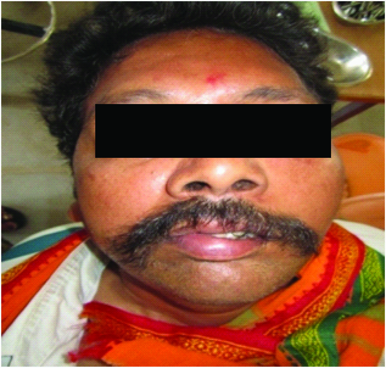
In March 2013, a 50-year-old male patient reported to the Department of Oral & Maxillofacial Pathology, G.P.R Dental College and Hospital, Kurnool, Andhra Pradesh, India with a complaint of ulcer in left side of the palate and severe halitosis from past 6months. History revealed that 6 months back, he noticed an ulcer on left side of the palate, for which surgical debridement was done. Two months later, he noticed change in the voice. There was watery discharge from left eye. He gave history of inability to close the eye lids for which the opthalomalogist did clamping of eyelids. There was diffuse swelling on the left side of the face which was firm on palpation [Table/Fig-1].
Clinical photograph showing swelling, clamping of the eye & deviation of the mouth to right
Intraorally an area of necrosis of the left palatal mucosa, exposing underlying bone with perforation at the junction of hard and soft palate was seen. The surface of the lesion was rough, covered by granulation tissue and surrounded by erythema, firm in consistancy and tender on palpation [Table/Fig-2]. Based on clinical findings provisional diagnosis was given as Chronic granulomatous infection, non healing ulcer. Hematological examination revealed hyperglycemic state and elevated ESR.
Intra-oral photograph showing ulcer covered by necrotic slough with perforation at the junction of hard and soft palate

Potassium hydroxide smear from lesional area showed thin walled broad non-septate fungal hyphae. An incisional biopsy was performed at the site of the ulcer. The Hematoxyllin & Eosin (H&E) stained soft tissue section showed orthokeratinized stratified squamous epithelium with ulcerations and connective tissue stroma which showed multiple granulomatous areas having lymphocytes, giant cells and epitheloid cells [Table/Fig-3]. Broad thin walled non-septate fungal hyphae with branching at right angles were seen in the connective tissue [Table/Fig-4]. Periodic Acid Schiff (PAS) stained sections showed magenta coloured nonseptate fungal hyphae with branching at right angles in the connective tissue [Table/Fig-5].
H&E stained section showing orthokeratinized stratified squamous epithelium showing granulomatous area with fungal hyphae

Higher magnification (40X) of H&E stained section showing fungal hyphae branching at right angles

Higher magnification (40X) of PAS stained section showing broad nonseptate fungal hyphae

Diagnosis of Mucormycosis granulomatous infection was confirmed by Haematoxyllin and eosin, PAS stains and growth of fungal hyphae on culture medium which showed rhizoids and branched sporangiophores.
Patient was referred to ophthalomalogist and otolaryngeologist, where he received surgical debridement of the lesion and was kept under insulin injection to control the glucose levels. Oral Amphotericin- B was given 1mg/kg/day for 2wks and Laceyl eye ointment and tear drops were given as eye management.
Discussion
Mucormycosis is a rapidly progressive, often fatal opportunistic infection caused by fungi belonging to class zygomycetes /phacomycetes order mucorales first described by Paultauf [1,2]. The most common genera are Absidia, Rhizomucor, Rhizopus and Mucor [3,4]. The organisms are found throughout the world growing in their natural state on variety of decaying material. Spores are liberated in to the air and inhaled by human host [4,5]. Mucormycosis is diagnosed histologically when broad, irregularly shaped, nonseptate hyphae with right angle branching are seen invading the tissue [1,4]. It mainly effects immunocompramised individuals such as with uncontrolled diabetes, long term steroid therapy, cytotoxic agents, leukemia, lymphoma, organ transplantation and severe burns [2]. Mucormycosis is the third most common infection in patients with haematologic malignancies after Candidiasis and Aspergillosis [1,6].
Primary sites of invasion are nasal sinuses and lung, when the spores are inhaled and gastrointestinal tract, when they are ingested [6]. Clinically mucormycosis occurs in four forms, rhinocerebral, pulmonary, gastrointestinal, and disseminated. Rhinocerebral form is most common representing 1/3rd to 1/4th of all cases of mucormycosis, which is further divided into two subtypes. The highly fatal rhino-orbito–cerebral form which is invasive and may involve ophthalmic and internal carotid arteries, the other one being the rhinomaxillary form which is seen most commonly in individuals with uncontrolled diabeties and involves sphenopalatine and greater palatine arteries resulting in thrombosis of turbinates and necrosis of the palate [3,7]. Once established in the paranasal sinuses, the infection can easily spread to the orbit via the nasolacrimal duct and medial orbit. Spread to brain can occur via orbital apex, orbital vessels or via cribriform plate. As the disease progress to the orbit or skull, the patient may become confused and comatose. Fungal invasion of globe or retinal artery leads to blindness [8].
Mucormycosis mainly effects immunocompromised people. Fungal hyphae produce a substance called Rhizoferrin which binds iron ardently. This Iron- Rhizoferrin complex is then taken up by fungus and becomes available for vital intercellular process [7]. The low pH, hyperglycemic state and iron rich environment in diabetic patients favours the fungal growth. Mucormycosis is also caused by Rhizopus arrihzus species, in diabetic patients, due to their ability to produce enzyme Ketoreductase which allows them to utilize patient’s ketone bodies for their nutrition [8]. Hematogenous spread occurs to other organs like brain or lungs leading to disseminated sepsis. The disease is non-contagious and does not spread from person to person [8].
Initially mucormycosis presents as per-orbital cellulitis, facial pain and unilateral facial swelling with variable grade fever [9]. Involvement of oral cavity usually appears as palatal ulceration or necrosis with denudation of bone and later perforation of palate. Visual disturbances with concurrent proptosis and symptoms related to cranial nerve involvement like facial paralysis is often present [4] which were also seen in our case. In long standing diabetics with poor glycemic conditions, there is atherosclerosis and microangiopathy of blood vessels which further compromise the vascularity and predispose the patient to osteomyelitis [7]. Clinical differential diagnosis of lesion should include squamous cell carcinoma, chronic granulomatous infection like tuberculosis, tertiary syphilis, midline lethal granuloma and other deep fungal infections [2].
Radiographically opacification of sinuses may be observed in conjunction with patchy effacement of bony walls of sinuses [4].Computed Tomography with contrast or magnetic resonance image (MRI) scan can demonstrate erosion or destruction of bone and help to delineate the extent of disease [2].
Potassium hydroxide smear of lesional area can reveal non septate fungal hyphae which was positive in our case [10]. Culture on sabouraud’s dextrose agar is preferred but histological examination of biopsy specimen is conclusive with H&E and PAP stains [9,2]. The H&E stained section of biopsy specimen demonstrates broad nonseptate fungal hyphae that branch at right angles [2,4,6]. Histopathologically Mucormycosis has close resemblance with Aspergillosis. The differentiating features are, that hyphae of mucor are non septate and branch at right angles where as hyphae of aspergillus species are septate, smaller in width and branch at more acute angles. Tugsel et al., observed that the initial culture of biopsy tissue may be negative and that histopathological examination is essential for early diagnosis [6]. In our case the diagnosis was confirmed by all the three methods.
Successful treatment of mucormycosis consists of rapid accurate diagnosis of the condition followed by radical surgical debridement of infected necrotic tissue with systemic administration of antifungal drugs [2,4]. Most commonly used drug is Amphotericin-B deoxycholate with 80% of cure rate [1–5,8–10]. A new triazole derivative pasaconazole an oral antifungal agent has been used recently either alone or in combination with Amphotericin-B. The underlying systemic disease should be controlled immediately [2].
Patients with localised invasive sinus disease without cerebral involvement can have the survival rates of about 50-80%. If the infection spreads to brain fatality rate exceeds more than 80%. Prognosis may improve with rapid diagnosis, early management and reversible underlying risk factors [8].
The case represented here showed all the features of Rhinomaxillary mucormycosis like swelling of the face, facial paralysis and ulceration along with perforation of palate in diabetic individual which were consistent with features described in the literature.
Conclusion
In dentistry this condition gains increasing interest because of its initial manifestation in orafacial tissues. So, we emphasise that mucormycosis should be included in differential diagnosis whenever a patient with impaired immune response presents with spreading sinusitis, facial swelling, cellulitis and palatal ulcer. A degree of clinical suspician, early histopathologic diagnosis and prompt aggressive surgical evaluation of inflammatory material along with systemic Amphotericin –B therapy can save lives.
[1]. McDermott Nancy E, Barrett John, Hipp Jason, Merino Maria J, Lee Chyi-Chia Richard, Waterman Paige, Successful treatment of periodontal mucormycosis: report of a case and literature reviewOOOOE 2010 109:e64-69. [Google Scholar]
[2]. Doni Bharathi R, Peerapur Basavaraj V, Thotappa Lathadevi Hassan, Hippargi Surekha B, Sequence of oral manifestations in Rhino- maxillary mucormycosisIJDR 2011 20(2) [Google Scholar]
[3]. Badiee P, Jafarpour Z, Alborzi A, Haddadi P, Rasuli M, Kalani M, Orbital mucormycosis in an immunocompetent individualIran J Microbiol 2012 4(4):210-14. [Google Scholar]
[4]. Neville Damm Allen Bouquot Oral and Maxillofacial Pathology 2009 3rd editionNoida , IndiaElsevier publications [Google Scholar]
[5]. Shafer Hine Levy Rajendran R, Shivapathasundharam B, Shafer’s textbook of oral pathology 2012 7th editionNoida, IndiaElsevier publications [Google Scholar]
[6]. Khan S, Jetley S, Rana S, Kapur P, Rhinomaxillary mucormycosis in a diabetic femaleJ Cranio Max Dis 2013 2:91-93. [Google Scholar]
[7]. Goel S, Palaskar S, Shetty VP, Bhushan A, Rhinomaxillary mucormycosis with cerebral extensionJ Oral Maxillofac Pathol 2009 13(1):14-17. [Google Scholar]
[8]. Mohanthy N, Misra SR, Sahoo SR, Misra S, Vasudevan V, Kailasam S, Rhinimaxillary Mucormycosis Masquerading as Chronic osteomyelitis: A Series of Four Case With Review of LiteratureJ Indian Aca Oral Med Radiol 2012 24(4):315-23. [Google Scholar]
[9]. Mallis A1, Mastronikolis SN, Naxakis SS, Papadas AT, Rhinocerebral mucormycosis: an updateEur Rev Med Pharmacol Sci 2010 14(11):987-92. [Google Scholar]
[10]. Mohanty D1, Dhar M, Dwivedi S, MucormycosisTrop Doct 2010 40(2):127-28. [Google Scholar]