Background: The bone marrow is frequently involved in variety of cases presenting with hematological and non-hematological disorders, which are diagnosed by two separate but interrelated techniques such as bone marrow aspiration (BMA) and bone marrow biopsy (BMB).
Aim: This study was aimed to assess the diagnostic value of the BMA and BMB and role of both the procedures to reach final diagnosis when done simultaneously.
Settings and Design: It was a prospective study. The findings of BMA smears were correlated with BMB sections and data obtained was analysed.
Materials and Methods: BMA and BMB were performed on 50 patients. Criteria of inclusion included the main indications for performing this procedure, the availability of full medical records and patient consent. The patients had a male to female sex ratio of 1.6:1 and a wide age range from 4 years to 74 years.
Results: In the present study, the main indications for bone marrow examination were categorized. Out of 50 cases studied, in 23 cases, a strong positive correlation between BMA and BMB was noted. However, it was found that in the cases of aplastic anaemia, different phases of myeloproliferative neoplasm (MPN), multiple myeloma, tubercular granulomas and hemato-lymphoid neoplasm, involvement of the marrow was detected better in bone marrow biopsies.
Conclusion: The study concludes that preparations of aspirate and trephine biopsy are easy, rapid and complementary to each other in majority of the lesions. The advantage of both the procedures done together enabled us to study the cytomorphology of the cells along with the pattern of distribution of the cells depending on the cases, hence help in making the diagnosis accurately.
Introduction
Bone marrow is involved in variety of hematological and non-hematological disorders. The hematological disorders include acute leukemia, myeloproliferative neoplasm (MPN), hemato-lymphoid neoplasm, nutritional deficiency diseases. On the other hand non-hematological disorders include infectious diseases infiltrating the bone marrow such as tuberculosis, parasitic infections and metastatic deposits [1].
Although, diseases of bone marrow present with various clinical symptoms and also involve the blood but peripheral blood picture alone does not reflect the nature of disease process. Depending upon diagnosis suspected from the clinical features and peripheral blood examination, indications for bone marrow examination can be summarised. Therefore, complete hematological evaluation of cases where bone marrow examination was indicated includes BMA smear and bone marrow trephine biopsy as they are complementary to each other [2].
Hence, we attempted to correlate both these parameters to arrive at a more conclusive final diagnosis.
Materials and Methods
The study has been conducted for bone marrow examination on 50 cases presenting with anaemia, fever and organomegaly. The patients had a male to female sex ratio of 1.6:1 and a wide range of age from 4 years to 74 years [Table/Fig-1]. It includes both indoor and outdoor patients, who were suspected of having their bone marrow involvement by any hematological or non-hematological disorders. The relevant history and bio-data of patients was recorded and informed consent was taken. Patients were investigated for complete blood count, coagulation profile, reticulocyte count and peripheral blood film (PBF) examination. BMA and BMB were done Sectionsimultaneously for these patients. In patients of thrombocytopenia, five minutes of firm pressure was applied at the end of the procedure. However, as a precaution, the patients were kept in lying down position on his/her back for a further 10–15 minutes to apply more prolonged pressure.
BMA was performed by Salah’s marrow puncture needle; smears prepared were stained with leishman stain. Prussian blue for iron demonstration was done in selected cases and iron grading was done [3]. BMB was taken by Jamshidi biopsy needle and specimens were fixed in 10% formalin fixative, decalcified in 10% formic acid–5% formaldehyde and processed with paraffin-wax embedding. Sections, 1μm-thick, were cut and were stained by Hemotoxylin and Eosin (H & E) stain. The staining for reticulin fibers with Gomori’s Silver impregnation method was done in selected cases and grading was done [4]. Bone marrow biopsy and aspiration findings were analysed in context of clinical signs, symptoms, other laboratory investigations and diagnosis reached.
Results
Total cases studied were categorized further on the basis of peripheral blood features and accepted indications were summarized [Table/Fig-2]. The results were analysed as per the extent of correlation between BMA and BMB.
Out of the 50 cases studied, in 23 cases both the procedures were comparable [Table/Fig-3] . BMA was reported as suggestive of aplastic anaemia in nine cases but BMB revealed hypo cellular marrow only in three cases where as aplastic anaemia in rest of the six cases [Table/Fig-4] .
There was only a single case which was diagnosed as MPN by BMA and the BMB revealed reactive marrow.
There were seven cases, where diagnosis was made only on BMB, BMA was not contributory [Table/Fig-5] .
There were seen 10 cases in which diagnosis were possible to make with or without complementary findings of BMA. But pattern of involvement of marrow was detected only on BMB [Table/Fig-6] .
Conclusion
The bone marrow examination is valuable investigation in hematology practice. BMA and BMB both are important procedures for the diagnosis of hematological and non-hematological conditions. These procedures are also useful for follow up of the patients undergoing chemotherapy [5,6].
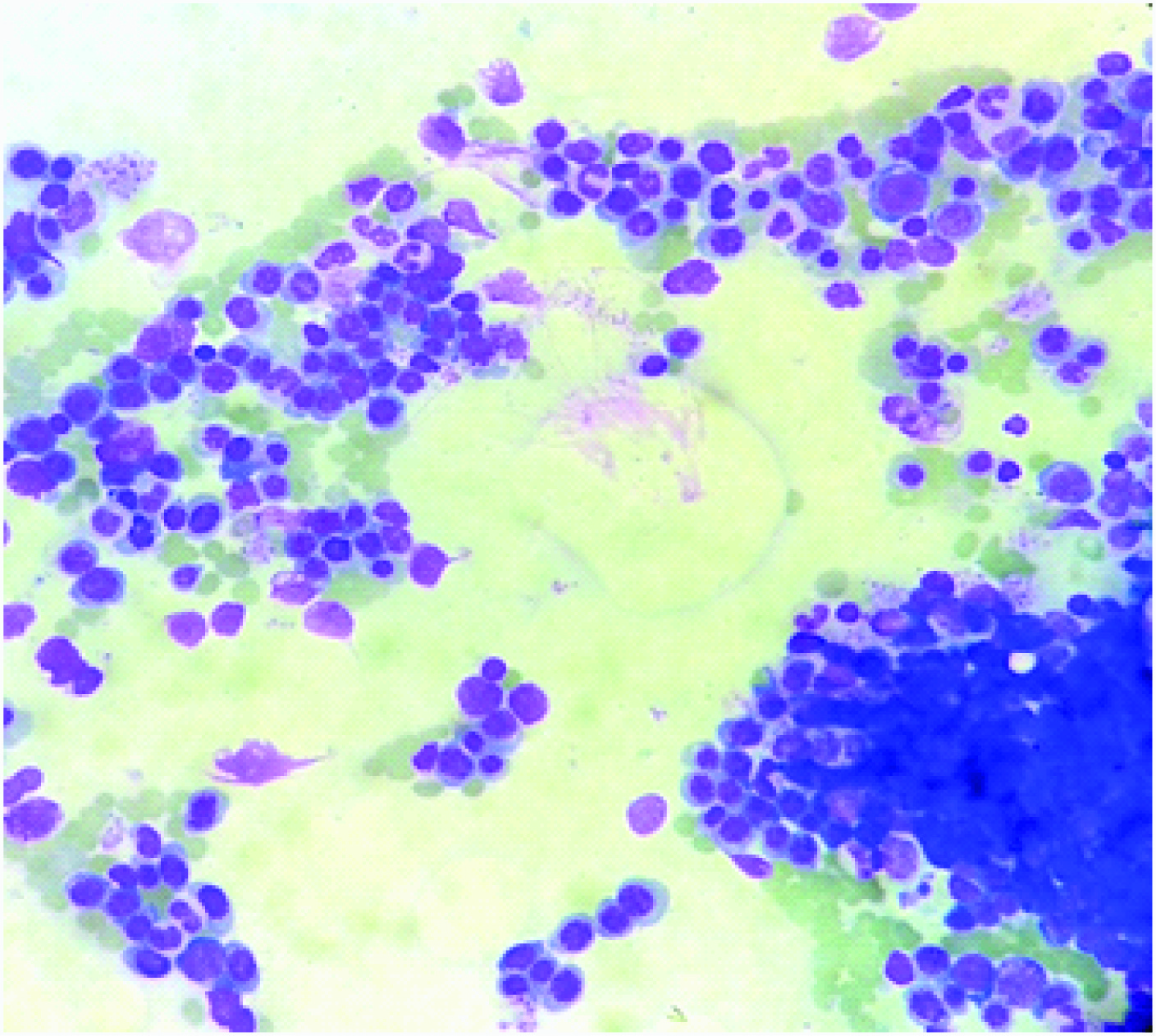
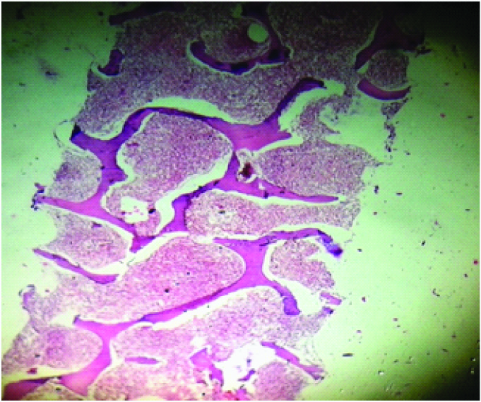
In the present study 23 out of total 50 cases showed comparable results between BMA and BMB. Out of these 23 cases, indication for bone marrow examination was anaemia in 16 cases and both BMA and BMB examination revealed erythroid hyperplasia with either micronormoblastic or megaloblastic proliferation [Table/Fig-7a,Table/Fig-7b]. These observations were nearly similar to the findings seen in a study conducted by Ch Toi P et al., [7] . But iron stained sections of BMB showed differences in iron content from that of BMA smears. Stuart-Smith SE et al., have also shown in a study that aspirate smears reflect bone marrow iron stores more reliably than formic acid decalcified trephine biopsy sections [8] . Rest of the 7 out of these 23 cases were diagnosed as acute leukemia by peripheral blood film examination in all, except two cases, where it was confirmed on bone marrow examination. Younus U and associates emphasized that although BMA confirms the diagnosis of acute leukemia, bone marrow biopsy specimen complements the peripheral blood and aspirate findings in providing additional information for the diagnosis and especially prognosis of acute leukemia [9]. Also various investigators have studied the diagnostic value of antibodies suitable for use on paraffin wax embedded sections in the diagnosis of acute leukemia in sections from bone marrow biopsy specimens [10].
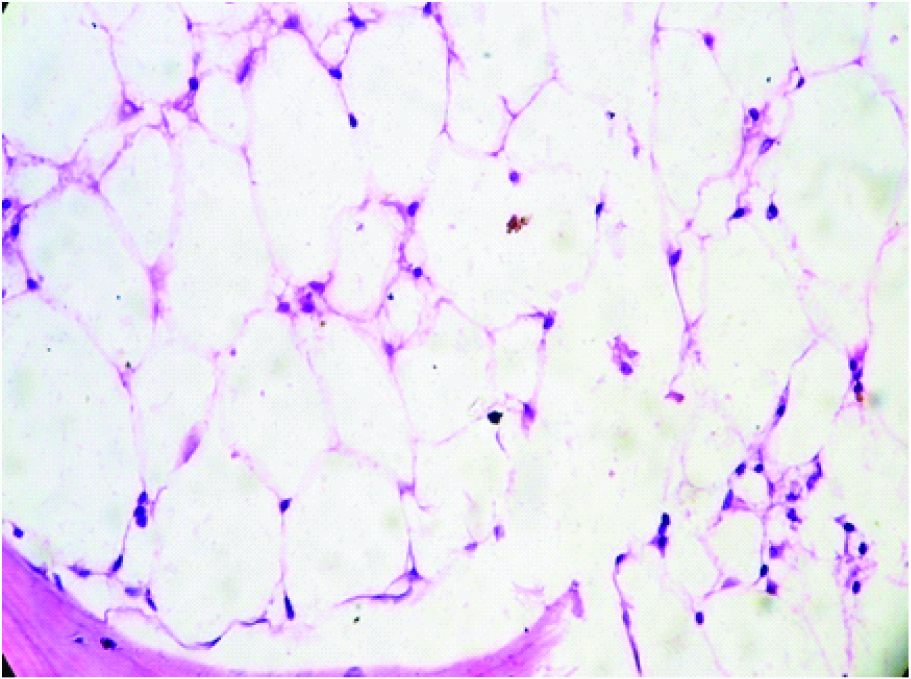
In 9 out of total 50 cases, BMA smear interpretation was suggestive of aplastic anaemia, where as BMB revealed hypocellular marrow with focal hyper cellular areas in 3 cases. Rest of the 6 cases were ultimately confirmed as aplastic anaemia. As BMB gives the qualitative and quantitative assessment of cellularity, therefore confirms the diagnosis and overcomes the limitation of BMA [Table/Fig-8] [8,11].
Due to variability of cellularity from one intertrabecular space to the next, a case which was wrongly diagnosed as MPN on BMA was turned out to be of normocellular marrow with patchy hypercellular areas on thorough examination of BMB. Thus, the use of the biopsy avoids misinterpretation of cellularity by smears [12].
An important limitation of bone marrow obtained by aspirate is the admixing of marrow and sinusoidal blood, which may not allow for reliable estimates of marrow cellularity. Also It is necessary that finding of a ‘dry tap’ should never be dismissed as being due to faulty technique and always needs a bone marrow biopsy for further evaluation [13]. In the present study, out of total 50 cases, 5 cases were reported as bloody tap/dry tap on BMA. Which were turned out to be hypocellualr/ hypercellular marrow on BMB. Also, one case of leukoerythroblastic blood picture, on repeated BMA attempts was found to be of dry tap. BMB showed myelofibrosis along with additional finding of few epithelioid cells. On correlation with clinical details possibility of Koch’s pathology was kept. Reticulin stained sections revealed grade-3 fibrosis. Ch Toi P et al., have mentioned that 80% cases of granulomatous lesions were diagnosed by BMB alone [7] . It was observed in a single treated case of multiple myeloma that marrow was normocellular on aspiration. Whereas, BMB examination revealed the focal collections of plasma cells. Babarovic E and fellows have mentioned the role of BMB for detection of minimal residual disease after treatment in case of multiple myeloma [14].
We have encountered six cases of MPN’s out of which diagnosis was possible to be made from PBF and BMA in 2 cases only. Role of trephine biopsy is not only in differentiation of MPN but also to assess the overall marrow cellularity, histotopography, morphology of megakaryocytes as well as blasts, and degree of Myelofibrosis [Table/Fig-9a,9b]. Rest of the four non diagnostic aspirates in patients who had grade-3 marrow fibrosis highlights the importance of trephine biopsy [Table/Fig-10] [15] . BMA does not have much role in diagnosis of primary myelofibrosis because diffuse osteomyelosclerosis, intrasinusoidal hematopoiesis and vascular proliferation as seen in present study, which is characteristic of primary myelofibrosis, can be confirmed and graded on BMB only. Also, reticulin stain which gives accurate platform for grading of fibrosis to get done is possible on BMB only [Table/Fig-11a,11b] [16] .
There were two cases of NHL in present study where BM biopsy renders information which cannot be determined from aspiration such as spatial distribution and extent of infiltrates, overall cellularity and fibrosis. This also implies that trephine biopsy may be more useful in post chemotherapy patients to assess the residual tumour cell burden and degree of chemotherapy response [17].
There was one case of Chronic Lymphocytic Leukemia (CLL) with diffuse involvement of marrow, which was seen on biopsy section while aspiration showed only that marrow is involved. Bone marrow examination in case of CLL should always include a trephine biopsy because bone marrow aspirate gives very little information beyond that already available from examination of blood [Table/Fig-12a]. Pattern of marrow involvement by leukemic cells could be only analysed by trephine biopsy. Also, trephine biopsy permits an accurate assessment of extent of infiltration and gives information of prognostic importance [Table/Fig-12b] [18] .
In one case of thrombocytopenia in present study BMA and BMB findings were consistent to each other. But an additional finding on BMB was normal arrangement of megakaryocytes which were seen increased in number on BMA [19].
Hence, it was observed from the above discussion that bone marrow evaluation is an important and effective tool in diagnosing and evaluating hematological and non-hematological disorders. Complete evaluation of bone marrow samples includes a brief patient history, hematological profile, BMA smears and biopsy sections [20]. A correlation of bone marrow aspiration and biopsy showed that both the procedure were complementary to each other. The BMA generally provides an excellent cytomorphological details which enables hematopathologist in recognising the abnormal hematopoietic cells or the non-native cells in case of non-hematological disorders. Whereas, a bone marrow trephine biopsy demonstrates the topographic arrangement of hematopoietic cells within the marrow framework and hence gives a more representative view of the cellularity of the marrow and allows infiltration to be recognized clearly. BMB examination has definite edge over BMA in the detection of minimal residual diseases, staging of lymphoma and for the diagnosis of acute leukemia in relapse cases which are otherwise clinically silent.
Showing age and sex distribution of patients
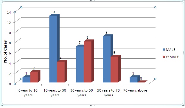
Table showing accepted indications of bone marrow examination
| Accepted indications | No. of cases | Percentage |
| Pancytopenia | 20 | 40 |
| Anaemia | 19 | 38 |
| Acute leukemia | 05 | 10 |
| Chronic leukemia | 04 | 08 |
| Leucoerythroblastic blood picture | 01 | 02 |
| Thrombocytopenia | 01 | 02 |
showing cases with similar findings on bone marrow aspiration and bone marrow biopsy
| PBF* | BMA† | BMB‡ | No of cases | percentage |
| AML§ | AML§ | AML§ | 03 | 06 |
| ALL|| | ALL|| | ALL|| | 02 | 04 |
| Pancytopenia | AML§ | AML§ | 01 | 02 |
| Pancytopenia | ALL|| | ALL|| | 01 | 02 |
| Anaemia | Anaemia | Anaemia | 16 | 32 |
| | Total | 23 | 46 |
*Peripheral Blood Film, †Bone Marrow Aspiration, ‡Bone Marrow Biopsy, §Acute Myeloid Leukemia, ||Acute Lymphoblastic leukemia
Showing cases where findings on bone marrow aspiration were suggestive only.
| PBF* | BMA | BMB | No of cases | percentage |
| Pancytopenia | Hypocellular | Aplastic anaemia | 06 | 12 |
| Pancytopenia | Hypocellular | Hypocellular | 03 | 06 |
| | Total | 09 | 18 |
*Peripheral Blood Film, †Bone Marrow Aspiration, ‡Bone Marrow Biopsy
showing cases where diagnosis was possible only on bone marrow biopsy
| PBF* | BMA† | BMB‡ | No. | Percentage |
| Pancytopenia | Bloody tap | Hypocellular | 03 | 06 |
| Pancytopenia | Dry tap | Hypocellular | 02 | 04 |
| Leucoery-throblastic blood picture | Dry tap | Epithelioid cells | 01 | 02 |
| Anaemia | Normocellular marrow | Focal collection of plasma cells | 01 | 02 |
| | Total | 07 | 14 |
*Peripheral Blood Film, †Bone Marrow Aspiration, ‡Bone Marrow Biopsy
Showing cases where patterns of involvement were seen on biopsy
| Peripheral blood film | Bone marrow aspiration | Bone marrow biopsy | No of cases | Percentage of cases |
| CML* | Bloody tap | Prefibrotic phase of MPN† | 02 | 04 |
| Anaemia | Dry tap | Myelofibrosis with myelosclerosis | 01 | 02 |
| Pancytopenia | MPN† | MPN† with MF‡ (prefibrotic phase) | 01 | 02 |
| Pancytopenia | Bloody tap | MPN† with MF‡ | 01 | 02 |
| Pancytopenia | Dry tap | MPN† with MF‡ | 01 | 02 |
| CLL§ | CLL§ | Diffuse involvement | 01 | 02 |
| Anaemia | NHL|| (> 40% lymphoblasts) | Nodular involvement | 01 | 02 |
| Pancytopenia | NHL|| (> 90%) | Difuse involvment | 01 | 02 |
| Thrombocytopenia | Megakaryocytosis | Megakaryocytosis | 01 | 02 |
| | Total | 09 | 18 |
*Chronic myeloid leukemia, †Myeloproliferative Neoplasm, ‡Myelofibrosis, §Chronic lymphocytic leukemia, ||Non-Hodgkin’s lymphoma
BMA smear of megaloblastic marrow (Leishman stain, 40x)
BMB sections of megaloblastic marrow (H & E, 10x)
BMB sections of Aplastic Anaemia (H & E, 10x)
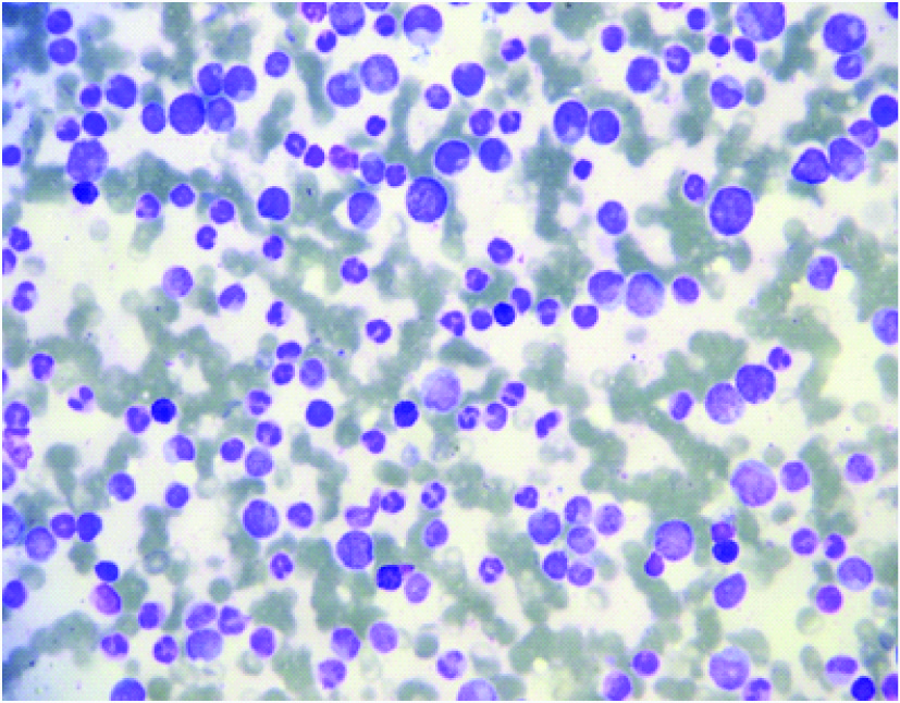
BMA smear of CML (Leishman stain,40x)
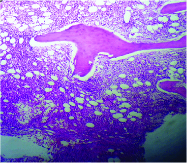
BMB sections of CML with mild degree of MF (H & E, 10x)
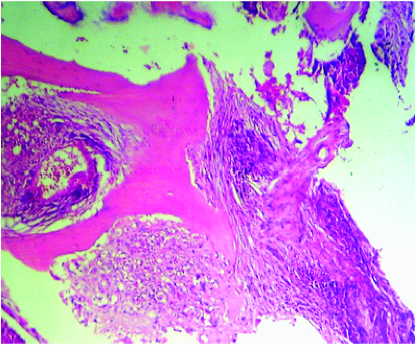
BMB section showing Myelofibrosis in Chronic Myeloid Leukemia (H & E 40x)
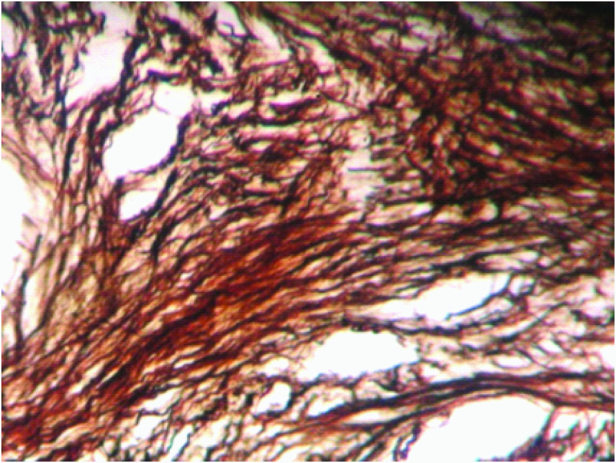
BMB section showing Myelofibrosis Grade2. (Reticulin Stain 100x)
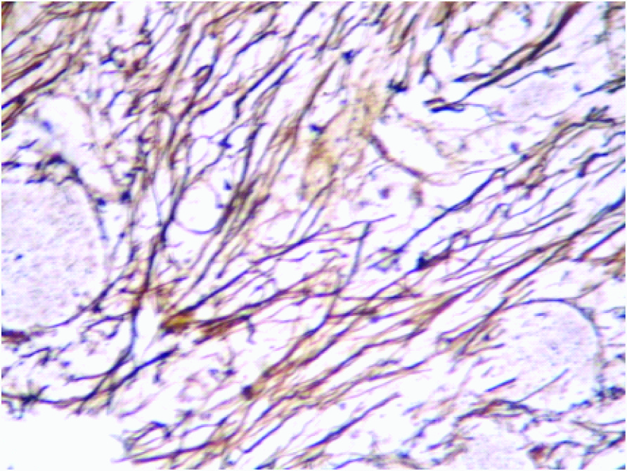
BMB section showing Myelofibrosis Grade-3 (Reticulin Stain 100x)
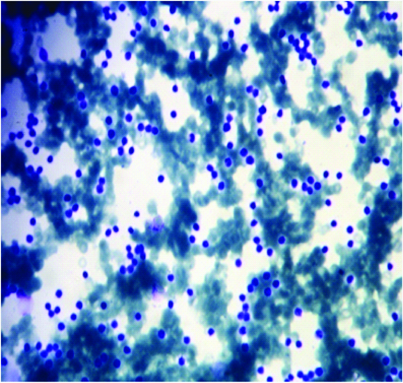
BMA smear showing Chronic Lymphocytic leukemia.(Leishman Stained Smear 40X)
BMB section showing diffuse infiltration of marrow spaces by mature lymphoid cells in Chronic Lymphocytic leukemia (H & E, 40X)

Conclusion
The present study showed that BMA and BMB are easy, rapid, costeffective and more or less are of equal value in various hematological and non-hematological disorders of bone marrow. Although, methods are complementary on correlation but in some cases one or other of these methods is more conclusive and also have important diagnostic value even by using basic standard fixation and embedding procedures, with the aspiration smears being primarily useful for cytological diagnosis and biopsy sections mainly helpful to identify histological features like architectural patterns, grading of fibrosis, pattern of infiltration with lymphomas and granulomatous conditions. Both of the procedures should be done simultaously as they play important role in providing findings, which are mandatory for making final diagnosis.
*Peripheral Blood Film, †Bone Marrow Aspiration, ‡Bone Marrow Biopsy, §Acute Myeloid Leukemia, ||Acute Lymphoblastic leukemia
*Peripheral Blood Film, †Bone Marrow Aspiration, ‡Bone Marrow Biopsy
*Peripheral Blood Film, †Bone Marrow Aspiration, ‡Bone Marrow Biopsy
*Chronic myeloid leukemia, †Myeloproliferative Neoplasm, ‡Myelofibrosis, §Chronic lymphocytic leukemia, ||Non-Hodgkin’s lymphoma
[1]. S Neal, Young Aplastic anaemia, myelodysplasia and related bone marrow syndromes. In: Kasper, Braunwald, Fauci, Hauser, Longo, Jameson, editors.Harrison’s Principles of Internal medicine 2005 1516th EditionNew DelhiMcGraw Hill:617 [Google Scholar]
[2]. JR Krause, An appraisal of the value of the bone marrow biopsy in the assessment of proliferative lesions of the bone marrow.Histopathology 1983 7(5):627-44. [Google Scholar]
[3]. E Gale, J Torrance, T Bothwell, EH Kass, The quantitative estimation of total iron stores in human bone marrow.J Clin invest. 1963 42(7):1076-82. [Google Scholar]
[4]. DE Bauermeister, Quantitation of bone marrow reticulin- a normal range.Am J Clin Pathol. 1971 56:24-31. [Google Scholar]
[5]. RS Riley, TF Hogan, DR Pavot, R Forysthe, D Massey, E Smith, A pathologist’s perspective on bone marrow aspiration and biopsy; Performing a bone marrow examination.J Clin Lab Anal. 2004 18(2):70-9. [Google Scholar]
[6]. A Islam, Bone marrow aspiration prior to bone marrow core biopsy using the same bone marrow biopsy needle. A good or bad practice.J Clin Pathol. 2007 60:212-15. [Google Scholar]
[7]. P Ch Toi, R Varghese G’Boy, R Rai, Comparative evaluation of simultaneous bone marrow aspiration and bone marrow biopsy. An institutional experience.Indian J Hematol Blood Transfus. 2010 26(2):41-44. [Google Scholar]
[8]. SE Stuart Smith, DA Hughes, BJ Bain, Are routine iron stains on bone marrow trephine biopsy specimens necessary?J Clin Pathol. 2005 58(3):269-72. [Google Scholar]
[9]. U Younus, K Saba, J Aijaz, MH Bukhari, S Naeem, Significance of Bone Marrow Histology in the Diagnosis of Acute Myeloid Leukemia.ANNALS. 2011 17(1):5-8. [Google Scholar]
[10]. SM Krober, A Greschniok, Acute lymphoblastic leukemia: correlation between morphological/immunohistochemical and molecular biological findings in bone marrow biopsy specimens.Mol pathol. 2000 53(2):83-87. [Google Scholar]
[11]. R Milosevic, G Jankovic, N Antonijevic, V Jovanovic, D Babic, M Colovic, Histopathologic characteristics of bone marrow in patients with aplastic anaemia.Srp Arh Celok Lek. 2000 128(5-6):200-04. [Google Scholar]
[12]. BJ Bain, Clark DM, Wilkins BS, editors. Bone marrow pathology.Histopathology 2010 4th EditionUK: Wiley Blackwell Publishing:11 [Google Scholar]
[13]. JE Humphries, Dry tap bone marrow aspiration: clinical significance.Am J Hematol. 1990 35(4):247-50. [Google Scholar]
[14]. E Babarovic, T Valkovic, S Stifter, I Budisavljevic, I Seili-Bekafigo, A Duletic-Nacinovic, Assessment of Bone Marrow Fibrosis and Angiogenesis in Monitoring Patients With Multiple Myeloma.Am J Clin Pathol. 2012 137:870-78. [Google Scholar]
[15]. KH Yookarin, D Basu, DK Dutta, Bone marrow trephine biopsy findings in chronic myeloid leukemia.Malaysian Journal of pathology. 2002 24:37-43. [Google Scholar]
[16]. V Mahajan, V Kaushal, S Thakur, R Kaushik, A comparative study of bone marrow aspiration and bone marrow biopsy in haematological and non haematological disorders –An institutional experience.JIACM. 2013 14(2):133-35. [Google Scholar]
[17]. S Goyal, UR Singh, U Rusia, Comparative Evaluation of Bone Marrow Aspirate with Trephine Biopsy in Hematological Disorders and Determination of Optimum Trephine Length in Lymphoma Infiltration.Mediterr J Hematol Infect Dis. 2014 6(1)e2014002. doi: 10.4084/MJHID.2014.002. eCollection 2014 [Google Scholar]
[18]. YS Kim, RJ Ford, JA Faber, B-cell chronic lymphocytic leukemia/small lymphocytic lymphoma involving bone marrow with an interfolicular pattern.Am J Clin Pathol. 2000 114(1):41-46. [Google Scholar]
[19]. J Steven, Jubelirer The role of the bone marrow examination in the diagnosis of immune thrombocytopenic purpura: case Series and Literature Review.Clinical and Applied Thrombosis/Hemostasis. 2002 8:173-76. [Google Scholar]
[20]. Bain B J, Bone marrow aspiration.J Clin Pathol 2001 54:657-63. [Google Scholar]