Vulvar Syringoma: A Rare Case Report
Sarita Nibhoria1, Kanwardeep Kaur Tiwana2, Ashish Yadav3
1 Associate Professor, Department of Pathology, Guru Gobind Singh Medical CollegeFaridkot Punjab, India.
2 Associate Professor, Department of Pathology, Guru Gobind Singh Medical CollegeFaridkot Punjab, India.
3 Junior Resident, Department of Pathology, Guru Gobind Singh Medical CollegeFaridkot Punjab, India
NAME, ADDRESS, E-MAIL ID OF THE CORRESPONDING AUTHOR: Dr. Kanwardeep Kaur Tiwana, H.No. 75 Medical College Campus Faridkot, Punjab-151203, India. Phone : 09815991588 , E-mail : kanwardeepjhajj@gmail.com
Syringoma is a benign eccrine sweat gland tumour affecting mostly females at puberty as multiple soft papules usually 1-2 mm in diameter. The sites of predilection are lower eyelids, cheeks. Syringoma of the vulva is a rare disorder. Only few cases have been reported in the literature. We report here a case of 46-year-old female who presented to gynecology department with vulvar papules associated with vulvar itching and burning since two years. Microscopic examination of the punch biopsy revealed numerous small ducts lined by two layers of epithelial cells embedded in a fibrous stroma. Some of the ducts showed small comma like tails of epithelial cells imparting them a tadpole shape. Despite being a rare diagnosis, vulvar syringoma should be kept in differential diagnosis with vulvar pruritus
Pruritus, Syringoma, Vulva
Case Report
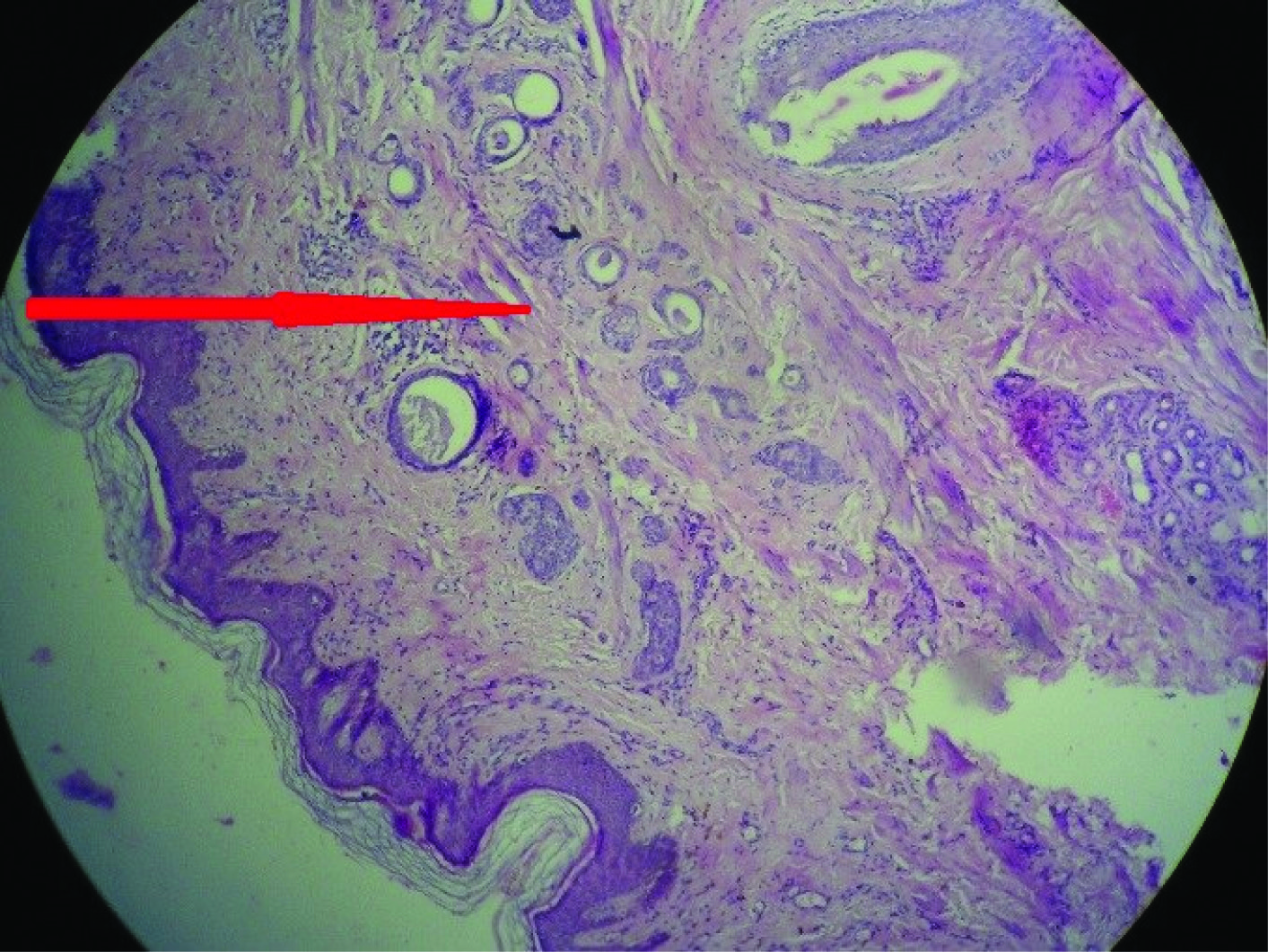
A 46-year-old female presented to gynecology department with complaints of severe vulvar itching and burning since two years. On examination, multiple small papules were present on the labia majora. Punch biopsy of the vulvar lesion was taken and histopathological examination revealed numerous small ducts lined by double layer of epithelial cells embedded in a fibrous stroma [Table/Fig-1]. The lumina of some of the ducts contained amorphous debris. Some ducts had tadpole like shape [Table/Fig-2]. Based on histopathologic findings, the diagnosis of vulvar syringoma was made.
Sections show numerous small ducts lined by two layers of epithelial cells embedded in a fibrous stroma (H& E X100)
Sections show ducts arranged as small comma like tails of epithelial cells imparting them a tadpole shape (H&E X100)

Discussion
Syringoma is a benign adnexal tumour which was first described by Kaposi and Biesiadeki in 1872 [1]. Based on histochemical and electron microscopic findings, the cell of origin of this tumour is intraepidermal eccrine sweat glands [2]. Syringoma is more common in females presenting during adolescence, and most commonly involving the lower eyelid of the face. Other sites of predilection are the cheeks, thighs, axillae and abdomen. Genital syringomas are rare and usually asymptomatic [3]. It can undergo cyclic changes in its size and symptoms presenting during the premenstrual period, pregnancy and with the use of oral contraceptives [1,4].
Syringoma occurs sporadically or it can, on rare occasion, have a hereditary aetiology. Syringoma appears to be more common in patients with Marfan, Down and Ehler’s- Danlos syndromes [4].
Conclusion
Vulvar syringoma although rare entity should be kept in differential diagnosis of pruritic lesions of the vulva.
[1]. Choi HJ, Lee YJ, Park SH, Kim HU, A case of vulvar syringoma with pruritusKorean J Dermatol 2005 43:291-93. [Google Scholar]
[2]. Lever’s histopathology of the skin10th Ed:884-85. [Google Scholar]
[3]. Tay YK, Tham SN, Teo R, Localized vulvar syringoma- an unusual cause of pruritus vulvaeDermatology. 1996 192(1):62-3. [Google Scholar]
[4]. Turan C, Vyur M, Kutlvay Vulvar syringoma exacerbation during pregnancyEur. J. Obstet. Gynaecol Reprod Biol 1996 64:141-42. [Google Scholar]