Fournier’s Gangrene in a Two Year Old Child: A Case Report
Satinder Pal Singh Bains1, Vikram Singh2, Manmeet Kaur Gill3, Amit Jain4, Vivek Arry5
1Senior Resident, Department of Surgery, SHKM, GMC Mewat, Haryana, India.
2Senior Resident, Department of Surgery, SHKM, GMC Mewat, Haryana, India.
3Senior Resident, Department of Pathology, SHKM, GMC Mewat, Haryana, India.
4Assistant Professor, Department of Surgery, SHKM, GMC Mewat, Haryana, India.
5Senior Resident, Department of Surgery, SHKM, GMC Mewat, Haryana, India.
NAME, ADDRESS, E-MAIL ID OF THE CORRESPONDING AUTHOR: Dr. Manmeet Kaur Gill, Department of Pathology, SHKM, GMC Mewat, Haryana, India. Phone : 09876610985,
E-mail: drmanmeet16@hotmail.com
Necrotizing fasciitis of the perineum and external genitalia is a life-threatening infective gangrene, primarily seen in adults. It may be seen at any age but it is relatively uncommon in children. Here, we report a case of Fournier’s gangrene in a two year old male child who was treated aggressively with broad spectrum antibiotics and early surgical debridement with hemodynamic stabilization. Even though no obvious precipitating cause was identified, hygiene was thought to be the inciting factor. Early surgical debridement with appropriate antibiotics and aggressive supportive care gave good results.
Child, Fournier’s gangrene, Hygiene
Case Report
A two-year-old male child was admitted to the General Surgical Unit, with complaints of progressively increasing scrotal swelling with discoloration of the scrotal skin 15 days prior to admission with fever and normal bladder and bowel habits. There was no history of any type of surgical intervention, injury to the perineum or lower abdomen, catheterization, insect bite, or other predisposing conditions.
On examination the child was dehydrated, in poor general condition, febrile and had decreased activities. Scrotal skin was found to be blackish in colour, parchment-like with sharp and clear demarcation with the normal skin. The surrounding normal skin was found to be erythematous and edematous. Other systemic examinations were within normal limits with the exception of mild gaseous abdominal distension.
Investigations revealed a leukocyte count of 16,000/mm3 with 78% neutrophils. Hemoglobin was 10.0 mg/100 mL, blood urea 74mg/100 mL, and serum creatinine 1.8 mg/100 mL. Serum electrolytes were normal. An abdominal X-ray showed the presence of distended bowel loops, whereas an ultrasonography of the scrotum and perineum demonstrated thickened fascial planes with oedema. Chest X-ray was normal.
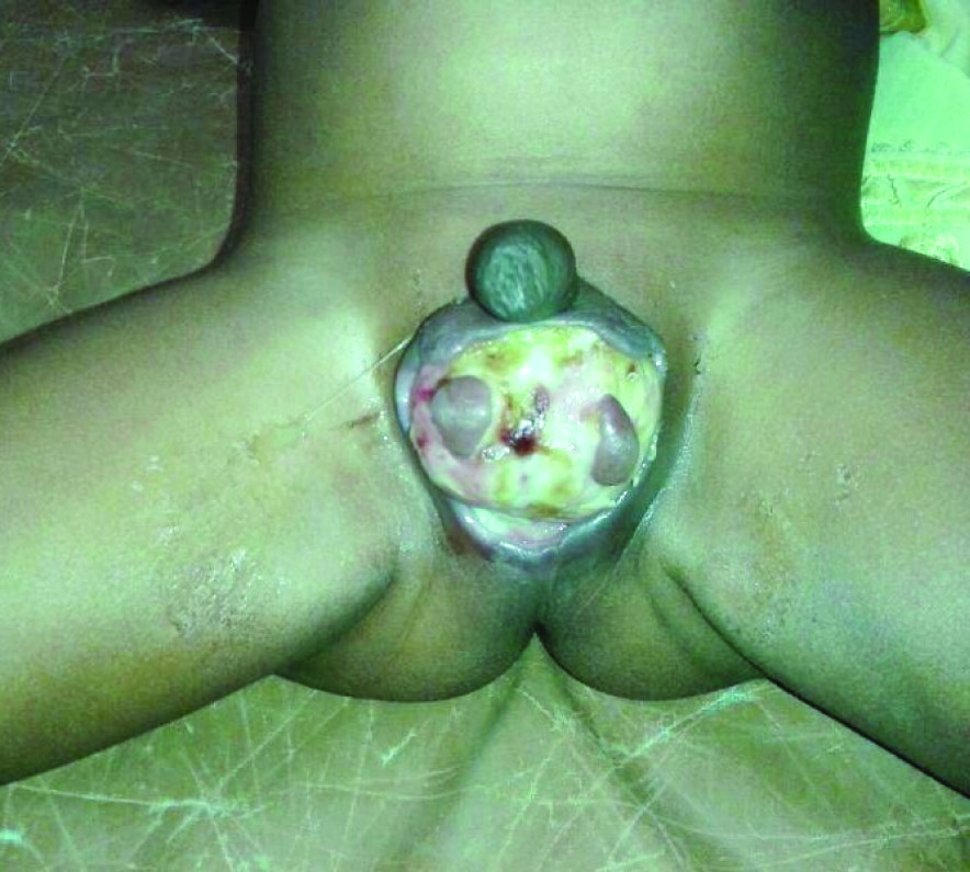
The child was vigorously resuscitated with intravenous fluids and broad spectrum antibiotics, which covered both aerobic and anerobic organisms, in addition to other supportive measures. Surgical debridement was undertaken under general anesthesia with endotracheal intubation and all devitalised and necrotic tissues were excised, up to the level of normal skin until active bleeding was encountered, thus exposing the unaffected testes. The wound was copiously irrigated with dilute hydrogen peroxide solution and normal saline; then packed with a povidone iodine soaked gauze pack. This dressing protocol was continued in the postoperative period [Table/Fig-1]. Wound swab showed growth of Staphylococcus aureus and Klebsiella species and antibiotics were continued according to the sensitivity report.
Subsequent investigations showed progressive fall in blood urea, serum creatinine and total leukocyte count. The blood culture report was negative. Antibiotics and other supportive treatment along with regular dressing were continued in the postoperative period which led to a fairly rapid contraction of the wound. The patient’s parents were properly counselled during the postoperative period regarding the maintenance of proper hygiene and its importance. Secondary repair of the wound was done on the 23rd postoperative Sectionday. Subsequently the child was discharged home after removal of sutures on the 8th day after secondary suturing.
Discussion
Fournier’s gangrene is a serious and aggressive form of infective necrotizing fasciitis involving the perineal region and genitalia due to poly microbial infection [1] . The bacteria act synergistically to produce enzymes such as collagenase and hyaluronidase that invade the fascial planes which leads to vascular thrombosis with subsequent gangrene of the overlying skin [2]. Bacteria further proliferate in these devitalised tissues. Infection from the superficial perineal fascia may spread to the penis and scrotum or to the anterior abdominal wall or vice versa. Testicular involvement is rare as it has a blood supply independent of the affected area, as evident in our case. The condition mostly affects males in the age group of 30-60 years. A 1997 literature review found only 56 paediatric cases, with 66% of those in infants younger than three months [3]. Although, originally described as idiopathic gangrene of the genitalia, Fournier’s gangrene has an identifiable cause in approximately 95% of cases [4]. The necrotizing process commonly originates from an infection in the anorectum, the urogenital tract, or the skin of the genitalia [5]. The reported aetiological factors in the paediatric age group include omphalitis, strangulated hernia, prematurity, diaper rash, varicella infection, circumcision, and perineal skin abscesses
Other causes in children include trauma, insect bites, surgeries or invasive procedures in the perineal region, urethral instrumentation, burns, and systemic infections. In children the causative organisms usually are Streptococci, Staphylococci, and anerobes [6] . In our case, there was no identifiable cause precipitating the condition. The only obvious feature was poor general hygiene.
Usually the diagnosis is clinical, even though a plain X-ray of the region may demonstrate gas in the subcutaneous and other tissue planes. Ultrasound may differentiate it from an intra scrotal pathology [4] .
The management of Fournier’s gangrene includes early and aggressive resuscitation with IV fluids, broad spectrum IV antibiotics and surgical debridement of the necrotic tissue[1] . Our patient also responded to this aggressive modality of treatment and skin coverage was achieved with secondary suturing. Mortality as reported by different authors ranged from 3% to 45% and was due to severe sepsis, coagulopathy and renal failure [7] . Adam et al., reported that paediatric cases have been successfully managed with a more conservative surgical approach and have had a significantly lower mortality rate than adult cases [8] .
Hygiene has a role to play in almost all skin infections and Fournier’s gangrene is no exception. Since this patient had poor hygiene and no obvious inciting factors were found.
Patient of Fournier gangrene on day 2 of post debridement

Conclusion
We have concluded that poor hygiene can be a cause of Fournier’s gangrene in paediatric patient. Proper health education regarding good hygiene of a child to the parents can probably prevent Fournier’s gangrene.
[1]. MC Safioleas, MC Stamatakos, AI Diab, PM Safioleas, The use of oxygen in Fournier’s gangrene.Saudi Med J. 2006 27:1748-50. [Google Scholar]
[2]. SS Lucks, Fournier’s gangrene.Sur Clin N Am. 1994 74:1339-52. [Google Scholar]
[3]. MT Marynowski, AA Aronson, Fournier gangrene.eMedicine. Available from: URL: http://emedicine.medscape.com/article/2028899-overview Accessed on Nov 18, 2013. [Google Scholar]
[4]. GL Smith, CB Bunker, MD Dinneen, Fournier’s gangrene.Br J Urol. 1998 81:347-55. [Google Scholar]
[5]. MD Clayton, JE Fowler, R Sharifi, RK Pearl, Causes, presentation and survival of fifty seven patients with necrotizing fasciitis of the male genitalia.Surg Gynecol Obstet. 1990 170:49-55. [Google Scholar]
[6]. S Dey, KL Bhutia, AK Baruah, B Kharga, PK Mohanta, VK Singh, Neonatal Fournier’s Gangrene. Archives of Iranian Medicine. 2010 13:360-62. [Google Scholar]
[7]. N Eke, Fournier’s gangrene: a review of 1726 cases.Br J Surg. 2000 87:718-28. [Google Scholar]
[8]. JR Adams, JA Mata, DD Venable, DJ Culkin, JA Bocchini, Fournier’s gangrene in children.Urology. 1990 35:439-41. [Google Scholar]