Abdominal Tuberculosis includes tuberculosis infection of gastrointestinal tract, mesentery, lymph nodes and omentum, the peritoneum and related solid organs such as liver and spleen [1,2]. The initial clinical presentations are nonspecific as the disease involves multiple sites with different morphology. No single laboratory investigation is pathognomonic [1]. Radiology often fails to reveal the classical changes described in surgical textbooks. Bacterial culture and tissue histopathology though confirmatory are time consuming, and immunological tests though rewarding are expensive. Moreover, Abdominal Tuberculosis with an acute abdomen presents as an enormous challenge to the surgeon. A surgeon has to rely on his clinical judgement and surgical acumen to determine the extent of surgical management in an unprepared, physiologically compromised patient in the emergency setting. While providing surgical care, the surgeon has to collect sufficient pathological tissue for histopathology and microbiology to overcome the diagnostic dilemma [1,2]. It has to be remembered that though emergency surgery may overcome the temporary crisis of an acute tuberculous abdomen, the permanent cure can only be achieved by a full course of antitubercular medication [1–5].
Materials and Methods
This prospective observational study was conducted in the surgical ward of Calcutta National Medical College over a period of three years from January 2009 till December 2011. The 70 patients included in the study were all adults. All of them presented with an acute abdomen. None of the patients had a previous history of tuberculosis.
A careful history taking and thorough clinical examination was carried out in each case. All the patients were rapidly resuscitated in the surgical ward with intravenous fluids, continous nasogastric aspiration and foley catherisation. Intravenous antibiotics & proton pump inhibitors were started. Blood samples collected from all patients were sent for routine haematology, blood biochemistry & serology to rule out HIV infection. Straight X-ray of abdomen in erect posture was done in all the patients.
Sixty-four out of 70 patients included in the study required surgery. The majority (55) of the patients were put up for emergency exploratory laparotomy after initial resuscitation. Another nine patients required surgical intervention after 24-48 hours of conservative treatment. During surgery macroscopic appearance of the intestine, mesentery, regional lymph nodes suggested the diagnosis of Abdominal Tuberculosis. Surgical intervention was performed depending on surgical pathology. A specimen of diseased tissue was sent for histopathology and tissue culture. In each case the diagnosis of Abdominal Tuberculosis was confirmed. Once abdominal tuberculosis was diagnosed, antitubercular drugs (ATD)were started. However, the remaining six patients responded favorably to conservative management and did not require surgery. Diagnostic studies (USG/CT scan/ guided FNAC for histopathology) carried on these patients confirmed the presence of Abdominal Tuberculosis.
After appropriate management the outcome of each case was studied. Each patient was followed up in the outpatient department for a year after discharge. In each case, it was mandatory to complete a course of ATD.
Results
Out of 718 patients who presented with an acute abdomen, 70 (10%) patients were found to be suffering from Abdominal Tuberculosis. Of the 70 patients studied, there were 27 males and 43 females. Most (64%) females were in the age group of 20-25 years and most (60%) males were in the age group of 35-40 years. Sixty-four out of 70 patients (92%) were from low socio-economic group with poor hygiene and malnutrition. None of the patients had a past history of tuberculosis, 45% of the patients had a positive family history with one or more family members having suffered from any form of tuberculosis in the recent past. The commonest mode of acute presentation in this series was intestinal obstruction (47%) followed by perforative peritonitis (31%), acute appendicitis (10%) and others (12%) [Table/Fig-1]. Diffuse abdominal pain was complained of by all patients. The pain was severe in intensity, acute in onset and accompanied by nausea, vomiting and constipation. All the patients complained of low grade fever, anorexia, disturbed bowel habits over last few weeks to months. More than 50% of female patients complained of menstrual abnormalities like oligomenorrhoea, polymenorrhoea, amenorrhoea and 20% of females complained of primary and secondary infertility. On clinical examination the patients had pallor of varying degrees (90%), abdominal tenderness (87%), muscle guard and rigidity (30%), abdominal lump (20%), ascites (10%) and hyperperistalsis (60%).
Straight X-ray of abdomen in erect posture revealed multiple fluid and gas levels in more than 60% while pneumoperitoneum was found in about 20% of cases. After initial resuscitation, 55 (78.5%) patients were put up for emergency surgery. Another nine patients (12.8%) required surgical intervention after 24-48 hours of conservative treatment. Remaining six patients (8.57%) responded favourably to conservative management. USG or CT scan of abdomen was performed on these patients. Ultrasonography showed thickened bowel loops, enlarged mesenteric lymph nodes, ascites, ileocaecal mass suggestive of Abdominal Tuberculosis. CT scan showed typical findings of mesenteric, omental and peritoneal thickening with dilated matted bowel loops, enlarged lymph nodes with peripheral rim-like enhancement and hypodense centre and adnexal (tuboovarian) mass. Tissue for microbiological or histopathological examination was obtained by US/CT guided fine needle aspiration cytology which later confirmed tuberculosis.
Blood investigations revealed haemoglobin values less than 8 gm/dl in 45% patients, between 8-10 gm/dl in 30% patients and over 10 gm/dl in 25% patients. ESR was raised in 80% of cases and leucocytosis was present in 100% of patients. Sputum was collected from all the patients and sent for detection of AFB. Sputum for AFB was found to be negative in all the cases. Chest X-rays also did not suggest the presence of pulmonary tuberculosis in any of the patients.
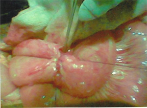
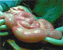
On laparotomy the predominant site of involvement was terminal ileum and ileo caecal region followed by proximal ileum and jejunum. The pathological changes demonstrated on laparotomy included hypertrophic variety of ileocaecal tuberculosis, single or multiple sites of intestinal perforation [Table/Fig-2], multiple small intestinal strictures [Table/Fig-3], mesenteric lymphadenopathy with presence of caseous tubercles, abdominal cocoon, appendicular inflammation, and others [Table/Fig-4].
Types of clinical presentation
| Clinical Presentation | No of Cases | Percentage of Cases |
|---|
| Intestinal obstruction | 33 | 47.1% |
| Perforative peritonitis | 22 | 31.4% |
| Acute appendicitis | 07 | 10% |
| Abdominal pain with lump | 03 | 4.3% |
| Abdominal pain with ascites | 05 | 7.2% |
| Total | 70 | 100% |
Exploratory laparotomy done in a case of acute abdomen with perforative peritonitis revealed tubercular perforation in small intestine shown with tip of dissecting forceps
Multiple tubercular strictures in small intestine in a case of acute intestinal obstruction
Operative findings on exploratory laparotomy
| Pathology Found | No of Operated Cases | Percentage of Cases |
|---|
| 1. Single/ multiple strictures in small gut | 15 | 23.4% |
| 2. Hypertrophic variety in ileocaecal region | 14 | 21.9% |
| 3. Small gut perforation with single or multiple strictures distally | 9 | 14.1% |
| 4. Small gut perforation in hypertrophic variety | 8 | 12.5% |
| 5. Small gut perforation with tubercles | 5 | 7.8% |
| 6. Acute appendicitis with abdominal rculosis | 7 | 10.9% |
| 7. Abdominal cocoon | 3 | 4.7% |
| 8. Mesenteric lymphadenopathy with caseous tubercles and ascites | 3 | 4.7% |
| Total | 64 | 100% |
The surgical procedures included resection of diseased segment and restoration of intestinal continuity (primary anastomosis), creation of a stoma (ileostomy), stricturoplasty, repair of perforation, appendicectomy etc. [Table/Fig-5].
Operative procedure performed
| Operative Procedure | No of Operated Cases | Percentage of Cases |
|---|
| 1. Resection of affected segment of bowel with primary anastomosis | | 9.3% |
| 2. Resection of affected segment of ileum & exteriorisation of ends in 1st stage- stoma reversal with ileoileal anastomosis in 2nd stage. | 19 | 29.7% |
| 3. Limited right hemicolectomy,exteriorisation of ends in 1st stage & stoma reversal with ileocolic anastomosis in 2nd stage. | 17 | 26.6% |
| 4. Repair of perforation (jejuna, ileal) | 2 | 3.1% |
| 5. Exteriorisation of perforated area/ loop | 3 | 4.7% |
| 6. Stricturoplasty | 4 | 6.25% |
| 7.Appendicectomy with biopsy from mesenteric lymph nodes | 7 | 11% |
| 8. Biopsy only and collection of caseous material for histopathology & microbiology with adhesiolysis | 6 | 9.4% |
| Total | 64 | 100% |
Resected portions of gut, mesenteric lymphnodes, omental fragments, caseous material were sent for histopathological and microbiological studies. All patients were proved to be suffering from Abdominal Tuberculosis.
Postoperative complications included respiratory tract infection (15%), wound infection (10%), septicaemia (7%), anastomotic leaks (4.6%), stomal retraction (2%), etc . Postoperative mortality was 5% due to septicaemia and multiorgan failure.
All 70 patients were prescribed ATD for six months (4 drugs:HREZ for two months and two drugs: HR for four months) and were counseled to complete the course. All patients were followed up regularly for a year in the outpatient department after discharge from hospital. Patients with an ileostomy had a stoma reversal after receiving ATD for 10-12 weeks. Out of all studied patients only three patients had an episode of subacute intestinal obstruction in the follow up period which could be managed conservatively. Others were symptom free and their general condition improved satisfactorily. At the time of completion of follow up, all the patients had completed antitubercular medication.
Discussion
In our study there was a female predominance (Female: Male=5:3).This study showed that abdominal tuberculosis is predominantly a disease of young adults with females being affected at an earlier age. These facts bear a marked similarity to the findings mentioned in the literature reviewed [1–3,6–10]. Almost all our patients(92%) were from low socioeconomic group with poor hygiene and malnutrition similar to cases reported in literature [1–9]. 45% of the patients had one or more family members who had suffered from any form of tuberculosis recently. The reviewed literature have reported variable percentages of positive family history in different case series [1–5].
The different modes of presentation as mentioned earlier with their relative frequencies of incidence closely resemble the presentations reported in other series [6–9]. Though all patients presented with symptoms & signs of acute abdomen, all of them also complained of low grade fever, weight loss, anorexia, disturbed bowel habits, abdominal distension, menstrual abnormalities over a variable period of several weeks to months. Symptoms and signs have been reported similarly by other authors with variable percentages of prevalence [3,6–8,10,11].
Blood investigations revealed haemoglobin values less than 10gm% in 75% patients. ESR was raised in 80% of cases and leucocytosis was present in 100% of patients. Results of haematology corroborates the findings of other authors [1–5,8,9]. Sputum for AFB was negative in 100% of cases. Chest x-ray in 100%of cases showed no lesion suggestive of pulmonary tuberculosis. None had an active chest lesion. None of the patients had a past history of tuberculosis. Associated pulmonary disease in Abdominal Tuberculosis has been variously observed in literature [6–8]. The radiological investigation like USG & CT scan all showed features suggestive of Abdominal Tuberculosis. The reviewed literature mentions similar radiological findings in different case series [1,2,5,7,9–11].
HIV infection was not present in any of our patients although the reviewed literature mentions the possibility of co-existence in about 10% of cases [2]. Commonest pathological change found were the presence of intestinal stricture, often multiple in number with or without presence of perforation situated proximally. On laparotomy the predominant site of involvement was terminal ileum and ileo caecal region similar to the findings of other study [1,2,5–12]. 20% of females showed the presence of tuboovarian mass, and tubercles on the serosal surface of tubes and uterus. Laparotomy findings have been variously reported in several series by other authors [6–11].
The choice of surgical procedure depended on site and extent of disease, status of the remaining gut, general condition of the patient, surgeon’s expertise and individual preference. In a considerable number of cases complicated with faecal peritonitis and intraabdominal sepsis, a two stage procedure with creation of a stoma (ileostomy) was preferred to primary anastomosis. Creation of a stoma followed by reversal of stoma in a well prepared gut after 10-12 weeks of ATD therapy reduces the risks of anastomotic leaks and septic complications [8–10]. The operative procedures of different authors varied in different series [6–11].
The most common postoperative complications were respiratory tract infection followed by wound infection which was treated with favourable response. However, three patients had an anastomotic leak which later lead to septicemia, multi organ failure & death. Mortality was nil among the patients who did not require surgery. Similar complications have been described by other authors. The mortality is more or less the same as reported by most authors [6–12].
Conclusion
Abdominal Tuberculosis constituted a significant percentage (10%) of all cases attending the emergency with an acute abdomen. Abdominal Tuberculosis is very difficult to diagnose and diagnosis is often delayed till an acute abdomen is presented with. The most common pathology was intestinal stricture with or without perforation. Almost all patients needed surgical intervention. Prompt surgical exploration, vigilant postoperative care and administration of ATD helped to treat the patients successfully with their complete cure and rehabilitation.