Unilateral Molariform Macrodont Mandibular Second Premolar: An Unusual Case Report in A Nonsyndromic Patient
Prashant Babaji1, Vishwajit Rampratap Chaurasia2, Vinay Kumar Masamatti3, Samarth Tiwari4, Sidharath Malik5
1Professor, Department of Pedodontics, Vyas Dental College, Jodhpur, Rajasthan, India.
2Post Gradute, Department of Conservative Dentistry & Endodontics, KLE’S Dental College, Belgaum, Karnataka, India.
3Senior Lecturer, Department of Conservative Dentistry & Endodontics, KLE’S Dental College, Belgaum, Karnataka, India.
4Senior Lecturer, Department of Periodontology, Bhabha College of Dental Science, Bhopal, Madya Pradesh, India.
5Reader, Department of Periodontics, PDM Dental College Bahadurgarh, Haryana, India.
NAME, ADDRESS, E-MAIL ID OF THE CORRESPONDING AUTHOR: Dr. Prashant Babaji, Professor, Department of Pedodontics, Vyas Dental College, Jodhpur-342001, Rajasthan, India.
Phone: 9460002862,
E-mail: babajipedo@rediffmail.com
Macrodontia is a rare dental anomaly that refers to teeth appears larger than normal. It can be generalized or isolated macrodontia. Isolated macrodontia involving premolar is very rare. This case report presents an unusual unilateral molarifrom macrodontia of mandibular second premolar.
Isolated, Macrodontia, Molariform, Second premolar, Unilateral
Case Report
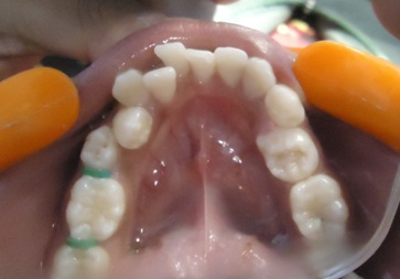
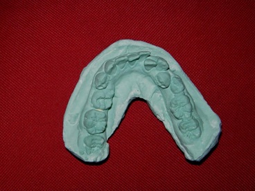
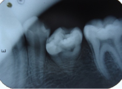
A 14-year-old boy reported for routine dental check up. Patient’s medical and family history was unremarkable. On intra oral examination, mandibular second premolar on left side showed macrodontia, which appear like molariform having multitubericular crown with 6 cusps, 3 on buccal and 3 on lingual sides which is extremely uncommon. The affected tooth was rotated due to large crown size and lack of space. There was a deep groove on occlusal table without any carious lesions. Both the mandibular first molars had 6 cusps i,e. 3 buccal and 3 lingual sides with normal crown and root shape and length [Table/Fig-1,2]. The macrodont premolar was measuring 10 mm mesiodistally and 8 mm bucco-lingually with total tooth length around 16 mm as compared to 22.5 mm for 1st premolar. Radiographic examination showed abnormal size and shape of crown with single tapering short conical root in relation to mandibular second premolar [Table/Fig-3].
Discussion
Anomalies in tooth structure, shape and size results by many factors during morphodifferentiation stage of tooth development [1]. Isolated bilateral macrodontia of mandibular second premolars is an extremely rare dental anomaly with only 10 reported cases till date where as unilateral macrodontia is also extremely rare as reported in our case. Dugmore proposed the term “macrodont molariform premolars” to macrodont premolars [2]. We suggest a term “molariform macrodont premolars” to teeth which are multitubericular” as most suitable one. Macrodontia is usually associated with systemic disturbances or certain syndromes such as, insulin-resistant diabetes, pineal hyperplasia with hyperinsulism, facial hemihyperplasia, KBG syndrome, Otodental syndrome, Ekman-Westborg and Julin trait syndrome, 47 XYY syndrome, Schinzel-Giedion syndrome, Duboitz syndrome and Cockayne’s syndrome [1,3]. The present case reports an isolated macrodont second premolar in nonsyndromic patient.
Macrodont teeth can create variety of functional and esthetic problems, depending on their size and morphology, which requires endodontic, prosthetic, surgical or orthodontic treatment [1]. Often macrodont teeth are unable to erupt due to lack of space for eruption[2,3]. Isolated macrodont premolar teeth are also present in multituberculism as seen in our case. Due to anatomic variation and deep grooves, these teeth may be prone to caries [1,3]. Most of the authors suggested extraction followed by orthodontic correction of malocclusion as a treatment strategy for isolated macrodontia with relation to premolars. Radiographic and clinical examination is helpful for early diagnosis and intervention [3].
Photograph of mandible showing macrodont molariform second premolar on left side
Photograph of mandibular cast showing macrodont molariform second premolar on left side and six cusps on permanent first molars
Intra-oral periapical radiograph showing macrodont mandibular second premolar with conical root
Conclusion
Isolated unilateral macrodontia of the mandibular second premolar is an extremely rare dental anomaly, which requires multidisciplinary treatment approach.
[1]. R Furentes, E Boriei, Bilateral macrodontia of mandibular second premolars: a case report.J Morhol Sci. 2011 28(3):212-15. [Google Scholar]
[2]. CR Dugmore, Bilateral macrodontia of mandibular second premolars: A case report.Int J Paediatric Dent. 2001 11:69-73. [Google Scholar]
[3]. E Canoglu, H Canoglu, A Aktas, ZC Cehreli, Isolated bilateral macrodontia of mandibular second premolars: A case report.Eur J Dentistry. 2012 6:330-34. [Google Scholar]