Tinea Barbae: In Released from Treatment (RFT) Hansen’s Disease Patient
Pugazhenthan Thangaraju1, VC Giri2, Hosanna Singh3, Vinod Kumar4, Showkath Ali5
1Medical Officer, Central Leprosy Teaching and Research Institute, Ministry of Health and Family Welfare, Government of India, Chengalpattu, Tamilnadu, India.
2Assistant Director (Epid), Central Leprosy Teaching and Research Institute, Ministry of Health and Family Welfare, Government of India, Chengalpattu, Tamilnadu, India.
3Medical Officer, Central Leprosy Teaching and Research Institute, Ministry of Health and Family Welfare, Government of India, Chengalpattu, Tamilnadu, India.
4HOD, Clinical Division,Central Leprosy Teaching and Research Institute, Ministry of Health and Family Welfare, Government of India, Chengalpattu, Tamilnadu, India.
5Director, Central Leprosy Teaching and Research Institute, Ministry of Health and Family Welfare, Government of India, Chengalpattu, Tamilnadu, India.
NAME, ADDRESS, E-MAIL ID OF THE CORRESPONDING AUTHOR: Dr. Pugazhenthan Thangaraju, Central Leprosy Teaching and Research Institute, Ministry of Health and Family Welfare, Government of India, Chengalpattu, Tamilnadu, India.
Phone: 9486279090,
E-mail: drpugal23@gmail.com
Hansen Disease (leprosy) is a chronic inflammatory infectious disease that primarily targets skin, nerves and other internal organs (testis, liver etc.) caused by the acid fast intracellular bacilli, Mycobacterium leprae. Clinical presentation occurs with a wide spectrum including hypo pigmented anaesthetic patches, raised erythematous plaques and nodules and thickened peripheral nerves showing tenderness. The most important complication is the disability and deformity. The diagnosis of leprosy is frequently delayed because of its similarity with other more common skin conditions prevailing in some non endemic areas. We present a rare case report of tinea barbae in an old treated case of leprosy. This case is one of the rarest fungal infection in leprosy patient searched in PUBMED as there were other more tinea infection involving various sites in body which sometimes misdiagnosed as leprosy.
Fungal infection, Multibacillary multidrug treatment
Case Report
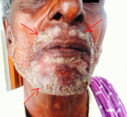
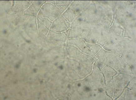
A 54-year-old, low socio-economic male from Kanchipuram presented to central leprosy teaching and research institute (CLTRI), Ministry of Health and Family Welfare, Goverment of India, Chengalpattu, Tamilnadu, India for the treatment of a symptomatic itching lesion that had been present for more than two months. He was previously treated for leprosy with multibacillary multidrug treatment adult (MBMDT -A) for 12 months in CLTRI and did not have any significant history related with any kind of reactions or toxicities. He had similar itching lesion in the same area a year back for which he was treated with drugs namely grisofulvin 250mg for 15 days from Chengalpattu Medical College and found normal thereafter. Physical examination revealed amputated right hand and scaly lesions distributed over the right mantle region and frenulum and some spots in the left mantle shown in [Table/Fig-1a]. As a routine investigation complete haemogram, biochemistry investigations (renal function test, lipid profile, liver function test, diabetic profile) were done and were within normal range. The clinical impression was suggestive of fungal infection and as tinea barbae on the basis of distribution of the lesion and the patient is given treatment with oral itraconazole 400 mg/day for two days in a week after confirming the diagnosis with light microscope [Table/Fig-1b]. Case was advised to return to the clinic after 15 days for follow up.
Discussion
Hansen Disease (leprosy), as the stigma attached with the word leprosy hence Hansen disease is used in remember of scientist Dr. Hansen who discovered bacilli in 1874, is a chronic gradually progressing infectious disease caused by an acid fast bacilli Mycobacterium leprae an obligate intracellular bacterium. It is endemic in tropical and subtropical regions with declining prevalence in India as of today [1]. As with other infectious disease, leprosy has a broad spectrum of clinical presentations. Because of its longer incubation period it is left out with the most fearful disability and deformity. Much stigma attached with disease involving the emotional background of patients and treated persons. The cardinal signs of leprosy are hypo-pigmented patches; thickened tender peripheral nerve, erythematous lesions, hypoesthesia and presence of other neurological deficits. Leprae has a predilection for cool areas of the body sparing the main body core explaining the tendency of leprosy lesions to occur on the extremities, nose, and ears. Present case report is a case of an old leprosy treated man with MBMDT A for 12 months and is under surveillance period, with an itchy lesion in and around mouth as a rare presented fungal infection.
The case which is presented in this report is one of the rarest presentations even though fungal infection as a whole is common in immunocompromised persons [2]. On search, with medical library site PUBMED, it was found that there were no reports on leprosy and tinea barbae. Another search in eHealth Me under the title “Review: could Leprosy cause Tinea barbae?” a study of 53 patients with leprosy for tinea barbae also revealed the same. Deep mycotic infections like chromoblastomycosis, mycetomas and rhinosporidiosis also have been reported in leprosy patients [3,4]. The mechanism behind the deep fungal infection is mainly due to the immunocomprised state occurred during active infection and steroid intake in reaction patients. No associations were found in literature regarding tinea barbae in leprosy patient who was released from treatment. In the context of the review for leprosy and barbae, this case might add knowledge of its occurrence in Indian population.
Showing the white scaly lesion distributed in and around mouth and beard bearing area
Showing branching filamentous structure evidence of superficial dermatophytes
[1]. World Health Organization. Global leprosy situation 2012.Wkly Epidemiol Record. 2012 34:317-28. [Google Scholar]
[2]. Thangaraju Singh Harmanjit, chakrabarti Amitava, Treatment of Deep and Superficial Infections of Candida-What we know and what is new.Int J Pharm Sci Res. 2013 4(7):2562-68. [Google Scholar]
[3]. K Pavithran, Chromoblastomycosis in a residual patch of leprosy.Indian J Lepr. 1988 60:444-47. [Google Scholar]
[4]. J Jayakumar, M Aschhoff, G Renuka, Mycetomas in leprosy.Indian J Lepr. 1993 65:229-33. [Google Scholar]