Emergency Diagnosis of Giant Cell Tumour (GCT) of Spine by Image Guided Fine Needle Aspiration Cytology (FNAC)
Prem Singh1, Manish Chaudhry2, Amitoj Singh3
1Associate Professor, Department of Pathology, MM Institute of Medical Sciences and Research, Mullana, Ambala, Haryana, India.
2Associate Professor, Department of Pathology, MM Institute of Medical Sciences and Research, Mullana, Ambala, Haryana, India.
3Resident, Department of Radiodiagnosis, MM Institute of Medical Sciences and Research, Mullana, Ambala, Haryana, India.
NAME, ADDRESS, E-MAIL ID OF THE CORRESPONDING AUTHOR: Dr. Prem Singh, Associate Professor, Deptartment of Pathology, MM Institute of Medical Sciences and Research, Mullana, Ambala, Haryana-133047, India.
Phone: 09896711727,
E-mail: premsingh011@rediffmail.com
Giant cell tumour (GCT) of spine is an extremely rare neoplasm accounting 0.5% to 1.5% of all cases. The patient usually presents with weakness of lower limbs. We describe a case of 25-year-old male who presented with sudden onset of paraplegia. On plain radiograph there was an osteolytic lesion in T9 vertebra. Computed tomography (CT) scan revealed expansile lytic lesion in T9 vertebral body with involvement of posterior elements on right side with associated soft tissue mass in the extradural location extending into the spinal cord. Further Magnetic Resonance Imaging (MRI) scan (T1 contrast) showed the enhancing extradural mass involving spinal cord from D 8-10 levels. A provisional radiological diagnosis of GCT was made. A CT guided FNAC of the mass was performed which revealed typical cytological features of Giant cell tumour. Role of image guided Fine Needle Aspiration Cytology (FNAC) of vertebral mass and its role in emergency situations with clear emphasis on differential diagnosis is highlighted.
Giant cell tumour, Paraplegia, Vertebral mass, Stromal cells
Case Report
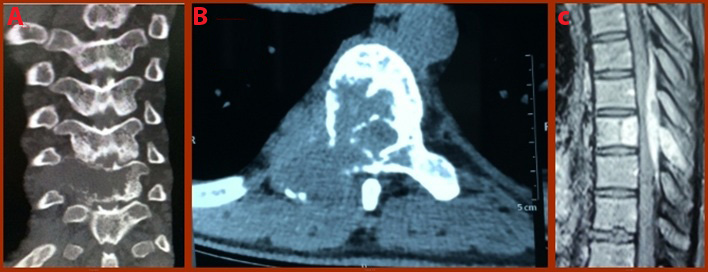
A 25-year-old male patient presented with sudden weakness of both lower limbs of 18 days duration in Department of Orthopaedics of a rural teaching and tertiary care hospital. The weakness was preceded by sudden pain in upper portion of back after lifting of heavy weight. Subsequently the weakness of both legs increased progressively. On general examination he was afebrile. Systemic examination was normal. On local examination of the spine no obvious swelling was noticed. Neurological examination revealed muscle power grade 1/5 in both lower limbs and hypoesthesia with sensory alteration below approximately T-8 dermatome. Laboratory investigations including complete blood count, erythrocyte sedimentation rate, serum calcium, renal and liver function tests were all normal. On plain radiograph there was an osteolytic lesion in T9 vertebra. Computed tomography (CT) examination revealed expansile lytic lesion in T9 vertebral body with involvement of posterior elements on right side with associated soft tissue mass in the extradural location extending into the spinal cord. Further MRI scan (T1 contrast) showed the enhancing extradural mass involving spinal cord from D8-10 levels [Table/Fig-1]. A provisional radiological diagnosis of GCT was made.
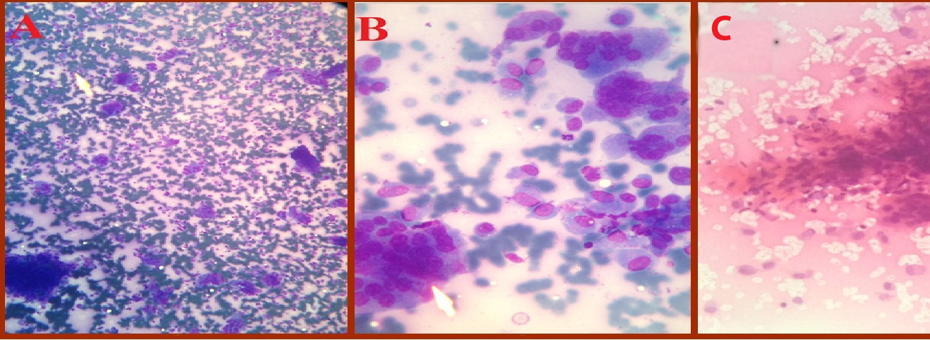
Patient was referred to Department of Cytopathology for FNAC. A written consent of patient for FNAC was taken. CT guided needle aspiration of the mass was performed in Department of Radiodiagnosis under all aseptic conditions with 18 gauge needle. Two passes were given. The aspirated material was smeared on glass slides, some air dried and remaining fixed in 95% alcohol. The smears were stained with May-Grunwald Giemsa (MGG) and Haematoxylin Eosin (H&E) stains. There were no complications like hematoma and sepsis following the procedure. The smears were cellular and showed cohesive as well as dispersed cell clusters. A double cell population consisting of mononuclear spindle (stromal) and giant cell of osteoclastic type attached to periphery of clustered spindle cells were seen. Both types of cell population, stromal cells and giant cells were mostly close intermixed. Mononuclear cells had moderate amount of dense ampophilic cytoplasm which was vacuolated at places and well defined borders. Nuclei were ovoid with bland chromatin, small nucleoli and exhibiting mild pleomorphism. The giant cells mostly intermixed with mononuclear cells, had 15-20 nuclei which resembled nuclei of stromal cells [Table/Fig-2].
A diagnosis of GCT of T9 vertebra was made without any cytological evidence of malignant transformation.
(A) CT image Coronal view of dorsal spine showing lytic expansile lesion in right half of T 9 vertebra with associated soft tissue mass. (B) CT image Axial view T9, shows involvement of vertebral body and posterior elements with associated soft tissue mass extending into spinal canal. (C) MRI scan T1 Contrast Saggital view: showing the enhancing extradural mass involving spinal cord from D 8-10 levels
(A) FNAC smear showing dual population of mononuclear cells and osteoclastic giant cells (MGG, X100). (B) Higher magnification showing bland nuclei of the stromal and osteoclastic giant cells (MGG, X400). (C) Figure show typical arrangement of clustered spindle cells with osteoclastic giant cells attached to the periphery of cluster (H&E,X200)
Discussion
Giant cell tumour (GCT) is relatively rare neoplasm constituting approximately 5% of bone tumours and is frequently found in long bones. Bone affection of the axial skeleton is extremely rare and is seldom reported in literature [1]. It commonly occurs in skeletally mature individuals between the ages of 20-40 years [2]. GCT in the vertebral column above the sacrum represents approximately 0.1% to 0.25% of the bone tumours [3].
GCT is a locally aggressive neoplasm, clinically possessing metastatic potential. It can also involve pelvic bones, vertebral bodies and small bones of digits [4]. GCT of the bone is a tumour of unknown histogenesis with distinct morphology. Whenever the patient presents with sudden onset of paraplegia with a radiological evidence of osteolytic lesion of vertebral body, an image guided FNAC of the lesion is likely to clinch the diagnosis [5].
The patients of GCT of the spine often complain of back pain in the beginning and as the tumour enlarges, a progressive weakness of the lower extremities occurs due to compression of spinal cord or nerve roots [3]. The diagnosis of osseous lesion anywhere in the body requires a combined clinical, radiological and histopathological correlation. On routine radiograph, GCT presents as an initially eccentric expanded lytic lesion, without a surrounding sclerotic halo, representing the cortical bone. As the lesion grows, it can encompass the entire cicumference of the bone, causing rupture of cortical bone, but a periosteal reaction is rarely seen [4]. In some cases of GCT clinical and radiological features are very typical suggestive of diagnosis, but in some these are inconclusive, where the diagnosis is made by open and needle biopsy. However, preoperative FNAC diagnosis is now well-documented with studies revealing clear benefits over both open and needle core biopsies [4]. The FNAC smears aided by clinical and radiographic parameters are fairly characterised to suggest the diagnosis of GCT of bone. Before concluding the diagnosis on FNAC, tumours with giant cells and tumour like lesions need exclusion. These include chondroblastoma, aneurysmal bone cyst, brown tumour of hyperparathyroidism, giant cell tumour of tendon sheath and giant cell rich osteosarcoma. In chondroblastoma smears reveal dispersed non-cohesive mononuclear cells having round to oval nuclei, with occasional nuclear grooves. The giant cells are sparser with fewer nuclei. Chondroid matrix noticeably is present in the background [6,7]. Aneurysmal bone cyst yields frank blood with little cellular material of occasional osteoclasts, osteoblasts and fibrous strands. The brown tumour smear show scattered mononuclear and giant cells. The giant cell tumour of tendon sheath is generally an extra osseous lesion with dispersed mononuclear and giant cells. The giant cells are dissociated from mononuclear cells, very scanty in number and may be small in size. Occasional collagen strands and macrophages are seen. Giant cell rich osteosarcoma shows giant cells of osteoclastic type and/or malignant tumour giant cells, but clue to the diagnosis is the striking atypia and malignant nature of the mononuclear tumour cells, irrespective of presence or absence of osteoid [5].
Management of giant cell tumour of spine is a great challenge and the surgical approach remains the best treatment option for GCT of spine, but the required surgical expertise, sophisticated and dedicated spine centres are not available everywhere in the world. The other alternative options include embolizations, cryosurgery and chemotherapy which should be explored intensively[8].
Conclusion
Sole presentation of GCT as paraplegia is very rare. Some cases require a diagnosis on emergency basis to initiate the treatment and in such cases FNAC is an infallible tool to clinch the diagnosis. It is an easy, rapid, relatively safe and cost effective diagnostic modality that can be used for both outpatients and inpatients.
[1]. CL Hunter, D Pacione, M Hornyak, R Murali, Giant-cell tumours of the cervical spine: case report.Neurosurgery. 2006 59:E1142-43. [Google Scholar]
[2]. M Balke, MP Henrichs, G Gosheger, H Ahrens, A Streitbuerger, M Kohler, Giant cell tumours of the axial skeletonSarcoma. 2012 Article ID 410973, 2012. Doi:10.1155/2012/410973 [Google Scholar]
[3]. D Refai, GP Dunn, P Santiago, Giant cell tumour of the thoracic spine: case report and review of the literature.Surg Neurol 2009 71:228-33. [Google Scholar]
[4]. R Aggarwal, U Bandlish, S Mahajan, N Gupta, R Rambani, Role of Fine Needle Aspiration Cytology in vertebral body lesionsIndian J Orthop [serial online]. 2002 36:46-8.[cited 2014 Feb 7] [Google Scholar]
[5]. AR Khan, BA Mir, FNAC- Giant cell tumour of bone: A study of 13 casesJK-Practitioner. 2003 10:236-38. [Google Scholar]
[6]. V Fanning, N Sneige, C Canrasco, A Ayala, J Marray, A Raymound, Fine needle aspiration cytology of ChondroblastomaCancer. 1990 69:1847-63. [Google Scholar]
[7]. S Kilpatrich, E Pik, K Geisinger, W Ward, Chondroblastoma of bone. Use of fine needle aspiration biopsy and potential diagnostic pitfallsDiagn Cytopathol 1997 16:65-71. [Google Scholar]
[8]. A Bakhsh, KM Ali Siddiqui, G Moumneh, T El Maghrabi, Giant cell tumour of C2 (axis): A case report and short review of literatureClin Cancer Investig J 2014 3:102-04. [Google Scholar]