Deep bite is a clinical problem not to be seen in terms of millimeters but to be seen in light of future changes in the aesthetics, function and health of the dentition [1]. Possible complications of deep bite include, temporomandibular joint disorders, unacceptable facial aesthetics, attrition of incisors, spacing of maxillary incisors, clenching of teeth, jaw stiffness, head ache and ringing in ears [2].
Methods to correct deep bite include extrusion of posterior teeth, relative intrusion of incisors and true intrusion of incisors [3,4]. True intrusion of incisors is primarily indicated in deep bite cases with a large vertical dimension, patients with excessive incision stomion distance and a large inter labial gap. Advantages of true intrusion of anterior teeth include achievement of lip competency, reduced incisal exposure without any increase in lower anterior facial height [3].
Appliances for incisor intrusion include utility arch by Ricketts, Burstone intrusion arch, Connecticut intrusion arch, and J-hook headgear. The major disadvantages with these appliances include extrusion and tipping of posterior teeth, complex wire bending and patient co-operation.
Mini screws have been successfully used as temporary anchorage devices for producing various tooth movements [5]. Recently, Mini screws as effective temporary anchorage devices have occupied a central role in a typical orthodontic setup, since anchorage control and patient cooperation are very critical. Many authors have documented the use of mini implants for intruding incisors and have reported statistically significant amounts of incisor intrusion with Minimplants [6–9]. This study aims at evaluating and comparing the intrusion effects on maxillary incisors by mini implant anchorage, j-hook headgear and utility arch.
Materials and Methods
The study was conducted on 30 patients (19 females and 11 males) and the average age range was 16-22 years. The study was proposed at the Institutional Ethical Committee of The Tamil Nadu Government Dental College and Hospital, Chennai, India with all the details regarding the study, and the approval was obtained. The study was conducted during the period of April 2009 to August 2010 at the Department of Orthodontics, The Tamil Nadu Government Dental College and Hospital, Chennai, India. Informed written consent was obtained from all the subjects who were willing to participate in this study. The Subject inclusion criteria were, 1) deep bite cases (overbite >4mm) with excessive incisal display at rest and at smile 2) subjects with average to vertical growth pattern and adequate bone support. Subject exclusion criteria were 1) Skeletal deep bite cases and Horizontal growers, 2) Cases with advanced alveolar bone loss and patients with systemic diseases which contraindicate orthodontic treatment.
The subjects were divided into three Groups:
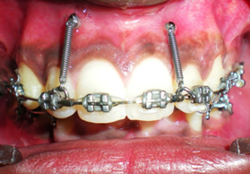
1. Group 1- consisted of 10 subjects, for whom intrusion of maxillary incisors was attempted with mini implant anchorage [Table/Fig-1].
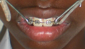
2. Group 2- consisted of 10 subjects, for whom intrusion of maxillary incisors was attempted with J-hook headgear [Table/Fig-2].
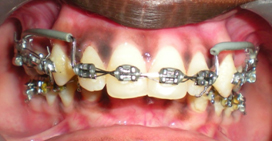
3. Group 3- consisted of 10 subjects, for whom intrusion of maxillary incisors was attempted with utility arch [Table/Fig-3].
The subjects were randomly allocated to each Group. Subject withdrawal criteria included 1) non reporting cases 2) subjects not wearing j hook headgear daily. None of the subjects were withdrawn in Group 1, one subject was withdrawn from Group 3 for not reporting and two subjects were withdrawn from Group 2 as they were not wearing j-hook head gear daily for the advised time period.
Group1
In this Group, two mini implants 6 mm length, 1.4 mm diameter, (Absoanchor by Dentos, Daegu Korea) were used. They were placed bilaterally between the maxillary central and lateral incisor under local anaesthesia with a long hand drive. The mini implant position was checked with an Islands Organic Producers Association (IOPA) after placement to rule out any root contact. The subjects were treated with pre adjusted edge wise mechanotherapy with first premolar extraction. The base arch wire 19×25 stainless steel was sectioned distal to the lateral incisor. Orthodontic load was applied by NITI closed coil springs (3M, Monrovia, California) of different sizes. One end of the spring was engaged on the implant and the other on the arch wire. Force was measured using a Dontrix guage and adjusted to 1.5 ounces on each side and subjects were reviewed once in three weeks.
Group 2
All subjects were treated with preadjusted edgewise mechanotherapy and maxillary first premolar extraction. The base arch wire was 19x25 S.S. J-Hooks were adapted on to the arch wire between the maxillary central and lateral incisors. Force was delivered by an elastic strap connected to an occipital pull headgear. The amount of force delivered was two ounces each side measured using a Dontrix gauge. Monthly appointments were given to recheck and adjust the amount of force applied, patient compliance and any appliance breakage. All subjects were requested to wear the headgear at night for at least 10h.
Group 3
All subjects were treated with pre adjusted edge wise appliance and maxillary first premolar extraction. Ricketts utility arch made of 19×25 Blue Elgiloy was used for intrusion of the maxillary incisors. The utility was sleeved to prevent any tissue irritation. It was also cinched back to prevent incisor proclination. The amount of force delivered was 1.5 ounces on each side. A Dontrix gauge was used to check the force applied and monthly appointments were given to adjust the amount of force applied.
Diagnostic Records
Lateral cephalogram, maxillary anterior occlusal radiograph, and intra oral periapical radiograph were taken before beginning intrusion of maxillary incisors in all the three Groups. Immediately after intrusion lateral cephalograms were taken to measure the amount of intrusion. All radiographs were taken by a single operator at the department of radiology. All lateral cephalometric radiographs were manually traced on an acetate paper with a sharp 3H pencil on a view box by same operator and rechecked randomly. Extra oral and intraoral photographs were taken before beginning the study and after completion. All the photographs were taken by a Nikon digital camera. Study duration was 120 days in all the three Groups.
Cephalometric Analysis
Cephalometric analysis was done to satisfy the selection criteria and to measure the amount of intrusion effects produced in all the three Groups. The parameters used to measure intrusion were.
Overjet, Overbite, Vertical distance from maxillary incisal edge to palatal plane (PP-U1), Vertical distance from maxillary molar cusp to palatal plane (PP-U6), Vertical distance from maxillary incisal edge to upper lip (UL-U1).
Results
The pre-treatment and post-treatment cephalograms were traced and the values were recorded. Arithmetic mean and standard deviation were calculated for all the pre and post-treatment cephalometric parameters in the three Groups. The arithmetic mean and standard deviation of the three Groups are given in [Table/Fig-4].
Arithmetic mean and standard deviation
| Treatment | Groups |
|---|
| Group 1 | Group 2 | Group 3 |
|---|
| Mean | SD | Mean | SD | Mean | SD |
|---|
| Over jet - Pre | 8.58 | 1.20 | 8.00 | 2.26 | 7.75 | 2.14 |
| Over jet - Post | 6.92 | .66 | 7.20 | 2.17 | 7.33 | 1.51 |
| Over bite - Pre | 6.25 | 1.08 | 6.20 | .84 | 7.08 | 1.96 |
| Over bite - Post | 3.92 | .92 | 5.40 | .55 | 5.08 | 2.01 |
| PP U1 - Pre | 31.33 | 2.58 | 29.80 | 2.14 | 30.25 | 2.54 |
| PP U1 - Post | 29.25 | 2.52 | 29.70 | 2.28 | 28.92 | 2.87 |
| PP U6 - Pre | 27.17 | 2.80 | 24.90 | 1.47 | 26.33 | 2.23 |
| PP U6 - Post | 27.00 | 3.02 | 25.10 | 1.43 | 27.08 | 2.13 |
| UL U1 - Pre | 7.33 | 3.09 | 8.90 | 1.29 | 6.08 | 2.06 |
| UL U1 - Post | 5.42 | 2.76 | 8.10 | .74 | 4.67 | 1.60 |
Student’s t- test was used to assess significance of difference in the pre and post-treatment changes in the individual Groups. ANOVA was done to assess the significance of difference in pre and post-treatment values among the Groups. p-value <.05 was considered significant. All the analysis was carried out with statistical analysis software (stat view, SPSS). Tukey HSD was done for multiple comparisons.
In Group1 significant reduction in over bite (p<0 .05), PP-U1 (p< 0.05) and UL-U1 (p<0.05) were noted, and no significant change in PP-U6 (p>0.05) was noted.
[Table/Fig-5] gives the student t-test values for all the three Groups.
Student’s t-test for all three Groups
| Group 1 | Group 2 | Group 3 |
|---|
| Treatment | T | Sig. (2-tailed) | T | Sig. (2-tailed) | T | Sig (2-tailed) |
|---|
| Over jet - Pre Over jet - Post | 3.953 | .011* | 3.138 | .035* | 1.112 | .317 |
| Over bite - Pre Over bite - Post | 11.068 | .000** | 4.000 | .016* | 3.873 | .012* |
| PP U1 - Pre PP U1 - Post | 25.000 | .000** | 1.000 | .374 | 5.394 | .003* |
| PP U6 - Pre PP U6 - Post | 1.000 | .363 | -1.633 | .178 | -4.392 | .007* |
| UL U1 - Pre UL U1 - Post | 9.550 | .000** | 1.725 | .160 | 5.222 | .003* |
Note: * denotes-p <.05, ** denotes – p<.001.
In Group 2, significant reduction in over bite (p<.05), over jet (p<.05) were noted and no significant changes in PP-U1 (p>.05), PP-U6 (p>.05) and UL-U1 (p>.05) were noted.
In Group 3 (utility arch Group), significant reduction in overbite (p<.05), PP-U1 (p<.05) and UL-U1 (p<.05) were noted. Also, significant increase in PP-U6 (p<.05) was noted.
Statistically significant reductions in over bite, PP-U1 (p<.05), PP-UL (p<.05) noted among the three Groups. Statistically significant increase in PP-U6 (p<.05) was also noted among the three Groups.
Greater reductions in overbite, PP-U1and UL-U1 noted in Group1 followed by Group 3 and least in Group 2. Greater increase in PP-U6 was noted in Group 3 followed by Group 2 and least in Group1. [Table/Fig-6] gives the results of ANOVA used for assessing the significance of difference in pre and post-treatment values among the three Groups.
ANOVA-for assessing the significance of difference in pre and post treatment values among the three Groups
| | F | Sig. |
|---|
| OVERJET_DIF | Between Groups | 3.160 | .074 |
| Within Groups | | |
| Total | | |
| OVERBITE_DIF | Between Groups | 4.784 | .026* |
| Within Groups | | |
| Total | | |
| PPU1_DIF | Between Groups | 33.778 | .000** |
| Within Groups | | |
| Total | | |
| PPU6_DIF | Between Groups | 10.697 | .002* |
| Within Groups | | |
| Total | | |
| ULU1_DIF | Between Groups | 12.189 | .001* |
| Within Groups | | |
| Total | | |
Note: * denotes- p <.05, ** denotes – p<.001.
Tukey test was used to do multiple comparisons among the three Groups.
Over bite reduction
Over bite reduction was statistically significant between Group1 and 2 (p<.05) but not significant between Group 1 and 3 (p>.05) and between Group 3 and 2 (p>.05).
PP-U1 reduction
PP-U1 measures true intrusion of the maxillary incisors, difference of PP-U1between pre and post treatment denotes the amount of true intrusion taken place. Statistically significant reduction in PP-U1 between Group 1 and Group 2 (p>.05), between Group 1 and 3 (p>.05), and between Group 3 and 2 (p>.05), were noted with the highest reduction in PPU1 seen in Group 1 followed by Group 3 and least in Group 2.
PP-U6 increase
PP-U6 measures the extrusion of molar teeth. No statistically significant increase in PP-U6 between Group 1 and 2 (p>.05) statistically significant difference between Group 3 and Group 1 (p<.05) and between Group 3 and Group 2 (p<.05) was noted.
UL-U1 reduction
UL–U1 denotes the incisal show at rest.
Statistically significant reduction of UL-U1 was noted between Group1 and 2 (p<.05) and between Group 3 and 2 (p<.05).
No significant reduction of UL-U1 was noted between Group1 and 3 (p>.05). Highest reduction in UL-U1 was seen in Group1 followed by Group 3 and least in Group 2. [Table/Fig-7] gives the results of tukey test done for multiple comparisons among the Groups.
Tukey HSD for multiple comparisons
| Dependent Variable | (I) Group | (J) Group | Mean Difference (I-J) | Sig. |
|---|
| OVERJET_DIF | Group 1 | Group 2 | .8667 | .268 |
| | Group 3 | 1.2500 | .067 |
| Group 2 | Group 1 | -.8667 | .268 |
| | Group 3 | .3833 | .756 |
| Group 3 | Group 1 | -1.2500 | .067 |
| | Group 2 | -.3833 | .756 |
| OVERBITE_DIF | Group 1 | Group 2 | 1.5333(*) | .025* |
| | Group 3 | .3333 | .779 |
| Group 2 | Group 1 | -1.5333(*) | .025* |
| | Group 3 | -1.2000 | .084 |
| Group 3 | Group 1 | -.3333 | .779 |
| | Group 2 | 1.2000 | .084 |
| PPU1_DIF | Group 1 | Group 2 | 1.9833(*) | .000** |
| | Group 3 | .7500(*) | .015* |
| Group 2 | Group 1 | -1.9833(*) | .000** |
| | Group 3 | -1.2333(*) | .000** |
| Group 3 | Group 1 | -.7500(*) | .015* |
| | Group 2 | 1.2333(*) | .000** |
| PPU6_DIF | Group 1 | Group 2 | -.2000 | .506 |
| | Group 3 | -.7500(*) | .001* |
| Group 2 | Group 1 | .2000 | .506 |
| | Group 3 | -.5500(*) | .019* |
| Group 3 | Group 1 | .7500(*) | .001* |
| | Group 2 | .5500(*) | .019* |
| ULU1_DIF | Group 1 | Group 2 | 1.6167(*) | .001* |
| | Group 3 | .5000 | .287 |
| Group 2 | Group 1 | -1.6167(*) | .001* |
| | Group 3 | -1.1167(*) | .012* |
| Group 3 | Group 1 | -.5000 | .287 |
| | Group 2 | 1.1167(*) | .012* |
Note: * denotes-p <.05, ** denotes – p<.001.
Discussion
Charles Burstone [10] stated, that every patient with deep bite requires a comprehensive treatment plan which establishes how the deep bite should be corrected either by i) extrusion of posterior teeth,ii) inhibition of eruption of anterior teeth or iii) genuine intrusion of anterior teeth. This decision is based in part on where the clinician desires to place the occlusal plane, the amount of mandibular growth anticipated and the vertical dimension desired at the end of the treatment. Extrusion of posterior teeth is commonly used to correct deep bite especially in growing patients, but it cannot be used in vertical growers and in adults.
‘Absolute intrusion of incisors to correct deep over bite is indicated in patients with excessive maxillary show at rest and a deep mandibular curve of Spee associated with a long lower facial height’ as stated by Bhavna et al., [11]. They also said that deep overbite correction by intrusion of anterior teeth affords a number of advantages including simplifying control of vertical dimension and allowing forward rotation of mandible to aid in class 2 correction. It also reduces i) torquing requirements, ii) need for class 2 elastics and iii) unfavorable tipping of the occlusal plane.
Significant amount of over bite reduction was achieved in all the three Groups. True intrusion of incisors was measured cephalometrically by the distance from palatal plane to the incisal edge of the upper incisor (PP-U1) and extrusion of the posterior teeth was measured cephalometrically by the distance from the palatal plane to the mesio buccal cusp of the upper molar (PP-U6) as stated by Deguchi et al., [12]. Statistically significant amount of true intrusion (PP-U1) of incisors was achieved in mini implant and the utility arch Group. The mean average true intrusion in the implant Group achieved was 2.1 mm with a standard deviation of 0.20 mm and in one subject highest intrusion of 3 mm was achieved. The mean average true intrusion in utility arch Group was 1.33 mm with a standard deviation of 0.6mm. Statistically significant amount of extrusion of molars was achieved only in the utility arch Group. The mean average upper molar extrusion in the utility arch was 0.75 mm with a standard deviation of 0.41 mm. Hence, upper molar extrusion has significantly contributed to overbite reduction in utility arch Group.
The maxillary incisal show was measured on the lateral cephalogram by the distance from the upper lip to the incisal edge of the maxillary incisor (UL-U1) both before and after the treatment as stated by Deguchi et al., [12]. The mean pre treatment values of (UL-U1) in all the three Groups were 7.33 mm, 8.9 mm and 6.08 mm respectively. Maxillary incisal show at rest was reduced in all the three Groups. But statistically significant amount of reduction was achieved only in the mini implant and the utility arch Group. The highest difference in the UL-U1values was noted in the implant Group (mean-1.91mm), this was followed by the utility arch (mean- 1.41mm) and least in ‘j’ hook headgear Group. Hence, of all the three methods for intruding the maxillary incisors, the mini implant assisted intrusion of maxillary incisors showed the most prominent results, attaining true intrusion without extruding the molars, and with no dependence on patient co operation. The j-hook headgear is highly dependent on patient co operation and this could be the primary reason for its failure. Utility arch is used for correction of deep overbite but it combines incisor intrusion along with molar extrusion for achieving the results, which may not be indicated in vertical growers. Measuring root resorption and long term follow up were not included in this study.
Conclusion
For bite opening both mini implants and utility arch were found to be effective.
The utility arch had resulted in extrusion of molars which prevents its use in high angle cases with deep bite and excessive incisal show.
Deep bite correction with mini implants resulted in effective bite opening through true intrusion of incisors with minimal or no changes in molars and also patient compliance was not required. Hence, mini implants are an ideal choice for bite opening in high angle deep bite cases with excessive incisal show.
Note: * denotes-p <.05, ** denotes – p<.001.
Note: * denotes- p <.05, ** denotes – p<.001.
Note: * denotes-p <.05, ** denotes – p<.001.