Adenomatoid odontogenic tumour is an uncommon, benign, hamartomatous lesion that commonly affects the anterior maxilla and has two radiographic variants, follicular and extrafollicular where the former is more common than the latter. Here, we report a case of 15-year-old female with midline swelling of the mandible. Radiographically, impacted right permanent mandibular canine was associated with the radiolucent lesion. Dentigerous cyst was given as provisional diagnosis. However, histologically the lesion represented the features of cystic variant of Adenomatoid odontogenic tumour.
Case Report
A 15-year-old female patient presented to our department with the chief complaint of swelling on the right side of the lower jaw since two months, which gradually increased to the present size of 5x4cm
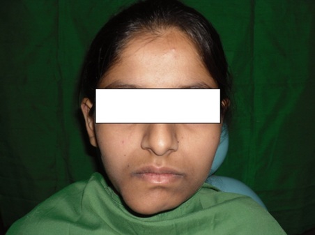
Extraoral examination revealed a large diffused swelling on the chin causing facial asymmetry [Table/Fig-1] . The swelling extended anteroposteriorly from right corner of the mouth crossing midline till left corner of the mouth and superioinferiorly extending from the vermillion border of the lower lip upto lower border of mandible. The overlying skin was smooth and normal in colour. The swelling was non-tender, firm consistency, non- fluctuant, non-compressible and fixed to underlying structures. The right and left submental and submandibular lymph nodes were nonpalpable.
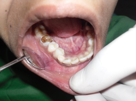
Intraoral examination revealed a single bony hard swelling measuring 4.5x3cm present in the anterior mandibular region obliterating the labial vestibule. Retained right deciduous mandibular canine was present and right permanent first molar was grossly carious [Table/Fig-2] Swelling extended anterioposteriorly from right deciduous mandibular canine to left permanent mandibular lateral incisor and superiorly from marginal gingiva of same teeth to vestibule inferiorly. Both buccal and lingual cortical plate expansion was observed. All present lower anteriors were vital on electric pulp testing. On palpation, the swelling was nontender, firm in consistency with well-defined borders.
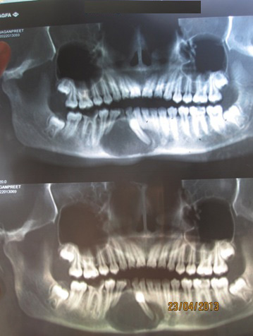
Orthopantamograph (OPG) revealed a well-defined unilocular radiolucency measuring about 4x3cm extending mesiodistally Dentistry Sectionfrom mesial aspect of right permanent mandibular first premolar, crossing the midline upto mesial aspect of left permanent mandibular canine & superioinferiorly extending from alveolar crest region w.r.t. right permanent mandibular incisors to lower border of mandible causing thinning of lower border of mandible. Impacted right permanent canine was embedded within the radiolucency. Root resorption was seen with all present mandibular anteriors [Table/Fig-3] .
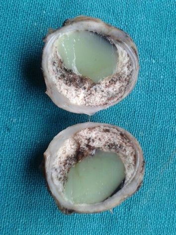
Surgical enucleation of the tumour was done under general anaesthesia and the resulting specimen was received for histopathological examination. The specimen was round in shape, brown colored, had variable consistency, measuring 4x3cm [Table/Fig-4] .The cut surface revealed a large cystic cavity filled with gelatinous greenish material. The wall was varying in thickness from 4mm to 12mm.
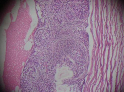
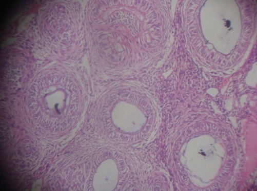
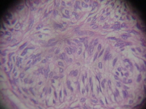
Microscopically, the tumour showed large cystic cavity [Table/Fig-5] . The cyst had an outer thick collagenous wall and an inner equally thick cellular wall. There were multisized solid nodules of cuboidal or columnar epithelial cells forming nests or rosette like structures. Between cell nests there was abundant hyaline eosinophilic material. Spindle shaped or polygonal, closely opposed epithelial cells with dark eosinophilic cytoplasm and round hyperchromatic nuclei fill in the spaces between epithelial nodules [Table/Fig-6] . Conspicuous within the cellular areas were structures of tubular or ductlike appearence. The duct like spaces were lined by single row of low columnar epithelial cells, the nuclei of which were polarised away from the luminal surface [Table/Fig-7] . The lumen contain variable amount of eosinophilic material. Calcification was frequently seen. The larger cyst cavity contain abundant eosinophilic colloid material.
The overall features were suggestive of “Adenomatoid odontogenic tumour”.
Discussion
Adenomatoid odontogenic tumour (AOT) is an uncommon, non invasive benign tumour that was first described by Steensland in 1905 as “epithelioma adamantinum”. Till date numerous terminologies, like adenoameloblastoma, ameloblastic adenomatoid tumour, adamantinoma, teratomatous odontoma, have been used to describe the entity currently known as adenomatoid odontogenic tumour. It was in 1971 when WHO adopted the term AOT which was introduced by Phillipsen and Brin in 1969 [1,2].
The 1992 WHO classification defines the AOT as “a tumour of odontogenic epithelium with duct like structures and with varying degree of inductive changes in the connective tissue. The tumour may be partly cystic and in some cases the solid lesion may be present as masses in wall of large cyst. It is generally believed that the lesion is not a neoplasm” [3].
AOT’s account for 2.2% to 7.1% of all odontogenic tumours. It has a wide age predilection i.e. between 3 and 82 years with maximum peak in the second decade of life. Females are affected more often than males, with a ratio of F: M = 1.9:1. Maxilla is the predominant site of occurrence and anterior maxilla is affected more often than the mandible by a factor of 2 [3]. Impacted, unerupted teeth mostly canines are associated with AOT in 60% of the cases [4].
The present case is of a 15-year-old female who had well defined anterior mandibular swelling with unerupted permanent right mandibular canine.
Clinically, AOT is a benign, non neoplastic lesion with a slow but progressive growth. It can occur in both intraosseous and peripheral forms. Radiographically, intrabony variants comprise of follicular and extrafollicular types. The follicular variant shows a well-defined unilocular radiolucency associated with the crown and often part of root of unerupted tooth, whereas extrafollicular type is not associated with unerupted tooth, here a unilocular radiolucency is found between, above or superimposed on the roots of erupted permanent teeth. Displacement of neighbouring teeth due to tumour expansion is much more common than root resorption. In peripheral variant there are hardly any radiographic changes to be noticed [5].
In the present case, the radiolucency was unilocular, well circumscribed involving the impacted canine in its periphery giving the picture of follicular variant.
Histopathologically, the tumour is made up of a multinodular proliferation of spindle, cuboidal and columnar cells in variety of patterns comprising of scattered duct like structures, eosinophilic material and calcifications in several forms. The duct like arrangement is lined by an eosinophilic rim of varying thickness called as hyaline ring. Anastomosing strands of basaloid epithelial cells may be arranged in plexiform, trabecular, cribriform or lattice like configuration. Cystic component can form a considerable part of the lesion. Some AOT contain varying amounts of dysplastic dentin, dentinoid and osteodentin. Irregular to round calcified bodies which may exhibit areas with concentric layered pattern may be seen in parenchymal or stromal zones. The supporting stroma is loose, hypocellular and fibrovascular that may show prominent vascular component [1].
The present case showed a large cystic cavity filled with eosinophilic coagulum, the lining of which showed characteristic features of adenomatoid odontogenic tumour.
Many histological variants have been described by various authors [6]. Marx & Stern have suggested the term adenomatoid odontogenic cyst (AOC) for such unilocular cystic lesion as is described here [7].
The origin of adenomatoid odontogenic tumour is controversial. Its occurrence in tooth bearing areas of jaws and close association with an impacted tooth in most of the cases favours odontogenic origin which can be through dental lamina, reduced enamel epithelium or their remnants. Some authors believe that they originate from odontogenic epithelium of dentigerous cyst [4,8]. According to this hypothesis, the lesion grows next to or nearby dental follicle leading to “envelopmental theory”. But many present as cystic lesions with only mural nodules of AOT [8].
Whether the origin of follicular variant occurs before or after cystic expansion has taken place is debateable. If it occurs after cystic expansion then this means origin might be from dentigerous cyst and if it occurs before the cystic expansion then the tumour tissue will fill the follicular space and AOT will present as solid tumour. But the question arises that if enough time is given to those AOTs originating from cyst, they may grow and fill the lumen completely [9].
Reports of AOT originating from an ameloblastoma, in the lining of unicystic ameloblastoma, in the wall of calcifying odontogenic cyst and in association with calcified epithelial odontogenic tumour have also been published. It has widely agreed that these are not hybrid lesions but may represent histological extremes in AOT [10].
Some authors are also of the view point that AOT represent a developmental outgrowth or hamartoma owing to limited size and lack of recurrence [11].
The differential diagnosis of AOT is crucial for surgical therapy. Differential diagnosis for intrabony variants of AOT are dentigerous cyst, residual cyst, radicular cyst, lateral periodontal cyst, ameloblastoma, CEOT, ameloblastic fibroma, central giant cell granuloma [12,13].
Dentigerous cyst and follicular variant of AOT may mimick similar radiographic features but both show their respective characteristic histological features. Dentigerous cyst is usually composed of a thin connective tissue wall with a thin layer of stratified squamous epithelium lining the lumen. Further in majority of cases, the unilocular radiolucency in dentigerous cyst is attached at CEJ while in AOT the unilocular radiolucency is attached beyond CEJ.
Again residual cyst, radicular cyst, lateral periodontal cyst share common radiographic features with extrafollicular AOT due to their location but histologically show a different picture [13].
Ameloblastic fibroma is a true mixed odontogenic lesion where both epithelial and mesenchymal tissue are neoplastic. CEOT is composed of tumourous polyhedral epithelial cells which have well outlined cell border with a finely granular eosinophilic cytoplasm and intercellular bridges are often prominent. Central giant cell granuloma is made up of a loose fibrillar connective tissue stroma with many interspersed proliferating fibroblasts and multinucleated giant cells [14].
Majority of the tumours of this variety have been treated by conservative surgical excision.
The case we have reported here is a rare follicular type of adenomatoid odontogenic tumour occurring in the mandible crossing the midline, radiographically mimicking dentigerous cyst and histologically showing a cystic variant laying the emphasis that AOT should be considered in the differential diagnosis of radiolucent swellings of the lower jaw.
Extra-oral swelling causing facial asymmetry
Intraoral photograph showing right mandibular swelling
OPG showing well defined unilocular radiolucency enveloping 43
Cut section of the specimen
Microscopic picture showing thick collagenous wall on the outermost followed by thick epithelial lining and cystic lumen filled with eosinophilic material. (H&E stain 10X)
Nests and duct like structures lined by low columnar epithelial cells. (H&E stain 10X)
Odontogenic epithelium arranged in form of whorls with rosette like pattern. (H & E stain 40X)
[1]. PA Reichart, HP Philipsen, Odontogenic tumours and allied lesions. 2004 1st EditionLondonQuintessence Publishing Co:69-77. [Google Scholar]
[2]. WG Shafer, MK Hine, BM Levy, Cysts and tumours of odontogenic origin.In: Rajendran R, Sivapathasundharam B, editors. Shafer’s Textbook of Oral Pathology. 2006 5th EditionNew DelhiElsevier:416-20. [Google Scholar]
[3]. IRH Kramer, JJ Pindborg, M Shear, EH Kass, WHO International Histological Classification of Tumours. Typing of Odontogenic Tumours. 1992 2nd EditionBerlinSpringer [Google Scholar]
[4]. KGJ Handschel, AR Depprich, CA Zimmermann, S Braunstein, RN Kubler, Adenomatoid odontogenic tumour of the mandible: review of the literature and report of a rare case.Head & Face Medicine. 2005 1:1-5. [Google Scholar]
[5]. B Shreedhar, I Ali, A Agarwal, S Alam, A huge Adenomatoid Odontogenic Tumour of Maxilla - Case Reports in Medicine. 2012 Hindawi Publishing Corporation. [Google Scholar]
[6]. K Vasudevan, S Kumar, Vijayasamundeeswari S Vigneswari, Adenomatoid odontogenic tumour, an uncommon tumour.Contemp Clin Dent. 2012 3(2):245-7. [Google Scholar]
[7]. RE Marx, D Stern, Oral and Maxillofacial Pathology. 2003 Illinois: Quintessence Publishing Co, Inc:877 [Google Scholar]
[8]. N Yilmaz, A Acikgoz, N Celebi, AZ Zengin, O Gunhan, Extrafollicular adenomatoid odontogenic tumour of the mandible: report of a case. 2009 3(1)European Journal of Dental Education.:71-4. [Google Scholar]
[9]. RE Friedrich, J Zustin, AH Scheuer, Adenomatoid Odontogenic Tumour of the Mandible.Anti Cancer Research. 2010 30:1787-92. [Google Scholar]
[10]. MS Archana, A Bagewadi, V Keluskar, A rare symptomatic swelling in the mandible. Int J Dent Reports. 2012 2(4):27-32. [Google Scholar]
[11]. V Jivan, M Altini, S Meer, F Mahomed, Adenomatoid Odontogenic Tumour (AOT) originating in a Unicystic Ameloblastoma: A case report.Head and Neck Pathol. 2007 1:146-9. [Google Scholar]
[12]. GT Shrihari, Adenomatoid Odontogenic Tumour with rare clinical and radiological presentation: A case report.Pacific Journal of Medical Sciences. 2011 9(1):3-8. [Google Scholar]
[13]. BW Neville, DD Damm, CM Allen, JE Bouquot, Oral and Maxillofacial Pathology. 2002 2nd EditionPhiladelphiaWB Saunders Co.:611-6. [Google Scholar]
[14]. JP Sapp, LR Eversole, GP Wysocki, Contemporary Oral and Maxillofacial Pathology. 1997 St Louis:Mosby:131-2. [Google Scholar]